
Advancement in Dairy Science Research(ADSR)


Research Article - (2023) Volume 1, Issue 1
Production of Polyclonal Antibodies (IgY) Against Foot-and-Mouth Disease (Serotype: O, A and Sat-2) in Ethiopia by Using Layer Hens
Received Date: Jan 18, 2023 / Accepted Date: Jan 25, 2023 / Published Date: Feb 03, 2023
Copyright: ©©2023 Ephrem Shimelis, et al. This is an open-access article distributed under the terms of the Creative Commons Attribution License, which permits unrestricted use, distribution, and reproduction in any medium, provided the original author and source are credited.
Citation: Ephrem, S., Tesfaye, S., Tadesse, F., Bilata, T., Paeshuyse, J. (2023). Production of Polyclonal Antibodies (IgY) Against Foot-and-Mouth Disease (Serotype: O, A and Sat-2) in Ethiopia by Using Layer Hens. Adv Dairy Sci Res, 1(1), 23-29.
Abstract
The use of chickens as experimental animals in laboratory for polyclonal antibody (IgY) production has increased in recent times, as its specificities to mammalian antigen, animal welfare (non-invasively extracted from egg yolk), high antibody per ml, very low cost and short turnover of production. The present study was aimed to produce IgY polyclonal antibody against the three Foot-and-Mouth disease viruses’ serotypes (O, A and SAT2) in Ethiopia by immunization of laying hens with the locally inactivated the same serotypes of Foot-and-Mouth disease viruses. At day 14, 21 and 28 post immunization, the eggs and serum were collected and the IgY polyclonal antibodies were extracted from the chicken egg yolk by using Polyethylene Glycol (PEG 6000g) precipitation method. The presence of the IgY polyclonal antibodies against the selected Foot-and- Mouth disease viruses’ serotypes were detected by the Prio- CHECK® FMDV NSP-ELISA kits), in which the mean percent of inhibition of the IgY (10 ± 8.9, 8.6 ± 6.9 and 13.46 ± 9.8, respectively at day 14, 21 and 28 post-immunization) and of the serum (0, 4.7 ± 3 and 6.7 ± 5.9, respectively at day 14, 21 and 28 post-immunization). In-vitro challenging were done by virus neutralization test of the three serotypes of FMDV (O, A and SAT-2) and the neutralization titers were measured with (TCID log10) of IgY (1.83, 2.1, and 2.07) and of serum (0, 1.9 and 1.84), respectively at day 14, 21 and 28 post immunization. The egg yolk IgY and serum antibody were significant difference only when compared by the FMDV serotypes (P-Value < 0.05) The study finding indicated that chicken egg yolk antibody (IgY) production could be the best alternative to the other mam- malian laboratory animals as egg yolk-based antibody production reduce the stress and bleeding during the blood collection, high antibody titer and low cost, easy to manage and handle chickens.
Keywords
Foot-and-Mouth Disease Virus, Antibody, Egg Yolk, IgY, Serum and Immune
Introduction
The foot-and-mouth disease virus (FMDV) is belonging under the family of Picornaviridae and genus of Aphthovirus also there are seven main serotypes of FMDV (A, O, C, Asia1, South African Territories (SAT) 1, SAT2, and SAT3), which resulted from the high mutation rate of FMDV with its rapid proliferation and extensive population and there is large number of subtypes evolved within each serotype [1-3]. In FMDV-endemic regions the major economic loss of the disease is associated with re- duced livestock productivity, regular mass vaccination and trade restrictions on animals and livestock products [4].
Four serotypes (A, O, SAT 1, and SAT 2) have been reported over the past ten years in Ethiopia. Sero-type O and A are the dominant serotypes out of for serotype reported in Ethiopia ; while serotype C was lastly reported in 1983 [5, 6]. In Ethio- pia, factors such as susceptibility of animals, wild and domestic animals indirect contact on the grazing pastures and watering points and uncontrollable animal movement within the country and transboundary contribute to the frequent occurrence of FMD outbreaks and making its controls and prevention difficult [7].
The properties of antibodies to recognize small specific struc- tures on the antigen to which it induced by immune cell of the animals makes it an important tool in laboratory for various di- agnosis and vaccination applications [3, 8, 9]. To produce the antibodies (polyclonal or monoclonal), the use of chickens as a laboratory animal has been increased in recent times because it has a major advantage including: antibody can be harvested from the egg yolk instead of serum reducing the stress and repeat bleeding of the animals, the antibody productivity of an egg-lay- ing hen is much greater than that of a similar sized mammal , specificities to mammalian antigen, animal welfare since the Ab are non-invasively extracted from egg yolk, high ab per ml and very cost-effective [10-12].
Using chicken as a host for generation of polyclonal antibod- ies (IgY) has valuable diagnostic and vaccine production ad- vantages over existing mammalian systems, however, it is an underused resource in Ethiopia as the most of the previous an- tibody production in Ethiopia uses laboratory animals such: as mice, rabbit, guinea pigs and other mammalian. Therefore; the current study was designed to produce antibodies of selected FMDV serotypes (A, O and SAT-2) in the way that ensure ani- mal welfare only by immunizing layer hens and provide a spe- cific IgY used for diagnosis and vaccination of selected FMDV serotypes from egg yolk.
Materials & Methods
Study Design
An experimental study design was conducted on polyclonal anti- body production (IgY) trials by using Egg-Yolk of a layers’ hens immunized with three FMDV serotypes (O, A and SAT-2) from January, 2020 up to August, 2020 G.C in Ethiopia. The three field isolates of the FMD viruses (01940/NAHDIC/O, 01940/ NAHDIC/A and 01940/NAHDIC/SAT-2) were provided by Sebeta National Animal Health Diagnosis and investigation cen- ter (NAHDIC) in Ethiopia. The selected serotypes were propa- gated and adapted on BHK-21. Then, titration of each serotype was determined according to the Spearmen- Karber formula. Purposively, eight 24 weeks age’ layer hens were assigned to each serotype and immunized by 50:50 FMD virus (serotype A, O and SAT 2) to oil adjuvants with boosting at day 14 post-im- munization. The immunized chickens were managed, handled and followed for clinical change in wire mesh cage separately. After two weeks of boosting the hens, the serums and eggs were collected at day 14, 21 and 28 post-immunizations. The bloods were collected in 5ml syringes and allowing to clot by leaving it undisturbed at room temperature for 30 minutes and remove the remained clot by centrifuging at 1,000x g for 10 minutes in a refrigerated centrifuge. Immediately, the serum transferred to the 1.5 ml cry vial tube and the collected eggs were coded by the code of chicken laid it and stored -20 "C until used. The IgY was extracted and purified with Polyethylene glycol (PEG-600mg) precipitation method from the egg yolk. The serum and extract- ed egg-yolk IgY were pooled into four samples for each sero- type. The antibody detection was conducted by Prio-CHECK® FMDV non-structural protein kit (© prionics AG, version 1.0_e, product No.: 7810770) and invitro challenging of samples show- ing inhibition Percent’s (%) were conducted by Viral neutraliza- tion tests (VNT).
Preparation and Adaptation of Fmdv in Bhk-21 Cell Culture
Foot and mouth disease virus serotypes (A, O and SAT-2) with high CPE were selected for this study from sebeta National an- imal diagnosis and investigation center of Ethiopia. And trans- ferred to 34th passage of BHK-21 (AU/PANVAC) monolayer cells with 90 percent cell confluence in three 25cm2 polystyrene TPP® tissue culture flasks (Sigma-Aldrich, Europe) for adapta- tion and harvesting to enough amounts for the study.
Fmdv Purification and Determination of Its Infectivity
The cell culture supernatant was harvested by repeated freezing for 20 minutes and thawing for 3 times and the cell suspension was clarified by centrifugation at 5,000 rpm for 15 min at 4°C [13, 14]. Then, according to, Fawzy, et al., the titer of the three FMDV serotypes were determined and expressed in log10TCID50 [15]. Virus titers in the samples were determined by inocula- tion of cell culture highly susceptible to FMDV (BHK-21) (AU/ PANVAC) in 96 well flat bottom cell culture plates. The titers were expressed as 50% tissue culture infectious doses (TCID50) per 100 µL calculated with the Spearman-Kärber-formula [16].
Immunization of Chickens
After 50% tissue culture infectious doses (TCID50) were cal- culated, the selected FMDV serotype was emulsified with equal volume of oil adjuvant. The hens were immunized two times (at initial day and at day 14 post immunization) [17].
Table 1: Determination of Cytopathic Forming Unit Particle of Selected Fmdv Serotypes (A, O and Sat-2).
|
IDNO |
FMDV Serotype |
Total Dilution Preparation |
Injection Per Chicken |
||
|
TCID50/ml |
CFU/ml |
TCID50/ml |
CFU/ml |
||
|
01940/NAHDIC |
O |
5.1×10⁴ TCID50/ ml |
3.57×10⁴CPU/ml |
2.55×10³ TCID50/ ml |
1.9 ×10³PFU/ml |
|
8450/NAHDIC |
A |
3.7×10⁴ TCID50/ ml |
2.59×10⁴CPU/ml |
1.85×10³TCID50/ ml |
1.3×10³PFU/ml |
|
31056/NAHDIC |
SAT-2 |
2.5×10⁵ TCID50/ ml |
1.75×10⁵CPU/ml |
1.25×10⁴ TCID50/ ml |
8.75 ×10³PFU/ml |
IgY Extraction and Purification from Hen’s Egg-Yolk
The method for IgY purification was adapted from Pauly. et al., [12]. The Extraction of chicken IgY antibodies from immunized chicken egg yolk was carried out by using Polyethylene Glycol (PEG 6000) precipitation method. Briefly, once the eggs were cleaned with 75% alcohol, the egg shell was cracked carefully and the yolk was transferred to a "yolk spoon" in order to re- move as much egg white as possible. The egg yolk was trans- ferred to and rolled on a filter paper to remove the remaining egg white. The egg yolk skin membrane was cut with lancet and by using the tea spoon the yolks was poured into a 50 ml tube to measure its volume. Twice the egg yolk volume of PBS (PH = 7.4) was added to the yolk tube and mixed by vertexing. Then 3.5% of PEG 600g of the total volume was added to the yolk tube, vortexed and rolled for 10 min by a rolling mixer before the tubes were centrifuged at 10,000 rpm for 20 min at 4oC. The supernatant was then poured through a folded filter paper and transferred to another tube before 8.5% PEG-6000g was added in relative to the new volume and then vortexed and centrifuged at 10,000 rpm for 20 min at 4oC. The supernatant was discarded and the pellet was dissolved in 1 ml PBS using a glass stick, mixed by vortexing before 9 ml of PBS was added to the tube to reach a final volume of 10 ml. The solutions were then mixed with 12% PEG 6000 and the tube centrifuged at 10,000 rpm for 20 min at 4oC. The supernatants were discarded and the pellet was dissolved in 800 µl PBS using a glass stick vortexed and the extracts was transferred to 2ml Eppendorf tubes and stored at -20oC until used.
Detection of FMDV-Specific Antibody
Antibodies against the capsid proteins are produced in both in- fected and vaccinated animals [18]. Detection and evaluation of antibody titer against FMDV in hen sera/Egg yolk and se- rum samples were carried out by the Prio- CHECK® FMDV NSP-ELISA kit (© prionics AG, version 1.0_e, product No.: 7810770). In order to enhance the antibody concentration serum and egg yolk, the sample of serum and egg yolk collected from 8 hens for each three FMDV serotypes were pooled in to four for test. Briefly, according to Prio-CHECK ® FMDV NS kit (© prionics AG, version 1.0_e, product No.: 7810770), first 80 µl of ELISA buffer was dispensed to all wells and 20 µl of negative control to (A1 and B1 wells), 20 µl of weak positive control (C1 and D1 wells), 20µl of positive control (E1 and F1 wells) and to the remaining wells, 20µl of Serum and egg yolk IgY were dispensed and incubated for 18 hrs. At room temperature (22 ±3 ºC). then, after six times washing with 200µl washing buffer, 100µl of diluted conjugate were dispensed to all wells and incubated for 1hr at 22 ±3 ºC, again after washing with 200 µl washing buffer, 100 µL of chromogen (TMB) substrate was dispensed to all wells and incubated for 20 minutes at 22 ±3 ºC, finally the optical density was measured at 450nm after adding stop solution and incubating for 20 minutes at 22 ±3 ºC. After the calculation of the mean OD-value of wells A1 and B1 (neg- ative control = OD450 max), the percentage of inhibition (PI) of the control and test antibody (egg yolk IgY and serum) was calculated.
Virus Neutralization Test
Virus neutralization test is to detect a specific reaction between antigen and antibody or neutralization of antigen to inhibit the viral infectivity. The 19 samples with titers ≥1% of inhibition in the Prio-CHECK® FMDV NS kit for antibodies against O, A and SAT- 2 were subjected to VNT test according to the protocol adapted from the procedure described in the OIE terrestrial man- ual [19]. Two-fold serially dilution of sera and IgY, starting from original dilution up to 10-10 in modified Eagle’s medium, from each dilution, 50μl serum and IgY samples were added in each well (duplicated). Next, 50μl containing 100 TCID50 FMDV (previously titrated) were added to each well and incubated at 37°C in CO2 incubator for 1hr to allow neutralization. Then,50μl of 106 cells/ml BHK-21 cells suspension was added to each well and incubated at 37°C of 5% CO2 incubator for 48 hrs.’ and ex- amined for the presence of CPE twice daily. After its incubation of 48 hrs. The presence of CPE was examined and neutralization titers in log10 were calculated according to Spearman-Kaerber based on FMDV induced cytopathogenic effect (CPE).
Data Analysis and Management
Data obtained from laboratory tests; virus titration, virus neu- tralization, and ELISA tests were entered into Microsoft Excel before being analyzed. Descriptive statistics was carried out us- ing Microsoft Excel spreadsheet to summarize the results. The log 10 of VNT were Calculated by Spearman-kärber method on Microsoft excel [16]. Analysis of variance (ANOVA) and unpaired t. test were performed to assess the significant differ- ence between IgY and serum induced by the three serotypes of FMDV between antiviral (IgY and serum) and between three post infection days by using R-Studio package.
Results
Determination of Antibody Response in Chickens
To characterize the polyclonal antibody–binding sites, their reactivity against the homologous FMDV (serotype: A, O and SAT-2) were examined using Prio-CHECK® FMDV NS-kit. Among the sixty pooled samples collected from the chicken im- munized with the locally inactivated FMDV; only 19 samples (32%) were shown the antibody against the selected serotype of FMDV (Figure 1). however, it was below the recommended Prio-CHECK® FMDV NS-kit standardized percentage of in- hibition; that indicating their binding sites were conformation- al. At day 14 of post immunization, the egg yolk IgY antibody mean inhibition percentage (PI) was (10 ± 8.9), while, the serum antibody inhibition percentage was not detectable and there was statistical significancy between egg yolk IgY and serum (P-Val- ue < 0.04) as described by paired t-test. The mean inhibition percentage of serotype A were highest as compared with O and SAT-2 serotypes (16.1, 10.24 and 3.15, respectively) with signif- icant difference between the group immunized by three different serotypes (P-value = 0.03).

Figure 1: The Egg Yolk IgY and Serum Antibody Mean Inhibition Percentage at Different Days Post Immunization (n = 19): Com- paration of IgY and Serum by Day Post Immunization (a) and Comparation of IgY and Serum by Serotypes of FMD Virus (b).
Virus Neutralization
During day 14 post immunization the mean of egg Yolk IgY and serum titer against the FMDV (serotype; O, A and SAT-2) was (1.83 ± 0.2 and 0, respectively) with statistical relationship of (P-Value = 0.03). the neutralizing titer of the IgY and the serum was evaluated in the association between the post immunization day, between the serotype and between antibodies (Serum and IgY) by using analysis of variance (ANOVA) and paired t-test analysis. The mean antibody titer induced by serotype A was the highest, while the SAT-2 was induced low (A = 2.25, O =2.07 and SAT-2 = 1.59, for FMDV serotype
A, O and SAT-2 respectively) It’s statically expressed in signif- icant of P-value. Statistically only the relationship between the serotypes had the significant impacts on the neutralizing anti- body titer against the three serotypes of FMDV (P-Value = 0.03).
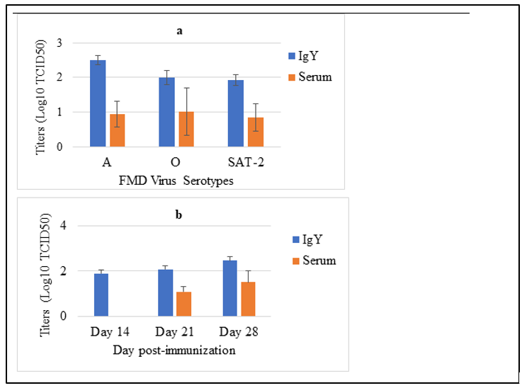
Figure 2: In Vitro Challenging of Produced Antibody with the Original Viruses (n = 19): Comparation of IgY and Serum by Day Post Immunization (a) and Comparation of IgY and Serum by Serotypes of FMD Virus (b).
Discussion
The FMDV antibody plays an important role in current specific diagnosis and vaccination of FMDV. These antibodies can be produced during primary infection or vaccination by the host animals and play a significant role in neutralizing the FMDV infection during post exposure [20]. The objective of the current study was to produce Foot-and-mouth disease (Serotype: O, A and SAT-2) polyclonal antibodies (IgY) by using layer hens in Ethiopia. However, the experimental chickens were immunized with the live virus, it did not show the sign usually observed in suspectable animals (fever, vesicle, loss in egg production and death of immunized chickens).
The antibodies detection for both the serum and egg yolk an- tibodies were carried out by Prio-CHECK FMDV NS ELISA kit. The Prio-CHECK® FMDV NS kit detection results were below the standard 0f the cut-off values (< 50%) of percentage of inhibition described on Prio-CHECK kits (PI > 50 were inter- preted as positive). At day 14, 21 and 28 post-immunizations of the chickens; the mean percentage of inhibition of Egg yolk IgY were ((10 ± 8.9, 8.6 ± 6.9 and 13.46 ± 9.8, respectively) with only a significant difference significancy between egg yolk IgY and serum (P-Value < 0.04) at day 14 of post-immunization and statistically difference three FMDV serotypes (P < 0.05). The obtained EISA results were agreed with the results of Ferris et al., and Bruderer et al. [21, 22]. According to, Ahmed Kamal et al, positive sera may not always give positive reaction with Prio- CHECK ELISA [23]. The heterogeneity in the 3ABC response is likely to reflect differences in the degree of the viral replica- tion and immunological predisposition to elicit anti-3ABC anti- bodies [22]. The time post immunization and FMDV serotypes had the effects on the antibody response by the chickens against the FMDV [24]. Aspects of the host cell including mutations, re- duced expression of viral receptors, obstacles to viral uptake af- ter receptor and changes in immune response including: cellular immunity, humoral immunity and cytokine response [25]. The majority of peptide molecules used for immunization are likely to present conformations that cannot be adopted by the cognate region in the virus, and it is thus not surprising that many of the antibodies that are induced will be unable to recognize the viral epitopes [21].
Despite to the Prio-CHECK® FMDV NS kit, when measured by standard neutralization test (100 TCID50 virus–serum di- lutions), as the day post immunization of chickens the FMDV immunized chicken had relatively neutralizing antibody titers ranging from 1.83 to 2.164 log10, that agreed the interpretation criteria of Pirbright laboratory manual (log10 titration > 0.3) and the result of Rodriguez et al., and Eschbaumer et al., on animals vaccinated with the commercial oil-based FMD viruses vaccine in which they had proven that protection did not depend on the reactivity in the peptide ELISA, rather the protection was asso- ciated with the presence of neutralizing antibody titers in vacci- nated animals [26, 27].
Also, the mean antibody titer in log10 of the antibody response of the chicken against the FMDV were compared in the study and the response of the chicken to the serotype O were high as compared its mean antibody titer against serotype (A, O and SAT-2), with significant difference between the three serotype (P-Value = 0.03) and agreed with the result of Daoud et al., (2013). As the mean antibody titer of egg yolk and serum show, the mean antibody titers of the IgY were exceed that of the se- rum antibody, however, its difference were not statistically sig- nificant (P-Value >0.05). The obtained results agreed with the result of Ibrahim et al., [9].
In general, in the current study there were poor correlation be- tween the ELISA antibody detection results and virus neutraliza- tion results of both egg yolk antibody and serum antibody. This difference in the two tests indicate that the epitopes of original antigen used for immunization of chicken were mutated during its adaptation to the chicken cells. This mutation can cause change the conformation of epitopes structure which impaired the antigen-antibody interaction during the ELISA tests, howev- er for antibody titers requires only the memory of the originally exposed antigen and different strains of FMDV vary consider- ably in pathogenicity, invasiveness, virulence spreading power and prevalence of occult, or in apparent infection [28]. The dif- ference between the VNT and Prio-CHECK® FMDV NS Kit perceived antigenic evolution of the virus in which the original virus for immunization of chicken may mutated in order to adapt to chicken cells, however, VNT assays implies the heterologous (cross-protection). According to Yang, poor correlation between ELISA and VNT might be of individual variation after FMDV inoculation, in which the ELISA detect only specific epitope of original antigen with its confirmation, however, the neutralizing antibody need only memory of original antigen. This Finding implied that the blocking ELISA results didn’t indicate the anti- body production which can be detected in post–exposure to the antigen by virus neutralization test. A possible explanation for the poor binding can be mutations that changed the conforma- tional structure of the monoclonal antibody binding sites and af- fecting the’ binding ability of antibody [29].
Conclusion and Recommendations
In the current study, chickens were experimental infected with three serotypes of FMD viruses (O, A and SAT-2) for production of FMDV polyclonal antibody. After infection on day 14, 21 and 28 the sample of egg and serum were collected. Both the se- rum and the egg yolk precipitate were detected and evaluated for their antibody’s titer by Prio-CHECK FMDV NS kit and virus neutralization tests respectively. In 35% of collected samples the antibody against the FMDV were detected with low percentage of inhibition (<50%), but the virus neutralization test results of those sample with low antibodies titer showed value comparable to the OIE standard of antibody titers against FMDV. The mean percentage of inhibition and antibody titer against FMDV in egg yolk (IgY) exceed the amount of antibody titer in the serum. This antibody can be used for any immunological tests applied for FMD diagnosis. Moreover, the study finding indicated that chicken egg yolk antibody (IgY) production could be the best alternative to the other mammalian laboratory animals as egg yolk-based antibody production reduce the stress and bleeding during the blood collection, high antibody titer and low cost, easy to manage and handle chickens [30, 31].
Based on the finding of this study, the following recommenda- tions are forwarded:
• The research institute and diagnostic laboratory should use the chicken as laboratory animal models for antibody pro- duction.
• Before immunization of chicken with virus to which it is not susceptible, in vitro adaptation of virus to the chicken origin cell should be done. ï?? Repeated immunization and monitoring for longtime duration should be done.
• Further research should be done in order to produce a highly purified antibody used for immunological test for FMDV.
• Sequencing analysis of antigen- antibody determinant epitopes should be done in order to validate the discrepancy be- tween antibody ELISA and VNT test result.
• For protein (antibody) quantification, SDS-PAGE and western blotting technique should be applied.
• This study should continue in such a way that the an- tibody produced can be used in house serotyping EISA test as a replacement of commercially available EISA kits.
Acknowledgements
I would like to express my heart-felt gratitude to National Ani- mal Health Diagnostic and Investigation Center, Sebeta and Ad- dis Ababa University. Lastly, I want to acknowledge, JOINT projects, Global Minds, International Master Programs (ICP), for provided me Short-term training related to my MSc. Thesis for six weeks at KU-Leuven and Sciansiano FMDV Diagnosis laboratory.
References
- Aftosa, F. (2014). Foot and Mouth Disease Foot and Mouth Disease, OIE Manual, Center for food security and Public health. Lowa State University.
- Gao, Y., Sun, S. Q., & Guo, H. C. (2016). Biological func- tion of Foot-and-mouth disease virus non-structural proteins and non-coding elements. Virology journal, 13(1), 1-17.
- Ateya, L. A., Ahmed, S. A., Ashraf, K. K., & Abdel-Hady,H. A. (2017). Evaluation of vaccination with local and im- portedvaccine against foot and mouth disease virus in Ka- lubeya governorate. J. Virol. Sci, 1(1), 20-26.
- Niedbalski, W. I. E. S. L. A. W., & Haas, B. E. R. N. D.(2003). Differentiation of infection from vaccination by detection of antibodies to the non-structural protein 3ABC of foot-and-mouth disease virus. Bulletin of the Veterinary Institute in PuÃÂ??awy, 47(1).
- Endris, A. (2018). Review on current status of FMD in Ethiopia. Spatiotemporal distribution, frequency and prev- alence, 10 (7).
- Sulayeman, M., Dawo, F., Mammo, B., Gizaw, D., & Shegu, D. (2018). Isolation, molecular characterization and sero-prevalence study of foot-and-mouth disease virus circulating in central Ethiopia. BMC veterinary research, 14(1), 1-10.
- Admassu, B., Getnet, K., Shite, A., & Mohammed, S. (2015). Review on foot and mouth disease: Distribution and economic significance.
- Chalghoumi, R., Beckers, Y., Portetelle, D., & Théwis, A. (2009). Hen egg yolk antibodies (IgY), production and use for passive immunization against bacterial enteric infections in chicken: a review. Biotechnologie, Agronomie, Société et Environnement, 13(3).
- Ibrahim, E. E., El Helw, H. A., Shafik, N. G., & Mekhail, M.A. (2017). Conjugation Of Foot And Mouth Disease IgY In Chicken Egg Yolk With Horse Radish Peroxidase For Typ- ing Of Foot And Mouth Disease Virus. Journal of Applied Veterinary Sciences, 2(1), 35-42.
- Munhoz, L. S., Vargas, G. D. Á., Fischer, G., Lima, M. D., Esteves, P. A., & Hübner, S. D. O. (2014). Avian IgY anti- bodies: characteristics and applications in immunodiagnos- tic. Ciência Rural, 44, 153-160.
- Rajeswari, S., Choraria, A., Antonysamy, M., & Zhang, X.Y. (2018). Applications of Chicken Egg Yolk Antibodies (Igy) in Healthcare-A Review. Biomedical Journal of Sci- entific & Technical Research, 2(1), 2161-2163.
- Pauly, D., Chacana, P. A., Calzado, E. G., Brembs, B., & Schade, R. (2011). IgY technology: extraction of chicken antibodies from egg yolk by polyethylene glycol (PEG) pre- cipitation. JoVE (Journal of Visualized Experiments), (51), e3084.
- Hassan, A. I. (2016). Effect of different culture systems on the production of foot and mouth disease trivalent vaccine. Veterinary world, 9(1), 32.
- Mahmud, M. S., Islam, E., Giasuddin, M., Samad, M. A., Karim, M. R., & Ali, M. Z. (2018). Biological Assay of Foot and Mouth Disease Virus (FMDV) Serotypes for Titrating BLRI Developed Trivalent FMD Vaccines Seed. Immunol- ogy and Infectious Diseases, 6(2), 23-26.
- Abu-Elnaga, H., Fawzy, H., Farouk, E., Ibrahim, E., Gam- il, M., & Zidan, S. (2015). Correlation between foot-and- mouth disease virus antigenic mass, titer and immune re- sponse in vaccinated sheep. Benha Veterinary Medical Journal, 28(2), 12-19.
- Dill, V., Hoffmann, B., Zimmer, A., Beer, M., & Eschbaum- er, M. (2017). Adaption of FMDV Asia-1 to suspension cul- ture: cell resistance is overcome by virus capsid alterations. Viruses, 9(8), 231.
- Veerasami, M., Singanallur, N. B., Thirumeni, N., Rana, S.K., Shanmugham, R., Ponsekaran, S., ... & Villuppanoor,S. A. (2008). Serotyping of foot-and-mouth disease virus by antigen capture-ELISA using monoclonal antibodies and chicken IgY. New Microbiol, 31(4), 549-554.
- FAO/IAEA. (2007). The Use of Non-Structural Proteins of Foot and Mouth Disease Virus, VIENNA, TECDOC-1546
- OIE. (2012). Foot and mouth disease, manual of diagnostic tests and vaccines for terrestrial animals (terrestrial manu- al).
- Reeve, R., Borley, D. W., Maree, F. F., Upadhyaya, S., Lukhwareni, A., Esterhuysen, J. J.. & Mahapatra, M. (2016). Tracking the antigenic evolution of foot-and-mouth disease virus. Plos one, 11(7), e0159360.
- Ferris, N. P., Bulut, A. N., Rendle, T., Davidson, F., & Mackay, D. K. J. (2000). A solid-phase competition ELISA for measuring antibody to foot-and-mouth disease virus.
- Bruderer, U., Swam, H., Haas, B., Visser, N., Brocchi, E., Grazioli, S., ... & Anderson, J. (2004). Differentiating in- fection from vaccination in foot-and-mouth-disease: evalu- ation of an ELISA based on recombinant 3ABC. Veterinary microbiology, 101(3), 187-197.
- Amro, W. A., Al-Qaisi, W., & Al-Razem, F. (2018). Produc- tion and purification of IgY antibodies from chicken egg yolk. Journal of Genetic Engineering and Biotechnology, 16(1), 99-103.
Copyright: ©2023 Ephrem Shimelis, et al. This is an open-access article distributed under the terms of the Creative Commons Attribution License, which permits unrestricted use, distribution, and reproduction in any medium, provided the original author and source are credited.
- Han, L., Xin, X., Wang, H., Li, J., Hao, Y., Wang, M., &Shen, C. (2018). Cellular response to persistent foot-and- mouth disease virus infection is linked to specific types of alterations in the host cell transcriptome. Scientific reports, 8(1), 1-13.
- Rodriguez, L. L., Barrera, J., Kramer, E., Lubroth, J., Brown, F., & Golde, W. T. (2003). A synthetic peptide con- taining the consensus sequence of the G–H loop region of foot-and-mouth disease virus type-O VP1 and a promis- cuous T-helper epitope induces peptide-specific antibodies but fails to protect cattle against viral challenge. Vaccine, 21(25-26), 3751-3756.
- Eschbaumer, M., Stenfeldt, C., Rekant, S. I., Pacheco, J. M., Hartwig, E. J., Smoliga, G. R., ... & Arzt, J. (2016).Systemic immune response and virus persistence after foot- and-mouth disease virus infection of naive cattle and cattle vaccinated with a homologous adenovirus-vectored vac- cine. BMC veterinary research, 12(1), 1-18.
- Vallce, U. (1961).Virus of Foot-and-Mouth Disease.
- Yang, M., Xu, W., Bittner, H., Horsington, J., Vosloo, W., Goolia, M., ... & Nfon, C. (2016). Generation of mAbs to foot–and–mouth disease virus serotype A and application in a competitive ELISA for serodiagnosis. Virology journal, 13(1), 1-9.
- Kamal, S. A., & Hassan, R. A. E. R. (2017). Advanced vi- rological and clinicopathological studies on cattle suffering from foot and mouth disease virus. Journal of Immuniza- tion, 1(1), 33.
- Khan K. H., Himeno A., Kosugi S., Nakashima Y., Rafique A., Imamura A., Hatanaka T., Kato D. (2017): IgY-binding peptide screened from a random peptide library as a ligand for IgY purification. Pp, 790–797.