


Research Article - (2021) Volume 6, Issue 2
Prevalence of some foodborne parasitic affection in slaughtered animals in local Egyptian abattoir
2Food Hygiene and Control Department, Faculty of Veterinary Medicine, Benha University, Egypt
3Food Hygiene Department, Animal Health Research Institute, ARC, Egypt
Received Date: Oct 26, 2021 / Accepted Date: Oct 30, 2021 / Published Date: Nov 04, 2021
Copyright: ©Fahim, et al. This is an open-access article distributed under the terms of the Creative Commons Attribution License, which permits unrestricted use, distribution, and reproduction in any medium, provided the original author and source are credited.
Citation: Omar, M Abd Elaziz., Fatin, S Hassanin., Fahim, A Shaltout., Othman, A Mohamed. (2021). Prevalence of some foodborne parasitic affection in slaughtered animals in local Egyptian abattoir, Egypt. Adv Nutr Food Sci, 6(2), 37-42.
Abstract
Fascioliasis, cysticercosis and hydatidosis were aimed to be investigated in some slaughtered animals (cattle, buffalo and camel) in local Egyptian abattoir located in Cairo governorate. 10317, 763 and 290 cattle, buffalo and camel carcasses, respectively were examined in the period of 2017-2018. Results of PM inspection along the investigation period revealed detection of Fasciola in 0.58 and 1.4% of the examined cattle and buffalo carcasses, respectively, while was not detected in camel carcasses. Also, cysticercosis was detected in 0.47 and 0.69% of the examined cattle and camel carcasses, respectively; while was not detected in buffalo carcasses. In addition, hydatidosis was detected in lung and liver of 0.038 and 0.096% of cattle carcasses; 3.4 and 1.03% of camel carcasses, respectively, while was not detected in the examined buffalo samples. Annual values of financial losses because of the condemned affected parts summed total of 11712.5, 32940.0 and 2410 LE due to fascioliasis, cysticercosis and hydatidosis, respectively along the investigation period (2017 and 2018). Referring to the obtained results, records of 2017 appeared to be more infection prevalent than 2018 with more financial losses, moreover, fascioliasis was the most prominent affection in the present study. In addition, the critical veterinary inspection has a great role in protecting human-being to be infected with zoonotic meat-borne parasites. So, magnification and great support should be given to training veterinary inspectors in slaughter houses in Egypt.
Keywords
Fascioliasis, Cysticercosis, Hydatidosis, Cairo Abattoirs, Meat producing animals
Introduction
Daily human requirement of protein was estimated to be 45-55 grams according to age and daily labor requirements [1]. Red meats provide consumers with high quality, excellent source of a high essential amino acids, fatty acids, B-complex vitamins and many minerals supplement [2]. Parasitic infections of slaughtered animals have a significance public health importance and cause great economic losses, that reflects the rearing environment. The most important parasites in meat inspection are those which are of zoonotic importance to human by consumption of under cooked meats of affected animals, while other parasites which are not transmissible to man may render the carcasses and organs repug¬nant and unmarketable [3]. Infection of human with such serious meat borne zoonotic parasites such as cysticercosis and/or hydati¬dosis may localized in muscle tissues and organs, causing epilep¬sy, anaphylactic shock, halazone disease and CNS affections [4, 5]. Meat-borne parasites have been considered as a major public health hazard, especially in areas with poor sanitation and tradi¬tionally bad habits populations that consumed raw or under cooked meat results in negative impact on livelihood and spreading of in-fection [6]. Therefore, the present study was conducted to detect the percentage of parasitic infection in some freshly slaughtered animal species (cattle, buffalo and camel) along two years from 2017 to 2018 in a local abattoir in Cairo, Egypt.
Material and methods
Area of study: the study was carried out in a public abattoir locat¬ed in Cairo governorate, Egypt.
Study design: the study was conducted through active survey by daily routine work in the slaughter house along the period from 2017 to 2018.
Animals included in the study: 10317 cattle, 763 buffalo and 290 camel carcasses were examined in the scope of the current study during the full length of the study period.
Procedures applied for detection of parasitic lesions in slaugh-tered animals
The routine PM of apparently healthy animals was carried out ac-cording to the method recommended by
FSIS/USDA (2019) including head region, different lymph nodes,pluck, and different internal organs [7].
Percentage calculation
Prevalence calculation was conducted according to as follows [8]:
The percentage of the parasitic diseases
The percentage of different parasites among examined animals was estimated by dividing the number of infected animals for each disease (animals with condemned organs or carcasses) by the total number of slaughtered animals then multiplies by 100.
Estimation of economic loss due to parasitic infection
It was calculated by weighing of condemned carcasses and organs by digital balance and multiplies it by current price in market ac¬cording to Table (1).
Table (1): Price of meat and offal/kg during the investigation period.
|
|
2017 price (LE) / Kg |
2018 price (LE) / Kg |
|
Cattle/buffalo (meat and heart) |
75 |
95 |
|
Camel meat |
50 |
70 |
|
Offal (Liver, lung, tongue, rumen, pancreas, spleen, intestine) |
30 |
340 |
N.B. the price of meat and offal/kg according to the Egyptian General Authority for Veterinary Services
Economic losses = weight of condemned organ × current price per Egyptian pound.
Results
Referring to the obtained results in Table (2), out of 6313 and 5057 examined carcasses along the investigation period of 2017 and 2018, 1.28 and 1.21% were recorded to have different parasitic infections in 2017 and 2018, respectively.
Table (2): Price of meat and offal/kg during the investigation period.
|
Years |
Cattle |
Buffalo |
Camel |
|
Total |
|||||||
|
No1. |
No2. |
% |
No1. |
No2. |
% |
No1. |
No2. |
% |
No1. |
No2. |
% |
|
|
2017 |
5598 |
70 |
1.25 |
603 |
5 |
0.83 |
112 |
6 |
5.36 |
6313 |
81 |
1.28 |
|
2018 |
4719 |
49 |
1.03 |
160 |
6 |
3.7 |
178 |
6 |
3.37 |
5057 |
61 |
1.21 |
|
Total |
10317 |
119 |
1.15 |
763 |
11 |
1.4 |
290 |
12 |
4.14 |
11370 |
142 |
1.25 |
No1: Number of the examined carcasses
No2: Number of the positive Fasciola affected carcasses.
%: percentage of infected carcasses.
Referring to the prevalence of fascioliasis in the examined carcass-es as recorded in Table (3) and Figure (1). Overall, 2017 recorded higher infection rates with Fasciola than 2018 with percentage of 0.73% and 0.49% during 2017 and 2018, respectively. In addi- tion, buffalo carcasses revealed higher infection rate (1.4%) than cattle carcasses (0.58%) along the investigation period, while fas¬cioliasis was not recorded in any of the examined camel carcasses during the current study.
Table (3): Percentage of fascioliasis in the examined carcasses
|
Year |
Cattles |
Buffaloes |
Camel |
Total |
||||||||
|
|
No1. |
No2. |
% |
No1. |
No2. |
% |
No1. |
No2. |
% |
No1. |
No2. |
% |
|
2017 |
5598 |
41 |
0.7 |
603 |
5 |
0.8 |
112 |
0 |
0 |
6313 |
46 |
0.73 |
|
2018 |
4719 |
19 |
0.4 |
160 |
6 |
3.7 |
178 |
0 |
0 |
5057 |
25 |
0.49 |
|
Total |
10317 |
60 |
0.58 |
763 |
11 |
1.4 |
290 |
0 |
0 |
11370 |
71 |
0.62 |
No1: Number of the examined carcasses
No2: Number of the positive Fasciola affected carcasses.
%: prevalence of Fasciola.

Figure 1: Cattle liver show Fasciola affection and cirrhosis of bile ducts
Annual financial assessment of condemned liver due to fasciolosis in the examined carcasses was recorded in Table (4). Total losses were summed to be 11712.5 LE along the investigation period; where 2017 recorded higher losses (7132.5 LE) than 2018 (4580 LE).
Table (4): The annual financial assessment of condemned liver due to fasciolosis in the examined carcasses
|
Animals |
Year |
Infected |
Condemned Liver (Kg) |
Value in EGP |
|
Cattle |
2017 |
41 |
215.75 |
6472.5 |
|
2018 |
19 |
85.5 |
3420.0 |
|
|
Buffalo |
2017 |
5 |
22 |
660.0 |
|
2018 |
6 |
29 |
1160.0 |
|
|
Total |
2017 |
46 |
237.75 |
7132.5 |
|
2018 |
25 |
114.5 |
4580.0 |
|
|
Sum of 2017 & 2018 |
71 |
352.25 |
11712.5 |
|
N.B. the price of meat and offal/kg according to the Egyptian General Authority for Veterinary Services
Moreover, cysticercosis, as recorded in Table (5) and Figure (2), was detected in 0.47% and 0.69% of the examined cattle and camel car-casses during the whole period of investigation, respectively. Although cysticercosis was not detected in 2017, camel carcasses harbored the highest incidence of cysticercosis infection in comparison with cattle carcasses along the investigation period (2017 and 2018); while cysticercosis was not reported in any of the examined buffalo carcasses.
Table (5): Percentage of cysticercosis in the examined carcasses
|
Animals |
|
No. of examined carcasses |
No. of infected carcasses |
% |
|
|
2017 |
5598 |
29 |
0.52 |
|
Cattle |
2018 |
4719 |
20 |
0.42 |
|
|
Total |
10317 |
49 |
0.47 |
|
|
2017 |
603 |
0 |
0 |
|
Buffalo |
2018 |
160 |
0 |
0 |
|
|
Total |
763 |
0 |
0 |
|
|
2017 |
112 |
0 |
0 |
|
Camel |
2018 |
178 |
2 |
1.1 |
|
|
Total |
290 |
2 |
0.69 |
|
Total |
2017 |
6313 |
29 |
0.46 |
|
|
2018 |
5057 |
22 |
0.44 |
|
Sum of 2017 & 2018 |
11370 |
51 |
0.45 |
|

Figure (2): Cattle heart show multiple cysts by C. bovis
Referring to the records of the affected parts with cysticercosis (Table 6) displayed that 25, 18 and 2 hearts of cattle during 2017, 2018 and camel during 2018, besides, 3 and 2 heads of cattle during 2017 and 2018. Only one cattle carcass’s total condemnation during 2017. The total economic losses was 32,940 LE along the investigation period (Table 7).
Table (6): Partial and total condemnation due to cysticercosis in the examined carcasses
|
Species |
Partial condemnation |
TC |
Total |
|
|
Heart |
Tongue & Masseter |
|||
|
Cattle2017 |
25 |
3 |
1 |
29 |
|
Cattle 2018 |
18 |
2 |
0 |
20 |
|
Camel 2018 |
2 |
0 |
0 |
2 |
T.C = Total condemnation.
Table (7): The annual financial assessment of condemned organs and carcasses due to cysticercosis
|
Species |
Heart (Kg) |
Head (Kg) |
Carcass (Kg) |
EGP (LE) |
|
Cattle 2017 |
34.5 |
75 |
225 |
25078.5 |
|
Cattle 2018 |
26.7 |
55 |
0 |
7761.5 |
|
Camel 2018 |
2 |
0 |
0 |
100.0 |
|
Total |
63.2 |
130 |
225 |
32940.0 |
N.B. the price of meat and offal/kg according to the Egyptian General Authority for Veterinary Services
Table (8) and Figure (3) revealed the prevalence of hydatidosis in the lung and liver samples of the examined carcasses. Records revealed that buffalo carcasses appeared to be free from hydatidosis along the investigation period. Among the examined cattle and camel’s or¬gans, the lung infected with hydatidosis were nearly similar to liver where the rates of infection were 0.12 and 0.11%, respectively; in addition, hydatidosis records in 2018 were higher than 2017.
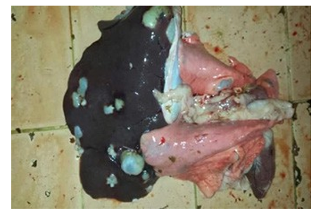
Figure (3): Cattle liver and the lung show heavy infection with hydatid cyst
Table (8): Percentage of hydatid cyst condemnation in the lung and liver of the examined carcasses.
|
species |
year |
Total No. of examined carcasses |
Lung |
Liver |
||
|
No. |
% |
No. |
% |
|||
|
|
2017 |
5598 |
1 |
0.01 |
0 |
0 |
|
Cattle |
2018 |
4719 |
3 |
0.06 |
10 |
0.2 |
|
|
Total |
10317 |
4 |
0.038 |
10 |
0.096 |
|
|
2017 |
603 |
0 |
0 |
0 |
0 |
|
Buffalo |
2018 |
160 |
0 |
0 |
0 |
0 |
|
|
Total |
763 |
0 |
0 |
0 |
0 |
|
|
2017 |
112 |
6 |
5.3 |
1 |
0.8 |
|
Camel |
2018 |
178 |
4 |
2.2 |
2 |
1.1 |
|
|
Total |
290 |
10 |
3.4 |
3 |
1.03 |
|
|
2017 |
6313 |
7 |
0.11 |
1 |
0.015 |
|
Total |
2018 |
5057 |
7 |
0.14 |
12 |
0.237 |
|
|
Total |
11370 |
14 |
0.12 |
13 |
0.114 |
Furthermore, the annual financial assessment for condemnation due to hydatidosis equaled 1145 and 1265 LE were the value of econom¬ic loss in lung and liver during the whole period of investigation, respectively.
Table (9): the annual financial assessment for partial condemnation due to hydatidosis.
|
Species |
Lung |
Total EGP (LE) |
Total EGP (LE) |
||||
|
No. of infected organs |
Weight (Kg) |
EGP (LE) |
No. of infected organs |
Weight (Kg) |
EGP (LE) |
||
|
Cattle |
|
|
|
|
|
|
|
|
2017 |
1 |
1.5 |
45 |
0 |
0 |
0 |
45 |
|
2018 |
3 |
7.0 |
280 |
10 |
29 |
1160 |
1440 |
|
Buffalo |
|
|
|
|
|
|
|
|
2017 |
0 |
0 |
0 |
0 |
0 |
0 |
0 |
|
2018 |
0 |
0 |
0 |
0 |
0 |
0 |
0 |
|
Camel |
|
|
|
|
|
|
|
|
2017 |
6 |
14 |
420 |
1 |
0.5 |
15 |
435 |
|
2018 |
4 |
10 |
400 |
2 |
2 |
80 |
480 |
|
Total |
|
|
|
|
|
|
|
|
2017 |
7 |
15.5 |
465 |
1 |
0.5 |
15 |
480 |
|
2018 |
7 |
17 |
680 |
12 |
31 |
1240 |
1920 |
|
Sum of 2017 & 2018 |
14 |
32.5 |
1145 |
13 |
31.5 |
1265 |
2400 |
Discussion
In the current study, a total number of 11370 carcasses (10317 cat¬tle, 763 buffaloes, and 290 camel carcasses) from January 2017 till December 2018 were slaughtered and PM examined for the presence of parasitic infection in a local abattoir in Cairo Gover¬norate. The overall prevalence of infected slaughtered animals in this study was 1.25%. This record was higher than other studies recorded in Egypt (0.7%), while less than (1.95%) [9, 10]. Among the detected parasitic infection during this study, fascioliasis was the most detected parasitic infection, followed by cysticercosis and hydatidosis, respectively. Percentage of parasitic infection in food animals varied according to the animal species, age and their resis¬tance to natural infection, grazing habits, as well as difference in the local climatic conditions [5].
The prevalence of fascioliasis in the examined carcasses was 62% [11, 12]. This result agreed with (0.7 and 0.8% for cattle and buf-falo, respectively) who reported higher prevalence of fascioliasis in buffalos than cattle, while differed with (0.07 and 0.14% for buffaloes and cattle carcasses, respectively) who reported that higher prevalence of fascioliasis in cattle than buffalo carcass sam¬ples. This may be explained by difference in the total number of slaughtered animals in each study. The recorded results of over¬all fascioliasis incidence came in agree with who recorded higher prevalence of fascioliasis in buffalo carcasses than cattle and cam¬els, respectively where the percent of Fasciola infection in cattle, camel and buffalo were 1.65, 2.26 and 0.04%, respectively [13].
Regarding to the prevalence of cysticercosis among slaughtered animals, our results revealed that cysticercosis was recorded in cattle during 2017 and 2018 as 0.52 and 0.42%, respectively. The overall percent of camel cysticercosis (0.69%) was higher than those recorded in cattle (0.47%) along the investigation period, while buffalo carcasses were free on infection. This result differed from those recorded by (0.13 and 0.44% for buffalo and cattle, respectively), who recorded that cattle carcasses were the most prominently had Cysticercus infection than buffalo and camel car¬cass samples, respectively (0.7, 0.59 and 0.09%).
The different percentage of Cysticercus infection between differ-ent studies may be due to growing intensive rearing of cattle than buffalos and camels in Egypt. Concerning the incidence of hydati-dosis in the examined carcasses, the present data revealed that hy¬datidosis was more observed in cattle’s liver than lung samples, but was more prevalent in camel’s lung than liver samples. In ad¬dition, the overall prevalence of hydatidosis in this study showed that camel carcasses were more prevalent infected with hydatid cyst than cattle samples, while buffalo carcasses were free from infection. Our results came in line with those recorded by who re¬corded that camel carcasses harbored the highest incidence of hy¬datidosis (2.33%) than cattle (0.91%) and buffalo (1.2%), and who recorded higher incidence of hydatidosis in the examined camel samples (8.63%) than cattle (1.15%) and buffalo (1.3%) carcasses. Variation between the current data and the previous recorded re¬sults can be attributed to variation in season of collection, age of the animal, rearing environment, and type of feeding [14].
Conclusion
Conclusively, the detected parasitic affections and demonstrated economic losses throw lights over the importance of strict well qualified meat inspection in slaughter houses to avoid the serious zoonotic meat-borne parasites to the consumers. Additionally, it recommended authorities of the scope to prepare qualified veteri¬nary inspectors to safeguard the public health of the human-being.
References
- Wu, G. (2016). Dietary protein intake and human health. Food & function, 7(3), 1251-1265.
- Williams, P. (2007). Section 2: key nutrients delivered by red meat in the diet. Nutr Dietet, 64(Suppl 4), S113-S119.
- Gupta, A., Gupta, J., Devkaran, B., & Gupta, A. (2017). Primary renal echinococcosis with gross hydatiduria. Case Reports, 2017, bcr-2017.
- sAHiN, N., Aydln, N. E., Akatll, A. N., Daglu, A. F., & sam-dance, E. (2019). Fascioliasis: a rare parasitic infection-mim-icking tumor in the liver: report of two cases.
- Tas, E. E., Akcay, G. F. Y., Yildirim, F., & Yavuz, F. (2018). Coexisting primary ovarian and omental hydatid disease mimicking an ovarian neoplasm: a case report. International Journal of Gynecological Pathology, 37(3), 301-304.
- Ekhlas Hamed, A. H., Amany Mohamed, K., Noha Hamed, A. G., & Mohamed Mahmoud, A. F. (2015). Parasites transmitted to human by ingestion of different types of meat, El-Minia City, El-Minia Governorate, Egypt.
- FSIS/USDA (2019): Animal disposition/food safety: Post-mortem inspection. http://www.fsis.usda.gov/wps/wcm/ connect/6d982860-3c8d-4685-8068- 6cffd00ae9ec/PHVt-Post_Mortem_Inspection.pdf?MOD=AJPERES.
- Thrusfield, M. (2007): Some general epidemiological concepts. In: Veterinary Epidemiology, 3rd Ed., Wiley-Blackwell,P. 20–29, ISBN: 978-1-118-71341-9.
- Elmonir, W., Mousa, W., & Sultan, K. (2015). The Prevalence of Some Parasitic Zoonoses in Different Slaughtered Animal Species at Abattoir in the Mid-Delta of Egypt; with Special Reference to its Economic Implications. Alexandria Journal for Veterinary Sciences, 47(1).
- El-Meleh, G. S., Elmeghnawy, R. A., Sabike, I. I., & Hassan,M. A. (2019). Parasitic affections of edible offales of slaughtered animals at El-Shohada abattoir, Monofia governorate, Egypt. Benha Veterinary Medical Journal, 36(2), 117-128.
- A Rasheed, S., & A Kadir, M. (2008). Prevalence of some parasitic helminths among slaughtered ruminants in Kirkuk slaughter house, Kirkuk, Iraq. Iraqi journal of veterinary Sciences, 22(2), 81-85.
- Taha, S. A. (2018): Studies on the parasitic Affection in slaughtered animals at Hurghada Abattoir. Thesis, Master of Vet. Med. (Food Hygiene and Control), Suez Canal University, Egypt.
- Yemisrach, A., & Mekonnen, A. (2012). An abattoir study on the prevalence of fasciolosis in cattle, sheep and goats in De-bre Zeit town, Ethiopia. Global Veterinaria, 8(3), 308-314