
Toxicology and Applied Pharmacology Insights(TAPI)
ISSN: 2641-0451 | DOI: 10.33140/TAPI

Research Article - (2021) Volume 4, Issue 1
Licorice (Glyccyrhiza Glabra) Accelerates the Burn Wound Healing in Rats and Inhibits Growth of Skin Pathogens In-Vitro
2School of Pharmacy, Shahi Sadoughi University of Medical Sciences, Yazd, Iran
3Corresponding author: Department of Pharmacognosy, School of Pharmacy, Shahid Sadoughi University of, Iran
4School of Pathology, Shahid Sadoughi University of Medical Sciences, Yazd, Iran
Received Date: Apr 02, 2021 / Accepted Date: Apr 05, 2021 / Published Date: Apr 07, 2021
Copyright: ©Copyright: ©2021 Mohsen Zabihi, et al. This is an open-access article distributed under the terms of the Creative Commons Attribution License, which permits unrestricted use, distribution, and reproduction in any medium, provided the original author and source are credited.
Citation: Mohsen Zabihi, Pooya Taherifard, Ali Mohammad Ranjbar, Fatemeh Shishehbor (2021) Licorice (Glyccyrhiza Glabra) Accelerates the Burn Wound Healing in Rats and Inhibits Growth of Skin Pathogens In-Vitro. Toxi App Phar Insig 4: 27-32.
Abstract
Background: Skin heat damage is considered as one of the most devastating condition and may cause some morbidities and mortalities. Licorice (Glycyrrhiza glabra) is used in traditional Iranian medicine to treat burns.
Methods: Second- and third-degree burns on the dorsal skin in rats were induced under general anesthesia by a metal plate (2 cm in diameter). The wounds were treated topically with 6 concentrations of licorice root hydroalcoholic extract, silver sulfadiazine cream (1%) or normal saline as control for 21 days. Subsequently, the macroscopic and the histopathologic parameters including epithelialization, neutrophil migration, angiogenesis, collagen formation and wound contraction were evaluated. The anti-bacterial effects of the extract on some pathogens of wound infections including Staphylococcus aureus, Pseudomonas aeruginosa and Acinetobacter spp were also studied in-vitro.
Results: Topical application of Licorice root hydroalcoholic extract at higher concentrations than 8% accelerated the wound healing process, and increased the contraction and shrinkage of the burn. Licorice root extract inhibited the growth of Staphylococcus aureus and Pseudomonas aeruginosa without any inhibitory effect on the growth of Acinetobacter baumannii.
Conclusion: Licorice is a promising remedy for accelerating wound healing in skin burns.
Keywords
Licorice, Glycyrrhiza Glabra, Wound Healing, Burn, Skin, Anti-Bacterial, Rats
Introduction
Burn is one of the most common injuries worldwide. Different types of burn by heat, electricity, chemicals, or radioactive materi- als cause damages to skin, muscles, mucosa, or air ways [1].
In the United States, more than one million burn victims require medical care, and about 45,000 of them need to be hospitalized each year. In the UK burns account for 1% of the workload and 0.014% of hospital admissions [2].
Burns are divided into four types based on the severity of skin damage. In the first-degree burn, only skin epidermis is involved and the burn site becomes red and swollen. While skin damage affects the dermis in addition to epidermis, second-degree burn occurs with painful blisters. All of the skin layers are damaged in third-degree burn and the muscles, tendons, or bone, involve in fourth-degree burn [3].
Management of burns includes non-pharmacological and phar- macological interventions including cooling, wound washing in chemical burns, wound dressing, prescribing analgesics, and so on [4].
The healing of burn wounds significantly depends on the bacterial infection of the burn site. Topical silver sulfadiazine commonly is used as a topical antimicrobial substance in burns. It improves in- fected wounds, nevertheless, it delays the establishment of epider- mis and prolongs the recovery period [5]. There are some botanical application of herbal medicines to improve (deleted) for wound healing in burns [6, 7].
Licorice (glycyrrhiza glabra) root and rootstock extract is used in Iranian traditional medicine to treat skin burns. Licorice root and rootstock contains glycyrrhizin, glycyrrhizic acid, potassium and calcium salts, coumarin, flavones, volatile oils, herbal sterols, high amounts of glucose, sucrose, asparagine, albumin, resins, essential oils, flavonoids, licoflavanol, glabrone, glabridin (anti-Oxidant), and lycoricidine [8]. Licorice root extract is effective against some species of streptococcus, staphylococcus, HIV, hepatitis A, and herpes infections [9]. Furthermore, glabridin (found in the plant roots) is utilized for anti-inflammatory, anti-scar and anti-microbi- al purposes [10]. We aimed to evaluate the effect of hydroalcoholic Licorice root extract on the histopathologic parameters in second- and third-degree burns in rats.
Materials and Methods
Licorice Root Extract Preparation
The dried Licorice root were obtained from medicinal plants mar- ket of Yazd, Iran. The scientific name and the quality were con- firmed with a quality assurance number was obtained from the Faculty of Pharmacy, Shahid Sadoughi University of Medical Sci- ences (SSU0042). Licorice root was pulverized by a mill and then the licorice hydroalcoholic root extract was obtained by percola- tion method. The dry extract was prepared by evaporating the sol- vent at 40oc and it was standardized. The different concentrations of the extract (8, 11, 14, 17, 20 and 23%) were prepared by normal saline as a solvent.
Animals
Ninety-six healthy male Wistar rats (200–250g) were purchased of the animal house of Shahid Sadoughi University of Medical Sciences, Yazd, Iran. The rats were housed in groups of 6 in tem- perature and humidity-controlled room (20–23°C, 50–60%) with a 12 hours light/dark cycle and free access to standard food and water. The animals were kept and handled according to the local guidelines of care and work with laboratory animals in Shahid Sadoughi University of Medical Sciences (IR.SSU.MEDICINE. REC.1398.055).
Burn Induction
Experimental burn was induced according to Shanmoga method with some modifications [11]. The dorsal skin in the rats was burned under general anesthesia with intraperitoneal (i.p.) injections of ketamine hydrochloride (50 mg/kg - ROTEXMEDICA- Germany) and xylazine (10 mg/kg -Westberg) by a metal plate (2 cm in diameter).
The hairs behind the rats were shaved and uniform second- (upper epidermis) and third-degree burns (Hypodermis) were induced. They were performed using a stainless-steel device at a tempera- ture of 100oC for 8- and 13-seconds skin contact, respectively ac- cording to the histopathological parameters (Figure 1).
The freshly prepared extract was applied to the burn site twice a day for 21 days since the first day of the burn induction.
Eight separate groups of rats (n=6) were considered for each type of burn. Control group: The rats were treated with normal saline topically. Positive control group: Treatment was performed by top- ical silver sulfadiazine cream 1%. Experimental groups: The rats were treated with the different concentrations of Licorice root hy- droalcoholic extract (8, 14, 17, 20 and 23%).
The animals were kept in separate cages with free access to food and water and treated twice a day for 21 days. They were weighted weekly. Eventually, the rats were sacrificed and the entire layer of burnt skin was removed and maintained in a 10% formalin for histopathological examinations.
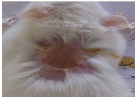
Figure 1: Burns induced on the skin behind the rats.
Histopathological Evaluation The skin samples were fixed in 10% formalin, and in the tissue processing system, different stages of dehydration, clarification with xylol and paraffinization were performed.
Sections with a thickness of 5-6 microns were prepared from the tissue, and placed in water and alcohol to remove the wrinkles. Then the specimens were drained off with a lam dipped in albu- min-glycerin glue.
Coloring was done by hematoxylin and eosin. Then the histo- pathologic criteria including epithelialization, angiogenesis, colla- gen formation, polymorphonuclears (PMNs) migration and wound contraction were scored (1 to 10 scores) under an optical micro- scope with a magnification of 10 and 40 times in blind condition.
Antibacterial Test
Staphylococcus aureus, Pseudomonas aeruginosa and Acineto- bacter baumannii were removed from freezing medium, set in room temperature, cultured on nutrient agar medium using lawn culture method and incubated at 37°C for 24 hours.
McFarland Standard is used to standardize the approximate num- ber of bacteria (1.5×10⁸) and cultured on Mueller-Hinton agar (MHA).
Ten wells were made on MHA culture using sterile Pasteur pipette. The bottom of the plates was covered with MHA medium. Differ- ent concentrations of the extract transferred to the wells contain- ing bacteria and MHA. The plates were incubated at 37°C for 24 hours.
Statistical Analysis
Data are expressed as mean ± S.E.M which were analyzed by one- way analysis of variance (ANOVA) followed by Tukey’s post hoc test. All statistical analyses were made by using SPSS software (version 19).
Results
The weight of the animals did not change significantly during the study. From the second day, their behavior was normal in all groups. Histological images have shown in the figures 2-5.
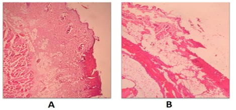
Figure 2: Histopathological presentation of second-degree (A) and third-degree (B) burnt skin in rats in the control groups. There are sever angiogenesis, thin epidermis or areas without epidermis and a large number of PMNs.
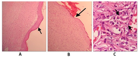
Figure 3: Histopathological presentation of burnt skin in rats in Licorice root hydroalcoholic extract groups after 21 days of treat- ment. A: second-degree burn group; thick and complete epithelial- ization. B: third-degree burn group; thin and complete epitheliali- zation. C: third-degree burn group; a large number of PMNs.
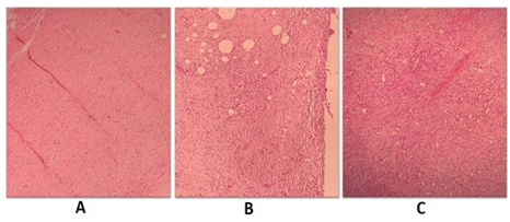
Figure 4: Histopathological presentation of burnt skin in rats in Licorice root hydroalcoholic extract groups after 21 days of treatment. A: second-degree burn group; mild angiogenesis. B: third-degree burn group; moderate angiogenesis. C: third-degree burn group; severe angiogenesis.
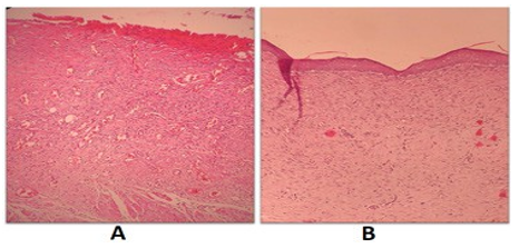
Figure 5: Histopathological presentation of burnt skin in rats after 21 days of treatment. A: third-degree burn group treated with nor- mal saline; without epithelialization, severe angiogenesis, a large number of PMNs and amorph collagen. B: second-degree burn group treated with Licorice root hydroalcoholic extract; complete epithelialization, mild angiogenesis, a low number of PMNs and disorganized collagen.
Polymorphonuclears Migration
Polymorphonuclears migration was scored according to: PMNs count>=40, score=0; 10=
Licorice root hydroalcoholic extract 11% showed a significant dif- ference compared to control group in second-degree burn. Lico- rice root extract in concentrations of 11, 17, 20 and 23% inhibited PMNs migration more than silver sulfadiazine 1% cream (P<0.05). In all the concentrations, Licorice inhibited PMNs migration in third-degree burn (P<0.05) (figure 6).
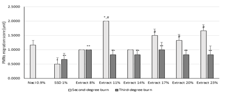
Figure 6: Effect of Glycyrrhiza glabra root hydroalcoholic extract on polymophonuclears (PMNs) migration. Data are analyzed as mean ± S.E.M, (n=6). *P<0.05 compared to control (Nacl 0.9%) in second-degree burn, #P<0.05 compared to silver sulfadiazine (SSD) 1% cream, **P<0.05 compared to control in third-degree burn. One-way ANOVA followed by Tukey’s post hoc test.
Collagen Formation Both Licorice in all applied concentrations and normal saline in- duced collagen formation in both second- and third- degree burns. In all of the groups, collagen formation was induced more than silver sulfadiazine (P<0.05) in second-degree burn (figure 7).
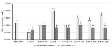
Figure 6: Effect of Glycyrrhiza glabra root hydroalcoholic extract on collagen formation. Data are analyzed as mean ± S.E.M, (n=6).*P<0.05 compared to control (Nacl 0.9%), #P<0.05 compared to silver sulfadiazine (SSD) 1% cream. one-way ANOVA followed by Tukey’s post hoc test.
Epithelialization
Licorice root hydroalcoholic extract (17, 20 and 23%) increased epithelialization in second-degree burn compared to both of nor- mal saline and silver sulfadiazine 1% cream. Concentration of 20% showed the best effect. There was no increase in epitheliali- zation in third-degree burn (P<0.05) (figure 8).
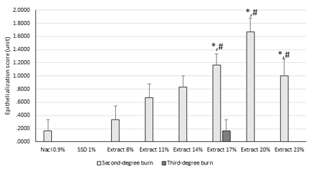
Figure 8: Effect of Glycyrrhiza glabra root hydroalcoholic extract on epithelialization. Data are analyzed as mean ± S.E.M, (n=6).*P<0.05 compared to control (Nacl 0.9%) group, #P<0.05 com- pared to silver sulfadiazine (SSD) 1% cream. one-way ANOVA followed by Tukey’s post hoc test.
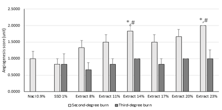
Angiogenesis
Licorice root hydroalcoholic extract 14% and 23% inhibited an- giogenesis in compared to normal saline in second-degree burn. There was no significant reduction in angiogenesis in third-degree burn in control and treatment groups (figure 9).
Skin Contraction
Skin contraction is the portion of epithelialization of burnt area. Some concentrations of Licorice root extract (14-23%) increased skin contraction compared to both control and silver sulfadiazine 1% cream (figure 10).
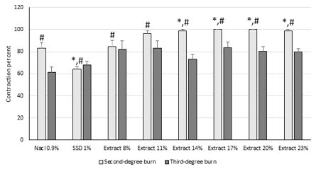
Figure 10: Effect of Glycyrrhiza glabra hydroalcoholic root ex- tract on skin contraction. Data are analyzed as mean ± S.E.M, (n=6). *P<0.05 compared to control (Nacl 0.9%) group, #P<0.05 compared to silver sulfadiazine (SSD) 1% cream. one-way ANO- VA followed by Tukey’s post hoc test.
Total Score
Total score was calculated from all of the histopathological scores. Licorice root extract (>11%) improved total score in second-de- gree burn. Silver sulfadiazine decreased total score compared to control group. There was no significant difference between the groups in third-degree burn (figure 11).
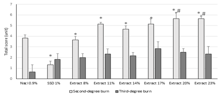
Figure 11: Effect of Glycyrrhiza glabra hydroalcoholic root ex- tract on total score of wound healing. Data are analyzed as mean± S.E.M, (n=6). *P<0.05 compared to control (Nacl 0.9%) group, #P<0.05 compared to silver sulfadiazine (SSD) 1% cream. one- way ANOVA followed by Tukey’s post hoc test.
Anti-Bacterial Effects
A brief evaluation showed Licorice root hydroalcoholic extract has anti-bacterial effects against some of the bacteria. It had no effect against Acinetobacter boumani (figure 12).
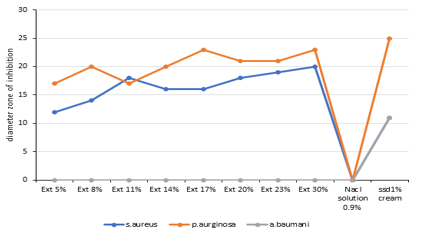
Figure 12: Effects of the various compounds against the bacteria. EXT, Licorice root extract; ssd, silver sulfadiazine.
Discussion
Skin injuries can be caused by a variety of causes, such as burns, surgery, trauma, and etc. The process of skin repair is similar in all of these skin lesions [12].
Wound healing is accomplished in three steps including inflamma- tory, cell proliferation, and remodeling. The inflammatory phase in which leukocytes, especially neutrophils, penetrate the damaged tissue, the cell proliferation phase in which fibroblasts, keratino- cytes, epithelial, and endothelial cells, grow, proliferate, and repro- duce the extracellular matrix and granular tissue at the site of the wound, and eventually, remodeling phase, in which angiogenesis, collagen accumulation and crosslinking, granulation and fibropla- sia formation, and then epithelial layer formation, contraction, and shrinkage occur. In the wound healing process, each of these steps does not begin with the completion of another step, but overlaps [13].
In general, the wound healing process lasts 3-4 months, and if completely healed, 70% of the skin will be repaired (not the hair follicles or blood vessels). Disorganized collagens will be seen af- ter 21 days, and linear collagens after 3-4 months [14]. The process of wound healing is directly related to the reduction of PMNs, mild angiogenesis, and increased collagen levels and epithelialization [15].
Penetration of microorganisms into deep layers of damaged skin and subsequent infections is one of the most important reasons for the delay in the healing process of burn wounds [16].
Glycyrrhizinic acid, is one of the most abundant compounds in licorice extract. Glycyrrhizinic acid belongs to the saponins, has a structure similar to adrenal secreted glucocorticoids, and in addition to its anti-inflammatory properties, also has antibacte- rial properties [17, 18]. This property allows it to accelerate the healing process of burn wound by inhibiting the proliferation of microorganisms [19]. The flavones in licorice extract are also an- ti-inflammatory compounds that can reduce wound inflammation and accelerate wound healing [20]. Isoliquiritigenin (ILTG), a fla- vonoid found in licorice, shows a clear inhibitory effect on COX-2 and iNOS and decreases PGE2 [21]. The licorice root derived iso- flavan glabridin increases the function of osteoblastic MC3T3-E1 cells, and induces collagen formation [22].
The results of this study show that topical application of licorice root hydroalcoholic extract in concentrations higher than 8% not only inhibit the growth of some skin pathogens in burn, but also accelerate the wound healing process in burns. Licorice increased skin contraction and shrinkage by affecting the wound healing fac- tors. Accordingly, the best results were observed at concentrations of 20% and 23%. The anti-inflammatory and antimicrobial effects of Licorice contents such as Glycyrrhizinic acid, Isoliquiritigenin and glabridin can accelerate the healing process of burn wounds.
Conclusion
The findings show that the hydroalcoholic extract of Glycyrrhiza glabra (Licorice) root has positive effects on the healing process of wound in second-degree burns. Considering the antibacterial effects of this herbal medicine, it can be a promise to clinical use in skin burns.
References
- Li H, Tan J, Zhou J, Yuan Z, Zhang J, et al. (2017) Wound management and outcome of 595 electrical burns in a major burn center. Journal of surgical research 214: 182-189.
- Pruitt B, Wolf SE, Mason A (2012) Epidemiological, demo- graphic, and outcome characteristics of burn injury. Total burn care 4: 15-45.
- Meckler G, Quereshi N, Al-Mogbil M, Kentab O (2016) Tin- tinalli's emergency medicine: a comprehensive study guide. New York: McGraw-Hill.
- Marx J, Walls R, Hockberger R (2013) Rosen's Emergency Medicine-Concepts and Clinical Practice E-Book: Elsevier Health Sciences.
- De Gracia C (2001) An open study comparing topical silver sulfadiazine and topical silver sulfadiazine–cerium nitrate in the treatment of moderate and severe burns. Burns 27: 67-74.
- Ayyanar M, Ignacimuthu S (2009) Herbal medicines for wound healing among tribal people in Southern India: Eth- nobotanical and Scientific evidences. International Journal of Applied Research in Natural Products 2: 29-42.
- Maver T, Maver U, Stana Kleinschek K, Smrke DM, Kreft S (2015) A review of herbal medicines in wound healing. Inter-national journal of dermatology 54: 740-51.
- Chin YW, Jung HA, Liu Y, Su BN, Castoro JA, et al. (2007) Anti-oxidant constituents of the roots and stolons of licorice (Glycyrrhiza glabra). Journal of agricultural and food chem- istry 55: 4691-4697.
- Gupta VK, Fatima A, Faridi U, Negi AS, Shanker K, et al. (2008) Antimicrobial potential of Glycyrrhiza glabra roots. Journal of ethnopharmacology 116: 377-380.
- RaÄková L, JanÄinová V, Petríková M, Drábiková K, Nosáľ R, et al. (2007) Mechanism of anti-inflammatory action of li- quorice extract and glycyrrhizin. Natural product research 21: 1234-1241.
- Priya KS, Gnanamani A, Radhakrishnan N, Babu M (2002) Healing potential of Datura alba on burn wounds in albino rats. Journal of ethnopharmacology 83: 193-199.
- Sharabiani MT, Aylin P, Bottle A (2012) Systematic review of comorbidity indices for administrative data. Medical care 2012: 1109-1118.
- Li J, Chen J, Kirsner R (2007) Pathophysiology of acute wound healing. Clinics in dermatology 25: 9-18.
- Gourdie RG, Potts JD (2013) Compositions and methods for tissue engineering, tissue regeneration and wound healing. Google Patents.
- Schreml S, Szeimies R-M, Prantl L, Landthaler M, Babilas P (2010) Wound healing in the 21st century. Journal of the American Academy of Dermatology 63: 866-881.
- Edwards R, Harding KG (2004) Bacteria and wound healing. Current opinion in infectious diseases 17: 91-96.
- Parvaiz M, Hussain K, Khalid S, Hussnain N, Iram N, et al. (2014) A review: Medicinal importance of Glycyrrhiza glabra L.(Fabaceae family). Global J Pharmacol 8: 8-13.
- Kim HK, Park Y, Kim HN, Choi BH, Jeong HG, et al. (2002) Antimicrobial mechanism of β-glycyrrhetinic acid isolated from licorice, Glycyrrhiza glabra. Biotechnology letters 24: 1899-1902.
- Cazander G, Jukema GN, Nibbering PH (2012) Complement activation and inhibition in wound healing. Clinical and De- velopmental Immunology.
- García-Lafuente A, Guillamón E, Villares A, Rostagno MA, Martínez JA (2009) Flavonoids as anti-inflammatory agents: implications in cancer and cardiovascular disease. Inflamma- tion Research 58: 537-552.
- Takahashi T, Takasuka N, Iigo M, Baba M, Nishino H, et al. (2009) Isoliquiritigenin, a flavonoid from licorice, reduces prostaglandin E2 and nitric oxide, causes apoptosis, and sup- presses aberrant crypt foci development. Cancer science 95: 448-453.
- Choi E-M (2005) The licorice root derived isoflavan glabridin increases the function of osteoblastic MC3T3-E1 cells. Bio- chemical pharmacology 70: 363-368.