



Research Article - (2018) Volume 3, Issue 1
Influence of Sago in Improving a Weight Gain of Rats and the Health Profile of the Small Intestine of Rats Infected by Enteropathogenic Escherichia Coli (O127:H6)
2Department of Pathologic, Faculty of Medicine, Sam Ratulangi University, Indonesia
Received Date: Apr 30, 2018 / Accepted Date: Jun 12, 2018 / Published Date: Jun 16, 2018
Copyright: ©Sarah M Warouw, et al. This is an open-access article distributed under the terms of the Creative Commons Attribution License, which permits unrestricted use, distribution, and reproduction in any medium, provided the original author and source are credited.
Abstract
Background: Sago is one potential source of traditional food contain carbohydrates and have benefits as an anti-diarrheal. Escherichia coli are a member of the normal intestinal flora. However, one of the serotypes of this bacterium, E. coli (O127: H6) can be an important cause of diarrhea in infants.
Objective: To analyze the sago effect on weight gain of rats and the profile morphology of small intestine that infected by E. coli (O127: H6).
Methods: An experimental research using 20 wistar rats from April to June 2015. Rats were divided into four groups; Negative control 14 days (A), giving sago for 14 days (B), giving E. coli (O127: H6) and body weight lowered 20% from baseline weight for 7 days (C), giving E. coli (O127: H6) and body weight lowered 20% from baseline weight for 7 days simultanly with giving sago for 14 days (D). Weight gain measured daily, tissue biopsy of the small intestine is processed by using paraffin embedding and stained with hematoxylin eosin. Data were analyzed using Analysis of Varian (ANOVA) and to determine the differences in respectively group continued with the Last Significant Different (LSD).
Results: Total body weight increase: (A) 19.80gr ± 1.64, (B) 12.80gr ± 1.64, (C) 27.40gr ± 2.40, (D) 14.20gr ± 0.44. Percentage villi damage (%): (A) 1.80 ± 0.44, (B) 1.60 ± 0.54, (C) 28.00 ±6.70, (D) 3.80 ± 0.83. Sago decreased percentage of small intestine villi damage 24.20%.
Conclusion: Sago can increase the body weight and protect the small intestine villi damage from E.coli.
Keywords
Escherichia coli, Sago, Small intestine
Background
Diarrheal disease is still one of the important public health issues because it is the third rank of morbidity and mortality rates in various countries including Indonesia. It is estimated that more than 1.3 billion attacks and 3.2 million deaths per year in infants are caused by diarrhea [1]. One of the pathogenic E. coli species is Enteropathogenic Escherichia coli (EPEC). Enteropathogenic Escherichia coli (EPEC) is an important cause of diarrhea in infants, especially in developing countries [2]. Enteropathogenic Escherichia coli (EPEC) (O127: H6) was the major cause of outbreaks of neonatal diarrhea in Chongqing in May 1987 [3].
Many people prefer medicinal plants as an alternative treatment, sago is the largest producer of starch (polymer glucose compound) rich in carbohydrates as an energy source, and cellulose that functions as fiber, sago also contains a tannin phytochemical substance that is useful as an anti diarrhea [4-7].
This study aims to analyze the influence of sago in rat weight gain and increase the health profile of infected rats of Enteropathogenic Escherichia coli (EPEC) (O127: H6).
Methods
The main ingredient used in this research is sago. For the invivo study, Wistar rats were used at 5-6 weeks old and given E. coli (O127: H6). After the tissue sampling is done then embedding in paraffin. The tissue pieces are further processed by eosin hematoxylin (HE) staining. After the rat was adapted for 1 week, then divided into 4 groups: negative control for 14 days (A), sago treatment for 50 gr / day for 14 days (B), E. coli (O127: H6) and BW treatment decreased 20% the initial weight at the same time with 10 g / day pellet delivery for 7 days was continued by administration of pellets 50 gr / day to day 14 (C), E. coli (O127: H6) and BW treatments were reduced by 20% of the initial weight together with administration sago 10 grams / day for 7 days then continued with sago 50 gr / day until day 14 (D). All rats were given standard ration and drinking water was given ad libitum, except for group C, D for day 1 to day 7 the amount of ration and drinking water was reduced. The rat treatment group can be seen in Table 1. The culture of E. coli (O127: H6) was used as much as 2 ml with a population of 106 cfu / ml for a single feed. E. coli was administered to mice using gastric sonde. The process of termination and intestinal organ sampling performed on day 14. Prior to the process of termination and sampling of the intestinal organs, weighed the final BW of the rats in all groups.
Table 1: Rats treatment groups
|
Group of Rats |
Treatment |
|
A |
Control rats, the rats given only 50 gr / day pellets from day 1 to day 14. |
|
B |
Rats given sago 50 gr / day starting day 1 to day 14. |
|
C |
The E. coli (O127: H6) and the rats BW were lowered by 20% of the initial weight together with the administration of pellets 10 gr / day starting day 1 - 7th day followed by pellet 50 gr / day until day 14. |
|
D |
E. coli-suppressed rats (O127: H6) and BW were decreased by 20% of the initial weight together with sago 10 gr / day from day 1 - 7th day followed by sago 50 gr / day until day 14. |
Intestinal tissue biopsy was treated using paraffin embedding and stained with eosin hematoxylin staining, tissue pieces (4 μm) subsequently processed and a percentage of damaged villous rat villi was observed. The percentage of intestinal villi damage was calculated based on the formula: the number of damaged villi divided by the total number of villi multiplied by 100%. The data were then analyzed using ANOVA and continued with LSD to determine the differences in each group.
The nutrient content of pellet consists of water 9.16%, ash 3.47%, crude fiber 6.65%, protein 20.22%, and carbohydrate 65.29% (Table 2) [8]. The largest content of sago in the form of carbohydrates. Sago nutritional content consists of moisture content of 14.00%, calories 343 cal, 0.70 gr protein, 0.20 g fat, carbohydrate 84.70 gr, mineral 0.40 gr, calcium 11.00 mg, phosphorus 13.00 mg, iron 1.50 gr, thiamine 0.01 mg (Table 3) [9].
Table 2: Content of pellet
|
Component |
Amount |
|
Water |
9.16% |
|
Ash |
3.47% |
|
Coarse Fiber |
6.65% |
|
Protein |
20.22% |
|
Carbohydrate |
65.29% |
Table 3: Content of sago
|
Component |
Amount |
|
Water content (%) |
14.00 |
|
Calories (Kal) |
343.00 |
|
Protein (g) |
0.70 |
|
Fat (g) |
0.20 |
|
Carbohydrates (g) |
84.70 |
|
Minerals (g) |
0.40 |
|
Calcium (mg) |
11.00 |
|
Phosphorus (mg) |
13.00 |
|
Iron (mg) |
1.50 |
|
Thiamine (mg) |
0.01 |
Results
Total weight gain
The result showed that the total weight gain (BW) of group A of 19,80 gr ± 1.64, group B was 12,80 gr ± 1,64, group C 27,40 gr ± 2,40, group D 14, 20 gr ± 0.44 (Table 4). In this study it was found that administration of sago 50 gr / day in group B resulted in a significant weight gain (p<0, 05). Similarly in group D, where all the rats lost their weight by 20% of their initial body weight, there was a significant weight gain (p<0.05). The same thing was also found in groups of A and C rats that got rations without sago (p<0.05).
Table 4: Average total of BW additions
|
Group |
Average total of BW additions (g) |
|
A |
19.80 ± 1.64 |
|
B |
12.80 ± 1.64 |
|
C |
27.40 ± 2.40 |
|
D |
14.20 ± 0.44 |
Percentage of villous damage
The percentage of villous damage to the small intestine was calculated from the histological picture of rat’s small bowel tissue, seen in Table 5 and Figure 1. The mean percentage of villus damage of large intestine occurred in rats who received E.coli (O127: H6) for 7 days without sago at 28.00 ± 6.70. While the percentage of small intestine damage at least found in rats that are only given sago alone is 1.60% ± 0.54%. The statistical analysis showed no significant difference between mean percentage of intestinal villous damage in control rats, sago-treated rats for 14 days, and rats who received E.coli (O127: H6) for 7 days simultaneously were given sago for 14 days (p>0.05). In rats who received E.coli (O127: H6) for 7 days without sago showed statistically significant differences with controls, sago-treated rats for 14 days or with rats who received E.coli (O127: H6) for 7 days simultaneously given sago for 14 days (p <0.05).
Table 5: Percentage of villous small intestine damage in various treatments
|
Treatment |
Damage to Small Intestinal Villi (%) |
|
Controls (-) |
1,80± 0,44a |
|
Sagu 14 days |
1,60± 0,54a |
|
E. coli 7 days |
28,00± 6,70b |
|
E. coli 7 days |
3,80± 0,83a |
|
Along with sago 14 days |
|
Description: numbers followed by different superscript letters show there was significant difference (p <0.05)
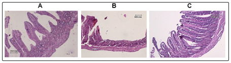
Figure 1:
(A) Microscopic picture of the small intestinal villi given sago 14 days (Group B)
(B) Microscopic images of the small intestinal villi given E. coli 7 days (Group C)
(C) Microscopic picture of the small intestine villus given E. coli 7 days simultaneously with 14 days of sago (Group D).
The average percentage of intestinal damage in healthy sago-treated rats (1.60% ± 0.54%) was lower than that of healthy sago-free rats (1.80% ± 0.44%) and mean percentage of intestinal damage given E. coli for 7 days along with sago for 14 days (3.80% ± 0.83%) lower than that given by E. coli for 7 days without sago (28.00% ± 6.70%).
Discussion
Sago is a starch producer; starch is a polymer glucose compound comprising amylose and amylopectin compounds. The biggest content of starch is carbohydrates as a source of energy, this substance is the source of energy from sago in rats given sago rations [5,10,11]. Glycogen is a nutrient polysaccharide in animals, a glycogen-like structure of amylopectin. Glycogen is stored in human and animal bodies in liver cells and muscle cells. Glycogen in cells will be hydrolyzed if there is an increase in the demand for sugar in the body. However, the energy reserves generated are so small that they can not be relied upon as long-term energy sources [11]. Sago also contains cellulose, which is a polymer glucose compound such as starch, for humans the function of cellulose as a fiber, the fiber in sago can serve as a prebiotic [11].
This result is in accordance with Ulfah et al. study which gained significant weight gain from non fermented sago ration and fermentation with no sago rations [12]. According to Antawidjaya et al. at a 5% concentration of sago dregs can be used in growing duck rations [13].
One of the benefits of sago is having anti-diarrhea effect [8,14]. Sago flour contains one chemical substance called tannin [6,15,16]. According to Bakhriansyah et al16 tannin is a chemical compound that effectively reduces diarrhea and works as an astringent by shrinking the surface of the small intestine mucosa and stimulating the absorption of water in the intestinal lumen [16]. Infusa root of sago proved to have anti-bacterial and anti-diarrhea effect. Infusa 10%, 20% and 40% can also decrease the frequency and duration of diarrhea [16]. The fibers contained in the sago play a role for prebiotics, prebiotics have a role to maintain intestinal microflora, boost immunity, anti-diarrhea effects, reduce the risk of colon cancer, reduce the risk of lung cancer [5,17]. Prebiotics are substrates, generally carbohydrates, which when consumed will stimulate the growth of probiotic germs [18]. In this study the provision of sago can reduce the percentage of intestinal villous damage. This is thought to be caused by the role of tannin substances. The results show that tannin content can inhibit the activity of extracellular protease enzyme from EPEC consequently EPEC can not be attached to intestinal epithelium [19]. Tannins are able to precipitate proteins resulting in decreased intestinal secretions that make the intestinal mucosa more resistant [16,19]. In a study conducted by Labrador et al. successfully isolated nine endophytic bacteria from sago plant tissue, which in its ninth has the effect of inhibiting the development of E. coli and has antibiotic activity against E.coli. In figure 1, the percentage of villous damage in group D rats was lower, presumably due to protective sago tannins in the intestinal mucosa and preventing the attachment of E. coli (O127: H6) to the intestinal epithelium [20].
Conclusion
Giving sago 50gr / day can significantly increase rat weight and can protect villous damaged of E. coli (O127: H6) infected rats.
References
- Widoyono Diare. In: Safitri A, Astikawati R (2008) Penyakit Tropis Epidemiologi, Penularan, and Pencegahan & Pemberantasannya. The twelfth edition. Jakarta: Erlangga H.145.
- Kusuma SAF (2010) Escherichia coli. Bandung: University of Padjadjaran H: 1-2.
- Wu SX, Peng RQ (2008) Studies on an outbreak of neonatal diarrhea caused by EPEC 0127:H6 with plasmid analysis restriction analysis and outer membrane proteindetermination.Accessedfrom http://onlinelibrary. wiley.com/doi/10.1111/j.16512227.1992.tb12207.x/ abstract;jsessionid=91362B17BAF7A775F250714F74AD5293. f02t04. Retrieved on June 19, 2015.
- Purwaningdyah YG, Widyaningsih TD, Wijayanti N (2014) Effectiveness of papaya seed extract (Carica papaya L.) as antidiarrheal in mice induced Salmonella typhimurium. Journal of Food and Agro-industry 3: 1283-1293.
- Umar A (2011) Pati Sagu. Makassar: Muslim University of Indonesia H. 6.
- Mahriani (2012) (RUMBIA) Metroxylon sagu. Accessed from Retrieved on July 08, 2015, http://biologitumbuhanlahanbasah. blogspot.com/2012/10/rumbia-metroxylon-sagu.html.
- Inez K (2015) Perbedaan pati dan selulosa. Accessed from http:// al-asror.blogspot.com/2015/02/perbedaan-pati-dan-selulosa. html. Retrieved on July 08, 2015.
- Anggraini R (2014) Pelet bulu ayam karya mahasiswa universitas sanata darma. Accessed from http://www.kompasiana.com/ ristianggraeni/pelet-bulu-ayam-karya-mahasiswa-universitas-sanata-dharma_54f698cda333116a7d8b50cc. Retrieved on July 08, 2015.
- Alfons JB Rivaie AA (2011) Sagu mendukung ketahanan pangan dalam menghadapi dampak perubahan iklim. Perspektif 10: 81-91.
- Almatsier S (2010) Prinsip Dasar Ilmu Gizi. Karbohidrat. The ninth edition Jakarta: Gramedia Pustaka Utama; 2010. H. 43.
- Safitri NS (2015) Polisakarida dan modifikasinya Accessed from http://k2castle.blogspot.com/2015/02/makalah-kimia-polimer-polisakarida-dan.html. Retrieved on July 08, 2015.
- Ulfah, Bamualim U (2002) Pemanfaatan ampas sagu (Metroxylon Sp) non fermentasi dan fermentasi dalam ransum terhadap pertumbuhan ayam buras periode grower. National Seminar on Livestock and Veterinary Technology 248-50.
- Antawidjaja T, Bintang IAK, Supriyati, Sinurat AP, Kompiang IP (1997) Penggunaan ampas kirai (Metroxylon sago) dan hasil fermantasinya sebagai bahan pakan itik yang sedang tumbuh. Journal of Animal and Veterinary Science 2: 175-180.
- Masketer. 2010. Manfaat sagu bagi tubuhmu. Accessed from http://masketer-zchual.blogspot.com/2010/12/sumber-karbohidrat-sagu-adalah-butiran.html. Retrieved on July 04, 2015.
- Rhama D, Astuti D, Lusiawati S, Julianty W, Avia S, et al. (2014) Bogor. Bogor Agricultural Institute. Accessed from http://www.academia.edu/9831377/Laporan_daya_cerna_pati. Retrieved on July 08, 2015.
- Bakhriansyah M, Febria A, Rahmah D (2011) Efek antibakteri in vitro dan anti diare in vivo infusa akar sago (Metroxylon sagu). Pharmaceutical Magazine Indonesia 22: 158-165.
- Alisha (2012) Sagu bahan makanan yang sehat untuk tubuh. Accessed from http://www.peterparkerblog.com/3643/sagu-bahan-makanan-yang-sehat-untuk-tubuh. Retrieved on July 06, 2015.
- Firmansyah A (2001) Terapi probiotik dan prebiotik pada penyakit saluran cerna anak. Sari Pediatri 2: 210-213.
- Dewi E, Sari W, Khairil (2014) Analisis potensi antibakteri the rosella terhadap histologi usus halus mencit akibat paparan Enteropathogenic Escherichia coli (EPEC). Science Research 4: 1-6.
- Labrador KL, Lustica EL, Novero AU (2014) Isolation and characterization of bacterial endophytes associated with sago palm (Metroxylon sagu rottb.) in tissue culture. Asian Jr. of Microbiol.Biotech. Env.Sc 16: 877-885.