

Archives of Epidemiology & Public Health Research(AEPHR)
ISSN: 2833-4353 | DOI: 10.33140/AEPHR

Impact Factor: 1.98


Research Article - (2022) Volume 1, Issue 1
Development of The Neuropil in The Dynamics of Postnatal Ontogenesis
Received Date: Jul 12, 2022 / Accepted Date: Jul 20, 2022 / Published Date: Jul 28, 2022
Copyright: ©Copyright: ©2022 Elizaveta I. Bon, et al. This is an open-access article distributed under the terms of the Creative Commons Attribution License, which permits unrestricted use, distribution, and reproduction in any medium, provided the original author and source are credited.
Citation: EI Bon, SM Zimatkin and NV Kokhan (2022). Development of The Neuropil in The Dynamics of Postnatal Ontogenesis. Arch Epidemiol Pub Health Res, 1(1), 17-20.
Abstract
This article describes the development of the rat isocortex neuropil in postnatal ontogenesis using the molecular marker of synaptic vesicles synaptophysin, as well as using electron microscopy methods. The study was conducted on outbred white rats. One animal was taken from each female when they reached 5, 10, 20, and 45 days after birth (24 rats in total) and decapitated. The dynamics of the formation of the rat neuropil in postnatal ontogenesis was established, which correlates with the formation of the morphological and functional characteristics of isocortex neurons.
Keywords
Neuropil, Rats, Synaptophysin, Synapses
Introduction
In addition to the bodies of neurons in the nervous tissue of the brain, neuropil is isolated, formed by the dendrites of cortical neu-rons and numerous axons of afferent neurons forming synapses. It is known that in pyramidal neurons of newborn rat pups, apical dendrites and rudiments of basal dendrites are determined, and the branching of apical dendrites is poorly developed. Dendrites have uneven contours and varicose-like thickenings [1]. Only single spines are found on the dendrites. Axons, going down, have al¬most no collaterals. Synaptic contacts in a newborn are extremely rare. Basically, these are axodendritic synapses on large dendrites with slight compaction of synaptic membranes and rare synaptic vesicles. The period of weak development of interneuronal con¬nections in the early stages of postnatal ontogenesis corresponds to the immaturity of the functional activity of the cerebral cortex. In newborn rat pups, neurons are not electrically excitable: up to 6 days of age there are no signs of electrical activity, or it is irregular and unstable [2,3].
On the 10th day after birth, on the apical dendrites of neurons of the 5th layer of the isocortex, three zones with different shapes and density of spines can be distinguished. In the first zone, close to the body of the nerve cell, the spines are sparse, wide and low, having the form of protrusions and thickenings. In the second zone, the number of spines increases and their shape changes: they become narrower and longer. On the terminal branches of the dendrites, the spines are sparse, narrow and long. Contacts of axon ramifications with the trunk and spines of dendrites are revealed. The ultrastruc¬ture of many synapses is becoming more complicated: the electron density of synaptic membranes, especially postsynaptic mem-branes, increases, and the number of synaptic vesicles increases [4,5,3]. However, synapses at this age are still small, their “active zones” in most cases have an insignificant length, the number of synaptic vesicles is small, and they are most often distributed quite diffusely at the end [5].
By the 20th day of life, the number and length of dendrites in-crease, and their branching becomes more complicated. The bas¬al dendrites of most neuron have primary, secondary, and tertia¬ry branches densely covered with spines, although there are still neurons with underdeveloped basal dendrites in the upper layers [6,7,8].
Changes in the bioelectrical activity reflect the complexity of the structural organization of the cortex during the first weeks of post¬natal ontogeny in rats [9]. At the age of 7-10 days in the cere¬bral cortex of rats develops a tendency to increase the regularity, rhythm, constancy and duration of electrical activity [10, 11].
However, of interest was a comprehensive study of the formation of the rat neuropil in the dynamics of postnatal ontogenesis using immunohistochemical and electron microscopic methods [12, 13].
The aim of this work is to study the development of the rat isocor¬tex neuropil in postnatal ontogenesis using the molecular marker of synaptic vesicles synaptophysin, as well as using electron mi¬croscopy methods.
Materials and Methods of Research
Immunohistochemical study:
One animal was taken from each female when they reached 5, 10, 20 and 45 days after birth and decapitated. To obtain comparable results from all animals, samples of the frontal cortex were pro¬cessed in parallel and under the same conditions. They were fixed in zinc formalin at +4°C (overnight) and then embedded in paraf¬fin. Paraffin sections 5 µm thick were prepared using a microtome. For immunohistochemical detection of synaptophysin, primary polyclonal rabbit antibodies from Thermo scientific (USA) Synap¬tophysin Antibody (PA5-27286) (at a dilution of 1:400, at +4ºC, 20 hours, in a humid chamber) were used. Bound primary antibodies were detected using the EXPOSE Rabbit specific HRP/DAB de¬tection IHC kit Thermo scientific (USA). Adjacent sections were stained with 0.1% thionine solution according to the Nissl method. In the fifth layer of the frontal cortex, the optical density of the chromogen deposit in the neuropil was measured [10,1].
Electron Microscopic Examination
Immediately after decapitation and quick extraction of the brain, sections of the frontal cortex were cut out with a blade and placed in a 1% osmium fixative in Millonig’s buffer (pH=7.4) for 2 hours at a temperature of +4°C. Then they were washed in a mixture of Millonig’s buffer (20 ml) and sucrose (900 mg), dehydrated in alcohols of increasing concentration, a mixture of alcohol and ac¬etone, in acetone; passed through a mixture of resins (araldite M + araldite H + dibutyl phthalate + DMR-30) and acetone and en-closed in this casting mixture of resins. Semi-thin sections (about 0.35 μm thick) were made on an MT-7000 ultramicrotome (RMC, USA), stained with methylene blue, and sections of the inner pyra¬midal layer necessary for study were cut out with a blade. Ultrathin sections (about 35 nm thick) were made on the same ultramicro¬tome, assembled on support grids, and counterstained with urani¬um acetate and lead citrate. To do this, meshes with slices were dipped into a drop of uranyl acetate and kept 20 minutes in the dark at room temperature, then washed in 3 portions of bidistilled water for 5 seconds and contrasted with lead citrate for 8 minutes, washed in 3 portions of bidistilled water for 5 seconds [4, 11,1].
The resulting preparations were studied using a JEM-1011 electron microscope (JEOL, Japan) and photographed with an Olympus MegaView III digital camera (Olympus Soft Imaging Solutions, Germany). Ultrastructural morphometry was performed using the Image Warp image processing program (Bit Flow, USA).
The study of histological preparations, their microphotography and cytophotometry were performed at different magnifications of an Axioskop 2 plus microscope (Zeiss, Germany), using a Leica DFC 320 digital video camera (Leica Microsystems GmbH, Ger¬many) and the Image Warp computer image analysis program (Bit Flow, USA). In each experimental group, 120–150 neurons were evaluated, which provided a sufficient sample size for subsequent statistical analysis.
Statistical processing of digital data. To prevent a systematic measurement error, brain samples from the compared control and experimental animals were processed in parallel under the same conditions.
Research Results
In our study, it was shown that in postnatal ontogenesis there is a progressive increase in the expression of synaptic vesicle marker synaptophysin. On the 5th and 10th days, the immunoreactivity was low, and from the 10th to the 20th day, a significant increase was observed - by 47% (p<0.05). Then, from the 20th to the 45th day, the expression of synaptophysin did not change significantly [Table 1; Figures 1, 2].
 A
A
 B
B
 C
C
Synaptophysin expression. Digital micrograph. SW. 400
Figure 1: Expression of synaptophysin in the neuropil of the 5th layer of the isocortex of rats a on the 5th (A), 20th (B) and 45th (C) days after birth

Notes: Me±IQR; * – p<0.05 compared to 10 days
Figure 2: Changes in the expression of synaptophysin in the 5th layer of the isocortex of rats at different times after birth
Table 1: Expression of synaptophysin in the neuropil of the 5th layer of the rat isocortex, (Me (LQ; UQ)), in units of optical density
|
Dates after birth |
Control |
|
5th day |
0,15 (0,14; 0,16) |
|
10th day |
0,165 (0,15; 0,17) |
|
20th day |
0,28 (0,27; 0,3)* |
|
45th day |
0,28 (0,27; 0,3)* |
Note - * - p<0.05 compared to the tenth day
At the electron-microscopic level, in the inner pyramidal layer in postnatal ontogenesis, an increase in the density of synapses was observed, they contained a greater number of synaptic vesicles, mainly associated with the presynaptic membrane [Figures 3, 4, 5, 6].
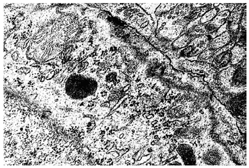
Figure 3: Axosomatic synapses on the internal pyramidal neurons of the isocortex of 20-day-old rats. Synapses are shown by arrows. Magnification: 50000. Electrongrams.
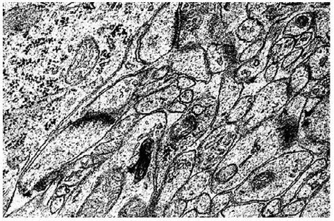
Figure 4: Axodendritic synapses in the neuropil of the inner py¬ramidal layer of the isocortex of 20-day-old rats. Synapses are shown by arrows. Magnification: 50000. Electrongrams
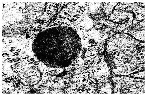
Figure 5: Axosomatic synapses on the internal pyramidal neurons of the isocortex of 45-day-old rats. Synapses are shown by arrows. Magnification: 50000. Electrongrams.
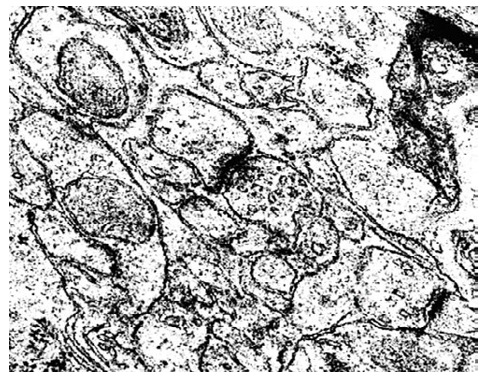
Figure 6: Axodendritic synapses in the neuropil of the inner py¬ramidal layer of the isocortex of 45-day-old rats. Synapses are shown by arrows. Magnification: 50000. Electrongrams.
Our data are confirmed by literature data, according to which the period of functional maturation of synaptic contacts coincides with the dynamics of the myelination process. By the 10th day of life, myelination covers the frontal cortex, by the 12th – the parietal, and from here it spreads to neighboring areas. The occipital cortex is the last to be myelinated.
Structural immaturity of the higher links of the motor system in newborns and rat pups during the first days of life correlates with functional immaturity of the brain and imperfection of motor re-actions. During this period of ontogenesis, rat pups lack a clear motor reaction to a sound stimulus and the development of a de-fensive conditioned reflex is impossible. 2 weeks after birth, the motor reactions of rat pups are already quite complex and coordi¬nated. Such a manifestation of the motor function corresponds to a sufficient differentiation of the isocortex.
It is by the 20th day after birth that the number of synapses sharply increases, their ultrastructure becomes more complex, and all three main types of interneuronal contacts are revealed. New forms of synaptic contacts appear on thin branches of dendrites, one axon with several postsynaptic processes, or, conversely, several axons with one dendrite. However, in contrast to the frontal cortex, in the occipital cortex, during the period of vision, there are only single axosomatic synapses in the upper layers.
The development of dendritic terminals goes hand in hand with a decrease in cell density.
Thus, a progressive increase in the expression of the synaptic ves-icle marker synaptophysin was observed. The development of the synaptic apparatus correlates with the formation of the ultrastruc¬ture and histochemical characteristics of rat isocortex neurons in the dynamics of postnatal ontogenesis [14].
References
- Swenarchuk, L. E. (2019). Nerve, muscle, and synaptogenesis. Cells, 8(11), 1448.
- Bourgeois, J. P. (2005). Brain synaptogenesis and epigenesis. Medecine Sciences: M/S, 21(4), 428-433.
- Hong, J. H., & Park, M. (2016). Understanding synaptogenesis and functional connectome in C. elegans by imaging technology. Frontiers in Synaptic Neuroscience, 8, 18.
- Benya-Aphikul, H., Pongrakhananon, V., Chetprayoon, P., Sooksawate, T., & Rodsiri, R. (2021). Neuronal growth and synaptogenesis are inhibited by prenatal methamphetamine exposure leading to memory impairment in adolescent and adult mice. Toxicology Letters, 351, 99-110.
- Bourgeois, J. P. (1997). Synaptogenesis, heterochrony and epigenesis in the mammalian neocortex. Acta paediatrica, 86(S422), 27-33.
- Faini, G., Del Bene, F., & Albadri, S. (2021). Reelin functions beyond neuronal migration: from synaptogenesis to network activity modulation. Current Opinion in Neurobiology, 66, 135-143.
- Jin Y. Synaptogenesis. WormBook. 2005 Dec 16:1-11.
- Kanemitsu A, Yoro T, Nakai J. [Synaptogenesis]. Shinkei Kenkyu No Shimpo. 1970 Mar;13(4):823-7.
- Fossati, G., Matteoli, M., & Menna, E. (2020). Astrocytic factors controlling synaptogenesis: a team play. Cells, 9(10), 2173.
- Chen, G., Wu, X., & Tuncdemir, S. (2007). Cell adhesion and synaptogenesis. Sheng li xue bao:[Acta physiologica Sinica], 59(6), 697-706.
- Huttenlocher, P. R., & Dabholkar, A. S. (1997). Regional differences in synaptogenesis in human cerebral cortex. Journal of comparative Neurology, 387(2), 167-178.
- Petzoldt, A. G., & Sigrist, S. J. (2014). Synaptogenesis. Current biology, 24(22), R1076-R1080.
- Ren, Y., & Palmer, A. A. (2019). Behavioral genetic studies in rats. Rat Genomics, 319-326.
- Shan, L., Zhang, T., Fan, K., Cai, W., & Liu, H. (2021). Astro-cyte-neuron signaling in synaptogenesis. Frontiers in Cell and Developmental Biology, 9, 1786.