



Case Report - (2026) Volume 9, Issue 3
Cortical Ribboning and Periodic EEG Discharges in Probable Sporadic Creutzfeldt-Jakob Disease
2Neurology, Clínica ARZ, Campinas, São Paulo, Brazil
Received Date: Aug 10, 2026 / Accepted Date: Sep 08, 2026 / Published Date: Sep 18, 2026
Copyright: ©2026 Raphael Zimmermann, et al. This is an open-access article distributed under the terms of the Creative Commons Attribution License, which permits unrestricted use, distribution, and reproduction in any medium, provided the original author and source are credited.
Citation: Zimmermann, R., Kibrit, S. Z., Zimmermann, A. (2026). Cortical Ribboning and Periodic EEG Discharges in Probable Sporadic Creutzfeldt-Jakob Disease. Adv Neur Sci, 9(3), 01-04.
Abstract
Background: Sporadic Creutzfeldt-Jakob disease (sCJD) is a rapidly progressive and fatal prion disease characterized by cognitive decline and variable neurological manifestations.
Case Presentation: We report a 62-year-old woman who developed progressive memory impairment followed by severe neurological deterioration, diffuse myoclonus, and pyramidal and extrapyramidal signs. Brain magnetic resonance imaging (MRI) demonstrated diffuse bilateral cortical signal abnormalities with restricted diffusion and basal ganglia involvement. Electroencephalography (EEG) subsequently revealed generalized periodic sharp-wave complexes. Prion- specific cerebrospinal fluid (CSF) testing was unavailable.
Conclusion: The combination of rapidly progressive cognitive impairment, characteristic neurological manifestations, MRI abnormalities, and typical EEG findings fulfilled diagnostic criteria for probable sCJD. The patient received palliative care and died during the same month. This case highlights the complementary diagnostic value of MRI and EEG in suspected sCJD, particularly when prion-specific biomarkers are unavailable.
Keywords
Prion Disease, Rapidly Progressive Dementia, Diffusion-Weighted Imaging, Electroencephalography, Neurodegenerative Disease
Introduction
Sporadic Creutzfeldt-Jakob disease (sCJD) is a rapidly progressive and invariably fatal prion disease characterized by cognitive decline and heterogeneous neurological manifestations, which may include myoclonus, cerebellar dysfunction, and pyramidal or extrapyramidal signs [1]. Contemporary diagnostic criteria allow a diagnosis of probable sCJD during life through integration of the clinical phenotype with characteristic findings on electroencephalography (EEG), magnetic resonance imaging (MRI), cerebrospinal fluid (CSF) biomarkers, or real-time quaking-induced conversion (RT-QuIC) [1,2]. Diffusion-weighted imaging (DWI) and EEG provide important diagnostic support in suspected sCJD. Restricted diffusion involving characteristic cortical regions or the basal ganglia constitutes an important neuroimaging finding, while generalized periodic sharp-wave complexes represent a characteristic electrophysiological pattern [1-3]. We present a case of probable sCJD demonstrating concordant clinical, radiological, and electrophysiological findings despite the unavailability of prion-specific CSF testing.
Case Presentation
A 62-year-old woman, previously functionally independent, developed progressive short-term memory impairment characterized by repetitive behavior and increasing cognitive decline. Over the following months, her neurological condition progressively deteriorated, with bilateral motor impairment initially involving the lower limbs and subsequently the upper limbs. She developed progressive dysphagia, initially for solid foods and later for liquids, as well as episodes clinically suspected to represent focal and generalized seizures. Sleep-wake cycle inversion and progressive loss of functional independence were also reported. An EEG obtained on December 12, 2022, demonstrated diffuse background slowing without the characteristic periodic pattern associated with CJD. As the disease progressed, diffuse myoclonic jerks emerged, accompanied by marked neurological deterioration.
CSF analysis performed on March 27, 2023, revealed 3 cells/ mm³, glucose of 58 mg/dL, protein of 87 mg/dL, and 5,760 red blood cells/mm³. CSF culture was negative. Prion-specific CSF testing, including RT-QuIC and 14-3-3 protein analysis, was not available. At neurological examination during the advanced stage of the disease, the patient was severely encephalopathic and poorly responsive. She exhibited diffuse myoclonus, tetrapyramidal spasticity with bilateral Babinski signs and clonus, prominent frontal release signs, and cogwheel rigidity. Brain MRI performed on March 31, 2023, demonstrated subtle diffuse bilateral cortical hyperintensity on FLAIR, with corresponding restricted diffusion on DWI, producing a cortical ribboning pattern. DWI additionally demonstrated bilateral hyperintensity involving the caudate nuclei and putamina (Figure 1).
A subsequent EEG performed on April 14, 2023, demonstrated generalized periodic activity occurring at intervals of approximately 1–1.5 seconds. The formal EEG report described high-amplitude biphasic and triphasic sharp waves associated with markedly slowed background activity and absence of a normal alpha rhythm and considered the pattern suggestive of CJD (Figure 2). Based on rapidly progressive cognitive impairment, diffuse myoclonus, pyramidal and extrapyramidal signs, and typical periodic EEG abnormalities, the patient fulfilled the International CJD Surveillance Network diagnostic criteria for probable sCJD [2]. The characteristic MRI abnormalities provided additional radiological support for the diagnosis. Given the advanced neurological deterioration and poor prognosis, supportive and palliative care was instituted. The patient continued to deteriorate and died later that month.
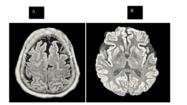
Figure 1: Brain magnetic resonance imaging findings in probable sporadic Creutzfeldt-Jakob disease
(A) Axial fluid-attenuated inversion recovery (FLAIR) image demonstrating diffuse bilateral cortical hyperintensity. (B) Axial diffusion-weighted imaging (DWI) demonstrating diffuse bilateral cortical hyperintensity consistent with cortical ribboning, with additional bilateral hyperintensity involving the caudate nuclei and putamina.

Figure 2: Electroencephalographic findings in probable sporadic Creutzfeldt-Jakob disease Electroencephalogram demonstrating generalized periodic sharp-wave complexes occurring at intervals of approximately 1–1.5 seconds on a diffusely slowed background.
Discussion
The present case demonstrates the convergence of clinical, neuroimaging, and electrophysiological findings in probable sCJD. Current International CJD Surveillance Network criteria permit classification as probable sCJD in patients with rapidly progressive cognitive impairment, characteristic neurological manifestations, and supportive EEG, MRI, CSF, or RT-QuIC findings [2]. Our patient presented with rapidly progressive cognitive impairment, diffuse myoclonus, and prominent pyramidal and extrapyramidal signs, together with typical generalized periodic EEG abnormalities, thereby fulfilling the clinical diagnostic criteria for probable sCJD. MRI is particularly valuable in the diagnostic evaluation of sCJD. Restricted diffusion involving characteristic cortical regions and the caudate and putamen represents an established neuroimaging feature and has been incorporated into diagnostic criteria [1-3]. DWI has demonstrated high diagnostic performance; a systematic review and meta-analysis including 1,144 patients with sCJD reported pooled sensitivity and specificity of 91% and 97%, respectively [4]. In our patient, MRI demonstrated diffuse bilateral cortical signal abnormalities, with cortical restricted diffusion and additional basal ganglia involvement, providing radiological support for the diagnosis.
EEG provides complementary diagnostic information in sCJD. Generalized periodic discharges, historically described as periodic sharp-wave complexes, represent a characteristic electrophysiological finding, although their sensitivity is limited. In a neuropathologically verified cohort, Steinhoff et al. (2004) reported a sensitivity of 64% and specificity of 91% for objective EEG criteria [5]. The serial electrophysiological findings in our patient were particularly informative. An earlier EEG demonstrated diffuse slowing without the characteristic periodic pattern, whereas a subsequent recording obtained during marked neurological deterioration showed generalized periodic sharp-wave activity. Characteristic periodic abnormalities may emerge during disease progression rather than being evident on earlier recordings. Steinhoff et al. (2004) demonstrated the diagnostic utility of periodic EEG complexes, while more recent work has characterized electrophysiological abnormalities that may precede typical generalized periodic discharges [5,6]. This evolution highlights the potential value of repeated EEG assessment when clinical suspicion for sCJD persists despite an earlier nondiagnostic examination.
Seizure-related manifestations may further complicate the interpretation of rapidly progressive neurological deterioration. CJD can occasionally resemble status epilepticus, and periodic EEG abnormalities may present diagnostic challenges when interpreted in isolation [7]. Accordingly, EEG findings should be interpreted together with the clinical trajectory and neuroimaging findings rather than as an isolated diagnostic marker. Prion-specific CSF testing, including RT-QuIC, was unavailable in this case. RT-QuIC has substantially improved the antemortem diagnosis of prion disease because of its high diagnostic accuracy and particularly high specificity [1,8]. Nevertheless, its absence does not preclude classification as probable sCJD when established clinical and supportive investigation criteria are fulfilled [1,2]. The convergence of rapidly progressive cognitive decline, myoclonus, pyramidal and extrapyramidal dysfunction, characteristic MRI abnormalities, and periodic EEG findings illustrates the diagnostic value of integrating clinical, radiological, and electrophysiological information when prion-specific testing is unavailable.
Conclusion
This case illustrates the complementary diagnostic value of MRI and EEG in probable sCJD, particularly when prion-specific biomarkers are unavailable. The evolution from an initially nonspecific EEG to characteristic periodic abnormalities further highlights the potential value of serial EEG assessment when clinical suspicion persists.
Declaration
Consent for Publication
Written informed consent for publication of this case and the accompanying clinical images was obtained from the patient’s family member.
Funding
The authors received no specific funding for this work.
Conflict of interest
The authors declare no conflicts of interest.
Author Contributions
the manuscript. Sheila Zimmermann Kibrit and Allan Zimmermann critically reviewed the manuscript. All authors read and approved the final manuscript.
Data Availability
Data supporting the findings of this case report are contained within the article. Additional patient information is not publicly available to protect patient confidentiality.
AI Usage Statement
Generative artificial intelligence was used for language editing and manuscript formatting. All clinical content, interpretations, references, and the final manuscript were reviewed and approvedby the authors. No generative artificial intelligence was used to create or modify the clinical images presented in this manuscript.
References
- Hermann, P., Appleby, B., Brandel, J.-P., Caughey, B., Collins, S., Geschwind, M. D., Green, A., Haïk, S., Kovacs,G. G., Ladogana, A., Llorens, F., Mead, S., Nishida, N., Pal, S., Parchi, P., Pocchiari, M., Satoh, K., Zanusso, G., & Zerr,I. (2021). Biomarkers and diagnostic guidelines for sporadic Creutzfeldt-Jakob disease. The Lancet Neurology, 20(3)235–246.
- Watson, N., Hermann, P., Ladogana, A., Denouel, A., Baiardi, S., Colaizzo, E., Giaccone, G., Glatzel, M., Green, A. J. E., Haïk, S., Imperiale, D., MacKenzie, J., Moda, F., Smith, C., Summers, D., Tiple, D., Vaianella, L., Zanusso, G., Pocchiari, M., … Pal, S. (2022). Validation of revised International Creutzfeldt-Jakob Disease Surveillance Network diagnostic criteria for sporadic Creutzfeldt-Jakob disease. JAMA Network Open, 5(1), e2146319.
- Zerr, I., Kallenberg, K., Summers, D. M., Romero, C., Taratuto, A., Heinemann, U., Breithaupt, M., Varges, D., Meissner, B., Ladogana, A., Schuur, M., Haïk, S., Collins, S. J., Jansen, G. H., Stokin, G. B., Pimentel, J., Hewer, E., Collie, D., Smith, P., … Sanchez-Juan, P. (2009). Updated clinical diagnostic criteria for sporadic Creutzfeldt-Jakob disease. Brain, 132(10), 2659–2668.
- Park, H. Y., Kim, M., Suh, C. H., Kim, S. Y., Shim, W. H.,& Kim, S. J. (2021). Diagnostic value of diffusion-weighted brain magnetic resonance imaging in patients with sporadic Creutzfeldt-Jakob disease: A systematic review and meta-analysis. European Radiology, 31(12), 9073–9085.
- Steinhoff, B. J., Zerr, I., Glatting, M., Schulz-Schaeffer, W., Poser, S., & Kretzschmar, H. A. (2004). Diagnostic value of periodic complexes in Creutzfeldt-Jakob disease. Annals of Neurology, 56(5), 702–708.
- Matsubayashi, T., Natsui, H., Satoh, K., Kitamoto, T., Yokota, T., & Sanjo, N. (2025). Specific early electroencephalogram for the diagnosis of sporadic Creutzfeldt-Jakob disease. Prion, 19(1), 17–24.
- Singh, K., Arora, N., Kakde, S., Chandershekran, P. N., & Merchant, R. (2026). An unusual case of Creutzfeldt-Jakob disease misdiagnosed as status epilepticus: A case study. Advances in Neurology and Neuroscience, 9(2), 1–9.
- Figgie, M. P., Jr., & Appleby, B. S. (2021). Clinical use of improved diagnostic testing for detection of prion disease. Viruses, 13(5), 789.