

Research Article - (2022) Volume 6, Issue 2
The Diagnosis of Sheep Cystic Echinococcosis by Native Antigen B Elisa Method
2Department of Proteomics and Biochemistry, Razi Vaccine and Serum Research Institute, Karaj, Iran
Received Date: Aug 16, 2022 / Accepted Date: Aug 23, 2022 / Published Date: Sep 12, 2022
Copyright: ©Copyright: Ã?©2022 Rasoul Madani. This is an open-access article distributed under the terms of the Creative Commons Attribution License, which permits unrestricted use, distribution, and reproduction in any medium, provided the original author and source are credited.
Citation: Shirazi, S., Hoghooghi-Rad, N., Madani, R., Golchinfar, F. (2022). The Diagnosis of Sheep Cystic Echinococcosis by Native Antigen B - Elisa Method. Stem Cell Res Int, 6(2), 78-82.
Abstract
Cystic echinococcosis (CE) is one of the most prevalent zoonotic diseases in some countries in the world. Cystic echinococcosis is considered a neglected disease. This disease increases economic damage via medical costs and loss of human and livestock productivity. The aim of the current study gains a better understanding of the prevalence of CE in sheep. Totally 250 sheep sera were collected. 25 serum samples from newborn lambs were used as negative, and 25 others were obtained from slaughterhouses as positive to infection with CE and 200 unknown sera. Antigen B Isolated from hydatid cysts fluid was used for designing ELISA methods. The first Method used anti-Sheep conjugate (SIGMA, at 1:3000 dilution). According to the results, the seroprevalence of CE in East Azerbaijan of Iran was 5.5% and sensitivity and specificity for the diagnosis of hydatidosis in sheep by AgB-ELISA methods was 92%. Using Antigen B in ELISA design for hydatidosis diagnosis has attracted researchers in recent years. During this study, an Iranian native B antigen was used to design the specific detection of hydatidosis in sheep using a specific ELISA technique. The results have shown that using Antigen B in ELISA design is so valuable.
Keywords
Echinococcosis, ELISA, Antigen B, Sheep.
Introduction
Cystic echinococcosis (CE) or Hydatidosis is a zoonotic parasitic infection of humans and domestic animals caused by larvae of the cestode Echinococcus granulosus [1]. Hydatidosis is common in sheep farming regions like Australia, New Zealand, China, South America, India, some African countries, the Middle East, and Iran [2]. This disease has been reported to be endemic in various parts of Iran [3]. World Health Assembly (WHA) noted CE as one of the neglected zoonosis diseases and recent reports indicate that there is a high rate of infection in humans in different parts of Iran and its health problem [4, 5]
In humans, the diagnosis of hydatidosis is mainly based on a posi- tive serological test along with an imaging finding as recommend- ed by WHO [6]. Among different serological methods, ELISA has been reported to be a useful test for the diagnosis of human hy- datidosis. Antigen B (EgAgB) is the main protein generated by the pathogenic larval stage of cystic echinococcosis, and this Ag is highly immunogenic and can be recognized in most sera of in- termediate hosts [7]. WHO recommends using specific serological methods with the specific Ag specially AgB [8]. In animals, CE seriously affected the production and growth of livestock, caus- ing a loss in livestock production and bringing enormous danger to public health [9]. Although, the prevalence of CE in sheep is very important because the highest prevalence of CE in humans is found in populations that raise sheep [10].
Slaughterhouse studies are known as useful references for evaluat- ing the epidemiological aspects of some diseases, especially para- sitic diseases [11]. Considering the presence of informal livestock slaughterhouses and unsanitary slaughter of livestock in some parts of Iran, and also the lack of systematic recording of parasitic disease information in slaughtered animals, Therefore, there is no accurate information on the prevalence of infection in slaughter livestock in different parts of Iran. Serological investigations seem to be necessary in some cases. This study aims to develop and evaluate a Native Antigen B for serodiagnosis of CE in sheep.
Method
Study Area
Tabriz is the fifth-most-populous city in northwestern Iran with latitude 38°04′47″ N and longitude 46°17′30″ E. This city is sur- rounded by mountains in the north, south, and east, with cold win- ters and temperate summers. Tabriz's elevation ranges between 1,350 and 1,600 meters above sea level (Fig 1).
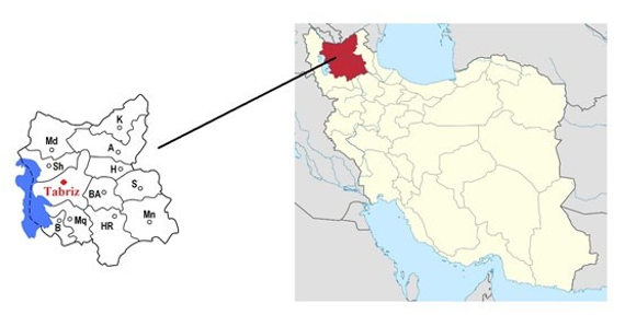
Figure 1: Map of Iran and Tabriz city as the studies area
Serum Samples
A total of 250 sheep sera were investigated in this study. 25 serum samples from newborn lambs were used as negative, and 25 others were obtained from slaughterhouses as positive to infection with CE and were entered the study. At last, 200 unknown sera were collected from the sheep herds. All sera were collected from Tabriz city, East Azerbaijan province of Iran. Sera were kept at -80°C until the experimental assay.
Extraction of Antigen B:
To extract antigen B from the hydatid cyst fluid, the modified method was implemented [12]. Hydatid cysts fluid was extracted from livers of sheep slaughtered at the abattoir of Tabriz, then it was transferred to a dialysis bag and was placed in a container containing polyethylene glycol (PEG) 4000 for 1 hour. This stage leads to the condensation of AgB in the hydatid cyst fluid. After that, the fluid was filtered using a 0.2 microfilter, and the resulting liquid was centrifuged at 1500g for 30 minutes. The extracted fluid was dialyzed for one night, and after that, the contents of the dial- ysis container were centrifuged with a refrigerated ultracentrifuge with 30000g in a vacuum condition and at a temperature of 4° C for 30 minutes. The resulting precipitate was dissolved in 10 ml of 0.2M phosphate buffer with pH 8 and the solution was saturated with 40% ammonium sulfate then the solution was centrifuged for 30 minutes at 3000g. The supernatant was placed in a boil-water bath for 15 minutes. In the next step, it was centrifuged for one hour using the ultracentrifuge, and finally, the supernatant-solu- ble antigen B was collected. After filtration with Millipore (0.2 microns) and the addition of 2% sodium azide (NaN3), it was stored at a temperature of -70 ° C until next use [12]. Finally, the Bradford protein analyzes method was used to measure the protein content of the prepared solutions. Also, the solution containing the prepared antigen B was evaluated using SDS-PAGE.
Designing the ELISA Method with Antigen B:
For design, the suitable level of serum dilution and the accept- able concentration of antigen B that should be coated was deter- mined in the first step. Thus, different levels of antigens and serum concentrations were tested. To prepare serum dilutions, a potent positive serum and a negative serum were used. Then, dilutions of 1:100, 1:200, and 1:400 were prepared. The used wells were polyester (Nunc, Denmark), and to achieve antigen binding, 100 μl of each antigenic concentration was added to each well and stored in the refrigerator for one night to complete the binding process of antigen to the wells.
In the next step, Skim Milk 5% was used to block the wells con- taining 100 μl of antigen solution, they were drained and washed three times using PBST and then, 250 μl of blocking buffer was added to the wells and they were placed in a humid condition for 75 minutes in a 37 ° C incubator. Wells were emptied and then, 100 μl of sera were transferred into the wells. They were incubated in a humid environment for 75 minutes at 37 ° C. Wells were emptied and 100 μl of the anti-Sheep conjugate (manufactured by SIGMA USA at 1: 3000 dilution) was prepared in wells for 75 minutes in a humid environment at 37 ° C.
In the next stage, 100 μl of BM Blue POD (Roche Company, Ger- many) was transferred to the wells and placed in a dark environ- ment for 12 minutes. The stop solution includes sulfuric acid 1M, of which 50 μl is transferred into each well, and then, the wells should be read immediately. To measure the optical absorbance of each well, an ELISA reader with a 450 nm filter was used and the absorbance of all wells was read. Due to the high sensitivity of the extracted antigen (AgB) in this study, a concentration of 0.5 μg / ml and a serum dilution of 1:400 were used. After determining the concentration of AgB and dilution of serum, in the last step, un- known sera were tested with the same concentration and dilution.
Results
Antigen B was measured by Bradford assay and its concentration was 0.7 (mg/ml) and the quality of AgB and presence of AgB sub- units was checked by SDS-PAGE and Coomassie blue staining. Fig 2 shows the profile of antigen B isolated from sheep hydatid cyst fluid (HCF).

Figure 2: SDS-PAGE bands of AgB, M3: The modified method using by polyethylene glycol (PEG) 4000
At first, 0.5 μg /ml AgB concentration and also 1:3000 the sheep's conjugate were used. To find the best serum dilution, 2 sera of sheep with hydatidosis and 2 sera without infection (newborn lamb) were used. According to the results, the best dilution of se- rum was determined at a dilution of 1:400. In the next step, for finding the cut-off, definitive positive and neg- ative sheep sera were examined by the ELISA method (Table 1).
Table 1: Positive and negative sheep sera results with Ag-B ELISA
|
Positive Serum |
No |
Blank |
1 |
2 |
3 |
4 |
5 |
6 |
7 |
8 |
9 |
10 |
11 |
12 |
|
OD |
0.265 |
1.946 |
1.457 |
1.318 |
1.455 |
1.201 |
2.147 |
2.321 |
1.482 |
1.195 |
2.093 |
2.095 |
0.355 |
|
|
No |
13 |
14 |
15 |
16 |
17 |
18 |
19 |
20 |
21 |
22 |
23 |
24 |
25 |
|
|
OD |
2.179 |
1.271 |
1.348 |
2.282 |
0.579 |
1.254 |
1.437 |
1.346 |
1.147 |
2.011 |
2.124 |
1.145 |
1.321 |
|
|
Negative Serum |
No |
Blank |
1 |
2 |
3 |
4 |
5 |
6 |
7 |
8 |
9 |
10 |
11 |
12 |
|
OD |
0.301 |
0.332 |
0.512 |
0.424 |
0.379 |
0.367 |
1.367 |
0.476 |
0.237 |
0.278 |
0.368 |
0.412 |
0.237 |
|
|
No |
13 |
14 |
15 |
16 |
17 |
18 |
19 |
20 |
21 |
22 |
23 |
24 |
25 |
|
|
OD |
0.285 |
0.312 |
0.278 |
0.376 |
0.507 |
0.389 |
0.217 |
0.329 |
1.378 |
0.391 |
0.403 |
0.327 |
0.202 |
According to the following formula, it was equal to one. In other words, the sera with up to one, positive and below one, optical absorption was considered negative.
Cut off=Average±A (Standard Deviation)
With the AgB-ELISA method two (8%) positive sera were found to be negative, and two (8%) negative controls were false positive. Also according to the results of this study, the sensitivity and spec- ificity of the ELISA designed with AgB for detecting hydatidosis in sheep determined 92% (Table 2). In the last step, unknown se- rum samples were evaluated and out of 200 serum samples, 11 (5.5%) were detected as positive.
Table 2: Sensitivity and Specificity of AgB-ELISA in sheep sera
|
Serum |
Negative |
Positive |
Case |
|
50 |
2 |
23 |
Infected |
|
50 |
23 |
2 |
Non infected |
|
100 |
25 |
25 |
Total |
|
92% |
Sensitivity |
|
|
|
92% |
Specificity |
|
|
Discussion, and Conclusion and Recommendations
Many Immunodiagnosis assays have been devised, including in- direct hemagglutination, latex agglutination, immunofluorescence, immunoelectrophoresis, western immunoblotting, and ELISA, based on various antigen preparations. Although, in the last two decades, many serological tests have been proposed, based mostly on crude HCF, Ag5, and AgB, but they have been less commonly used to diagnose sheep hydatidosis. But despite much research, Immunodiagnosis of CE is still a challenge [13]. Some investiga- tions were showed that an antibody detection assay is still superior and is more sensitive than an antigen detection assay, especially in diagnosing an active infection in which hydatid cysts are predom- inant [14].
Hydatid cyst fluid has a high sensitivity for immunological diag- nostic assay, ranging typically from 75% to 95%. Although, its specificity with infected sera with other parasite species (cross-re- action) is unreliable. Hence, the crude HCF is recommended for mass serological screening, and today the main component of HCF is antigen B that use for the serodiagnostic assay [15]. Antigen B is the most promising antigen used in various serology meth- ods for human infections and animals. In humans, several reports found high diagnostic sensitivities and specificities for this antigen for patients with CE [16]. AgB derived from sheep hydatid cyst fluids has potential for the serodiagnosis of ovine hydatidosis but Differences in the performance of diagnostic tests may be due to Different strains of the Echinococcus granulosus and isolation and purification of AgB that can be affected antigens action in serolog- ical assays [14, 17].
In the current study, an Iranian native B antigen was used to de- sign the detection of CE in sheep using an ELISA technique and the sensitivity and specificity of this ELISA method for detecting hydatidosis in sheep was 92%, which compared to similar stud- ies, can be said that the results of this study, in general, have a high sensitivity and specificity. Serological diagnostic methods such as ELISA for hydatidosis in sheep have been performed us- ing different antigens in hydatid cyst fluid and have had different results in the rate of sensitivity and specificity. The sensitivity and specificity of the crude cyst fluid antigen were determined as 32% and 90%, respectively [18]. In another study based on identifying serodiagnosis of hydatidosis that the sensitivity was 60%, and its specificity was 94% [19]. Using the total cyst fluid, purified cyst fluid and purified lipoprotein antigens in ELISA applied on sheep, obtained the highest sensitivity value from total cyst fluid antigen (77.8-89.2%) and the highest specificity from purified cyst fluid antigen (93.9-99.4%) [20].
The use of the ELISA method for the diagnosis of Echinococco- sis in sheep has resulted in cross-reactions against T. ovis and T. hydatigena, especially when crud hydatid antigens purified were used but previous serodiagnosis researches of sheep hydatidosis showed that using the family of antigen B (8, 16, and 20 and 24 kDa) leaded high levels of specificity therefore, in this study, the presence of other parasites was not considered [21, 22]. In previ- ous studies on human by native AgB-ELISA method, the average of sensitivity and specificity was 92.9% and 91.2% respectively [8, 23-25].
In the present study, the prevalence of CE in East Azerbaijan of Iran was relatively the same reports from neighboring countries in the Middle East such as Iraq: 7.3% Syria: 2.8% Saudi Arabia: 2.33% but differs from those reported in other studies in Turkey [26-29]. There are a clear need and demand for research into the development of diagnostics programs. World Organization for An- imal Health recommendations includes sequential testing based on a screening test model such as enzyme-linked immunosorbent as- says (ELISA) [30]. But in Iran, there is no specialized commercial ELISA kit for diagnosing CE in sheep. Using the ELISA method can be very efficient because of the precision and rapid results it provides [31, 32].
Acknowledgment: We would especially like to thank Dr. Tara Emami and Mr. Arash Ghanizadeh.
Funding: None.
Ethical approval: Authors were not involved with animal han- dling and the sera samples were obtained directly through the vet- erinary laboratories and veterinarian of the slaughterhouse.
Conflict of interest: The authors declare that they have no conflict of interest.
References
- Dabaghzadeh, H., Bairami, A., Kia, E. B., Aryaeipour, M., & Rokni, M. B. (2018). Seroprevalence of human cystic echi- nococcosis in Alborz Province, central Iran in 2015. Iranian Journal of Public Health, 47(4), 561.
- Fomda, B. A., Khan, A., Thokar, M. A., Malik, A. A., Fazili, A., Dar, R. A., ... & Malla, N. (2015). Sero-Epidemiological survey of human cystic echinococcosis in Kashmir, North In- dia. PLoS One, 10(4), e0124813.
- Mehrabani, N. G., Kousha, A., Khalili, M., Oskouei, M. M., Mohammadzadeh, M., Alizadeh, S., ... & Hamidi, F. (2014). Hydatid cyst surgeries in patients referred to hospitals in East Azerbaijan province during 2009-2011. Iranian journal of par- asitology, 9(2), 233.
- Fasihi Harandi, M., Budke, C. M., & Rostami, S. (2012). The monetary burden of cystic echinococcosis in Iran. PLOS ne- glected tropical diseases, 6(11), e1915.
- Mableson, H. E., Okello, A., Picozzi, K., & Welburn, S. C. (2014). Neglected zoonotic diseases—the long and winding road to advocacy. PLoS neglected tropical diseases, 8(6), e2800.
- Eckert, J., Gemmell, M. A., Meslin, F. X., Pawlowski, Z. S., & World Health Organization. (2001). WHO/OIE manual on echinococcosis in humans and animals: a public health prob- lem of global concern. World Organisation for Animal Health.
- Zhang, W., Li, J., Jones, M. K., Zhang, Z., Zhao, L., Blair, D., & McManus, D. P. (2010). The Echinococcus granulosus an- tigen B gene family comprises at least 10 unique genes in five subclasses which are differentially expressed. PLoS neglected tropical diseases, 4(8), e784.
- Sadjjadi, S. M., Abidi, H., Sarkari, B., Izadpanah, A., & Ka- zemian, S. (2007). Evaluation of enzyme linked immuno- sorbent assay, utilizing native antigen B for serodiagnosis of human hydatidosis. Iranian journal of immunology, 4(3), 167- 172.
- Tianli, L., Xifeng, W., Zhenzhong, T., Lixia, W., Xingxing, Z., Jun, Q., ... & Xuepeng, C. (2019). Multi-Epitope Fusion Protein Eg mefAg-1 as a Serodiagnostic Candidate for Cystic Echinococcosis in Sheep. The Korean Journal of Parasitology, 57(1), 61.
- Merino, V., Westgard, C. M., Bayer, A. M., & García, P. J. (2017). Knowledge, attitudes, and practices regarding cystic echinococcosis and sheep herding in Peru: a mixed-methods approach. BMC Veterinary Research, 13(1), 1-9.
- Azami, M., Anvarinejad, M., Ezatpour, B., & Alirezaei, M. (2013). Prevalence of hydatidosis in slaughtered animals in Iran. Turkiye Parazitol Derg, 37(2), 102-6.
- Shirazi, S., Madani, R., Hoghooghi Rad, N., & Ranjbar Ba- hadori, S. (2016). Isolation and purification of Echinococcus granulosus antigen B from hydatid cyst fluid using three dif- ferent methods. Archives of Razi Institute, 71(2), 103-108.
- Pagnozzi, D., Biosa, G., Addis, M. F., Mastrandrea, S., Ma- sala, G., & Uzzau, S. (2014). An easy and efficient method for native and immunoreactive Echinococcus granulosus an- tigen 5 enrichment from hydatid cyst fluid. PLoS One, 9(8), e104962.
- Bauomi, I. R., El-Amir, A. M., Fahmy, A. M., Zalat, R. S., & Diab, T. M. (2015). Evaluation of purified 27.5 kDa protos- colex antigen-based ELISA for the detection of circulating an- tigens and antibodies in sheep and human hydatidosis. Journal of helminthology, 89(5), 577-583.
- Jeyathilakan, N., Basith, S. A., John, L., Chandran, N. D. J., & Raj, G. D. (2011). Development and evaluation of flow through technique for diagnosis of cystic echinococcosis in sheep. Veterinary Parasitology, 180(3-4), 250-255.
- Kittelberger, R., Reichel, M. P., Jenner, J., Heath, D. D., Ligh- towlers, M. W., Moro, P., ... & O’Keefe, J. S. (2002). Evalua- tion of three enzyme-linked immunosorbent assays (ELISAs) for the detection of serum antibodies in sheep infected with Echinococcus granulosus. Veterinary Parasitology, 110(1-2), 57-76.
- Ibrahem, M. M., Craig, P. S., McVie, A., Ersfeld, K., & Ro- gan, M. T. (1996). Echinococcus granulosus antigen B and seroreactivity in natural ovine hydatidosis. Research in veter- inary science, 61(2), 102-106.
- Ibrahem, M. M., Rafiei, A., Dar, F. K., Azwai, S. M., Carter,S. D., & Craig, P. S. (2002). Serodiagnosis of cystic echino- coccosis in naturally infected camels. Parasitology, 125(3), 245-251.
- Simsek, S., & Koroglu, E. (2004). Evaluation of enzyme-linked immunosorbent assay (ELISA) and enzyme-linked immuno- electrotransfer blot (EITB) for immunodiagnosis of hydatid diseases in sheep. Acta tropica, 92(1), 17-24.
- Gatti, A., Alvarez, A. R., Araya, D., Mancini, S., Herrero, E., Santillan, G., & Larrieu, E. (2007). Ovine echinococcosis: I. Immunological diagnosis by enzyme immunoassay. Veteri- nary parasitology, 143(2), 112-121.
- Moro, P., Verastegui, M., Gilman, R. H., Falcon, N., Bernal, T., Gavidia, C., ... & Dueger, E. (1997). Enzyme-linked im- munoelectrotransfer blot assay for diagnosis of hydatidosis (Echinococcus granulosus) in sheep. The Veterinary Record, 140(23), 605-606.
- Kesik, H. K., Simsek, S., Kilinc, S. G., & Koroglu, E. (2019). Identification of antigen B (AgB) Gene polymorphism in cat- tle and sheep isolates of Echinococcus granulosus and inves- tigation of effects on serological diagnosis. Acta tropica, 199, 105099.
- Rahimi, H., Sadjjadi, S. M., & Sarkari, B. (2011). Perfor- mance of antigen B isolated from different hosts and cyst lo- cations in diagnosis of cystic echinococcosis. Iranian journal of parasitology, 6(1), 12.
- Mohammadzadeh, T., Sako, Y., Sadjjadi, S. M., Sarkari, B., & Ito, A. (2012). Comparison of the usefulness of hydatid cyst fluid, native antigen B and recombinant antigen B8/1 for se- rological diagnosis of cystic echinococcosis. Transactions of the royal society of tropical medicine and hygiene, 106(6), 371-375.
- Savardashtaki, A., Sarkari, B., Arianfar, F., & Mostafavi-Pour,Z. (2017). Immunodiagnostic value of Echinococcus granulo- sus recombinant B8/1 subunit of antigen B. Iranian journal of immunology, 14(2), 111-122.
- Abdulhameed, M. F., Habib, I., Al-Azizz, S. A., & Robertson,I. (2018). Cystic echinococcosis in marketed offal of sheep in Basrah, Iraq: Abattoir-based survey and a probabilistic model estimation of the direct economic losses due to hydatid cyst. Parasite epidemiology and control, 3(1), 43-51.
- Seimenis, A. (2003). Overview of the epidemiological situ- ation on echinococcosis in the Mediterranean region. Acta tropica, 85(2), 191-195.
- Abdel-Baki, A. A. S., Almalki, E., & Al-Quarishy, S. (2018). Prevalence and characterization of hydatidosis in Najdi sheep slaughtered in Riyadh city, Saudi Arabia. Saudi Journal of Bi- ological Sciences, 25(7), 1375-1379.
- Miambo, R. D., Afonso, S. M. S., Noormahomed, E. V., Pond- ja, A., & Mukaratirwa, S. (2020). Echinococcosis in humans and animals in Southern Africa Development Community countries: A systematic review. Food and Waterborne Parasi- tology, 20, e00087.
- Agudelo Higuita, N. I., Brunetti, E., & McCloskey, C. (2016). Cystic echinococcosis. Journal of clinical microbiology, 54(3), 518-523.
- Asghari, M., Mohebali, M., Kia, E. B., Farahnak, A., Aryae- ipour, M., Asadian, S., & Rokni, M. B. (2013). Seroepidemi- ology of human hydatidosis using AgB-ELISA test in Arak, central Iran. Iranian journal of public health, 42(4), 391.
- Tamarozzi, F., Legnardi, M., Fittipaldo, A., Drigo, M., & Cas- sini, R. (2020). Epidemiological distribution of Echinococcus granulosus s. l. infection in human and domestic animal hosts in European Mediterranean and Balkan countries: A systemat- ic review. PLoS neglected tropical diseases, 14(8), e0008519.