


Research Article - (2021) Volume 6, Issue 2
Prevalence of some zoonotic parasitic affections in sheep carcasses in a local abattoir in Cairo, Egypt
2Food Hygiene and Control Department, Faculty of Veterinary Medicine, Benha University, Egypt
3Food Hygiene Department, Animal Health Research Institute, ARC, Egypt
Received Date: Oct 10, 2021 / Accepted Date: Oct 25, 2021 / Published Date: Oct 25, 2021
Copyright: ©Fahim, et al. This is an open-access article distributed under the terms of the Creative Commons Attribution License, which permits unrestricted use, distribution, and reproduction in any medium, provided the original author and source are credited.
Citation: Omar, M Abd Elaziz., Fatin, S Hassanin., Fahim, A Shaltout., Othman, A Mohamed. (2021). Prevalence of some zoonotic parasitic affections in sheep carcasses in a local abattoir in Cairo, Egypt. Adv Nutr Food Sci, 6(2), 25-31.
Abstract
Meat-borne zoonotic parasites raised from consumption of undercooked and/or crossly contaminated meats can infect human by direct and/or indirect ways causing many mild to serious diseases; so, in the present study, some meat-borne transmissible parasites were investigated in 5239 freshly dressed sheep carcasses in local Egyptian abattoir located at Cairo governorate along the period of 2017-2018. Results of Post- Mortem inspection revealed the detection of Fasciola, Cysticercus and Hydatid cyst in 3.47, 1.06 and 1.83% of the total examined carcasses with total economic losses of 9306.78 L. E, 2892 L.E and 4380 LE, respectively. It is worthily noted that fascioliasis recorded the highest incidence of infection, followed by hydatidosis and cysticercosis in the examined carcasses, respectively. Cysticercus ovis and C. tenuicollis were detected in 32.14% and 67.85% with total economic losses of 714 L.E and 2178 L.E because of infected heart and liver condemnation during 2017 and 2018, respectively. Furthermore, hydatid cyst was detected in 0.47% and 1.35% of the examined lung and liver samples with total economic losses of 1188 L.E and 3192 L.E because of infected lung and liver condemnation during 2017 and 2018, respectively. Referring to the obtained results, it was obvious that parasitic infection of sheep meat and offal constrains a lot of economic losses, threatens the meat production industry and throws lights over the health importance of veterinary inspection great role in protecting human-being from be infested with zoonotic meat-borne parasites. So, magnification and great support should be given to training veterinary inspectors in slaughter houses in Egypt.
Keywords
Fascioliasis, Cysticercosis, Hydatidosis, Sheep carcasses, Parasitic infection.
Introduction
Parasitic infection of any food animals causes direct or indirect losses related to retardation of growth, body weight, immune-sup-pression with increased infection susceptibility leading to financial losses because of partial or total condemnation of carcasses after slaughtering [1]. Zoonotic parasites can be classified into four cat¬egories, direct zoonotic parasites which infect human directly from animals and include Entamoeba histolytica, Cryptosporidium par-vum, Toxoplasma gondii and Trichinella spiralis. Meta-zoonotic parasites, which include Fasciola spp. and Schistosoma spp., can infect humans from invertebrate intermediate hosts. Cyclo-zoo-notic parasites have vertebrate intermediate hosts and include Echinococcus granulosus, Taenia saginata and Taenia solium, fi¬nally saprozoonotic parasites can infect human from soil or water and include Ancylostoma caninum and Strongyloides stercoralis [2]. Several recorded reports revealed that zoonotic transmissible foodborne parasites have been emerged as significant causes of human illness and ranged from mild discomfort to debilitation and possibly death; from which, hydatidosis (human echinococcosis) and cysticercosis (human taeniasis) are known to be one of the most important zoonotic parasitic diseases, also constitute one of the common problems of medical and veterinary public health im¬portance [3]. In addition, fascioliasis is known globally to be an important parasitic disease of ruminants caused by hepatic fasciola fluke species, and it is one of the most neglected tropical zoonotic. Human infection occurs mainly because of consumption of un-dercooked infected and/or contaminated meats. Symptoms appear within 15 days after undercooked meat ingestion accompanied by mild gastroenteritis, watery diarrhea, abdominal pain, nausea and vomiting; while may be more severe nervous and skeletal af¬fections [4]. In Egypt, detection of parasitic infection in freshly slaughtered sheep carcasses were recorded [5]. recorded detection of hydatidosis, Fascioliasis and cysticercosis in 0.44%, 0.54% and 0.54% of the examined sheep carcasses in Menoufiya governorate during 2017. Moreover, detected hydatidosis in 7.57% of the examined sheep carcasses during the period of 2012-2013 in Qalu-biya governorate, which were recorded in lung and liver with the incidence of 57.1 % and 71.4%, respectively. So, regular investi-gation of the incidence of parasitic infection in food animals, espe-cially zoonotic one, is of significance; therefore, the present study aimed to detect fascioliasis, hydatidosis and cysticercosis in fresh sheep carcasses slaughtered in local abattoir in Cairo governorate during the period of 2017-2018.
Material and methods
Area of study: the study was carried out in a public abattoir locat-ed in Cairo governorate, Egypt.
Study design: the study was conducted through active survey by daily routine work in the slaughter house along the period from 2017 to 2018.
Animals included in the study: 5239 sheep carcasses were ex-amined in the scope of the current study during the full length of the study period.
Procedures applied for detection of parasitic lesions in slaugh- tered animals
Routine meat inspection of the slaughtered animal was carried out by a well- trained veterinarian meat inspector which assigned by the Egyptian Veterinary Organization. The routine PM of ap-parently healthy animals was carried out according to the method recommended by including head region, different lymph nodes, pluck, and different internal organs. In details, head region was examined for the healthy status of lymph nodes, masseter muscles, and tongue without making exploratory incisions; pluck (esoph-agus, lung and heart) were examined apparently health status by naked eyes, palpation of lung tissue, and deep incisions in car¬dia muscle; liver was examined by visual inspection followed by palpation then finally by diagonal longitudinal incision or more through the bile ducts if necessary; finally, diaphragm examination has been performing after removal of the peritoneum, it is exam¬ined as additional inspection in case of Cyesticercus by making multiple thin cross sections in its muscles. Moreover, prefemoral, prescapular, renal and mediastinal lymph nodes were incised and explored for its normal texture and color.
Prevalence calculation
Prevalence calculation was conducted according to as follows [6].
The prevalence of the parasitic diseases
The prevalence of different parasites among examined animals was estimated by dividing the number of infected animals for each disease (animals with condemned organs or carcasses) by the total number of slaughtered animals then multiplies by 100.
Estimation of economic loss due to parasitic infection
It was calculated by weighing of condemned carcasses and organs by digital balance and multiplies it by current price in market ac¬cording to Table (1).
Table (1): Price of meat and offal/kg during the investigation period.
|
|
2017 price (LE) / Kg |
2018 price (LE) / Kg |
|
Sheep meat and heart |
85 |
110 |
|
Offal (Liver, lung, tongue, rumen, pancreas, spleen, intestine) |
60 |
90 |
N.B. the price of meat and offal/kg according to the Egyptian General Authority for Veterinary Services
Economic losses = weight of condemned organ × current price per Egyptian pound
Results
Referring to the obtained results in Table (2), out of 2729 and 2510 examined sheep carcasses, 5.86 % and 4.74% of the examined car¬casses were recorded to have different parasitic infections in 2017 and 2018, respectively to reveal that 2017 reports recorded higher prevalence of parasitic infections than 2018 recorded cases.
Table (2): Prevalence of parasitic infection in the examined sheep carcasses
|
Year |
Number of the examined carcass- es |
Positive affected carcasses |
|
|
No. |
% |
||
|
2017 |
2729 |
160 |
5.86 |
|
2018 |
2510 |
119 |
4.74 |
|
Total |
5239 |
279 |
5.32 |
Referring to the incidence of Fasciola infection in the examined sheep carcasses as recorded in Table (3) and Figure. (1), 2017 recorded higher infection rates than 2018 with incidences of 4.6 % and 2.2 % of the examined sheep carcasses, respectively
Table (3): Prevalence of Fascioliasis in the examined sheep carcasses
|
Year |
Positive affected carcasses |
||
|
No1 |
No2. |
% |
|
|
2017 |
2729 |
126 |
4.6 |
|
2018 |
2510 |
56 |
2.2 |
|
Total |
5239 |
182 |
3.47 |
No1: Number of the examined carcasses
No2: Number of the positive fasciola affected carcasses. %: prevalence of fasciola.
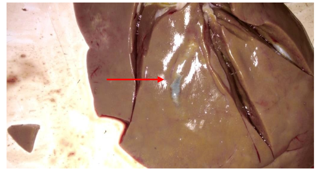
Figure 1: Sheep liver show Fasciola migratory tracts in liver tissues
Annual financial assessment of condemned liver due to Fascioliasis in the examined carcasses was recorded in Table (4), 5778.78 and 3528 L.E. were the values of economic loss because of affected liver condemnation during 2017 and 2018, respectively, with total losses of 9306.78 Egyptian pounds
Table (4): The annual financial assessment of condemned liver due to Fascioliasis in the examined sheep carcasses
|
Year |
Positive affected carcasses |
Condemned Liver (Kg) |
Value in EGP (LE) |
|
2017 |
126 |
96.313 |
5778.78 |
|
2018 |
56 |
39.200 |
3528 |
|
Total |
182 |
235.513 |
9306.78 |
N.B. the price of meat and offal/kg according to the Egyptian General Authority for Veterinary Services
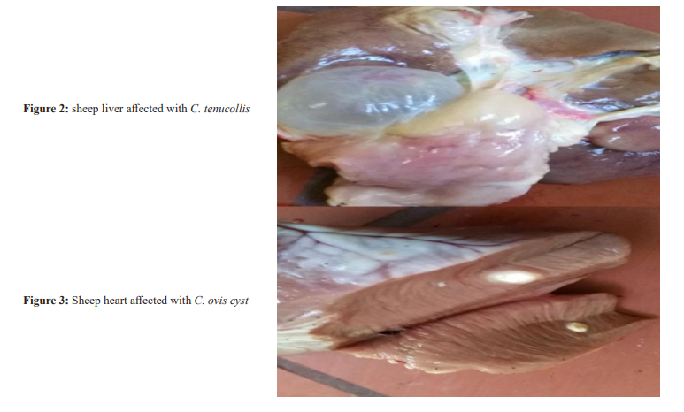
Moreover, cysticercosis, as recorded in Table (5) and Figs. (2 and 3), was detected in 0.8% and 1.35% of the examined carcasses during 2017 and 2018, respectively. In addition, C. ovis and C. tenuicollis were detected in a total of 18 (32.14%) and 38 (67.85%) examined heart and liver samples during the period of investigation, respectively, which revealed the higher liver cysticercosis than heart affection during the study as recorded in Table (6).
Table: (5): Prevalence of Cysticercosis in the examined sheep carcasses
|
Year |
No. of examined carcasses |
Positive affected carcasses |
% |
|
2017 |
2729 |
22 |
0.80 |
|
2018 |
2510 |
34 |
1.35 |
|
Total |
5239 |
56 |
1.06 |
Table (6): Prevalence of C. ovis and C. tenuicollis in the infected sheep carcasses
|
Year |
Total No. of positive affected carcasses |
C. ovis |
C. tenuicollis |
||
|
+ve |
% |
+ve |
% |
||
|
2017 |
22 |
5 |
22.7 |
17 |
77.2 |
|
2018 |
34 |
13 |
38.2 |
21 |
61.7 |
|
Total |
56 |
18 |
32.14 |
38 |
67.85 |
Referring to the condemned affected parts with cysticercosis as were recorded in Table (7); 714 and 2178 L.E. were the total value of economic losses after the affected hearts and liver condemna-tion because of C. ovis and C. tenuicollis infection during the peri-od of investigation, respectively.
Table (8) revealed the prevalence of Hydatidosis in the examined lung and liver samples of the examined sheep carcasses. Liver samples appeared to be the prominent affected organ with the inci¬dence of 1.35% along the investigation period; on the other hand, 0.47% of the examined lung samples appeared to be affected with hydatidosis. Furthermore,
Table (9) showed that the annual eco¬nomic losses for total or partial condemnation of the affected parts were 978 and 3402 L.E. due to hydatidosis during 2017 and 2018, respectively.
Table (7): The annual financial assessment for cysticercosis in the affected sheep carcasses
|
Year |
C. ovis |
|||
|
|
positive affected carcasses |
Heart (Kg) |
Liver (Kg) |
EGP (LE) |
|
2017 |
5 |
1.8 |
0 |
153.0 |
|
2018 |
13 |
5.1 |
0 |
561.0 |
|
Total |
18 |
6.9 |
0 |
714.0 |
|
Year |
C. tenucollis |
|||
|
|
positive affected carcasses |
Heart (Kg) |
Liver (Kg) |
EGP (LE) |
|
2017 |
17 |
0 |
12 |
720 |
|
2018 |
21 |
0 |
16.2 |
1458 |
|
Total |
38 |
0 |
28.2 |
2178 |
|
Total economic losses |
56 |
6.9 |
28.2 |
2892 |
N.B. the price of meat and offal/kg according to the Egyptian General Authority for Veterinary Services
Table (8): Prevalence of hydatidosis in the examined sheep carcasses’ lung and liver
|
year |
Total No. of examined carcasses |
Lung |
Liver |
||
|
|
|
No. |
% |
No. |
% |
|
2017 |
2729 |
5 |
0.18 |
22 |
0.81 |
|
2018 |
2510 |
20 |
0.79 |
49 |
1.95 |
|
Total |
5239 |
25 |
0.47 |
71 |
1.35 |
|
Total * |
5239 |
96 |
1.83 |
|
|
Total*: total incidence of lung and liver affections along the investigation period.
Table (9): The annual financial assessment for partial condemnation due to Hydatidosis.
|
|
Lung |
Liver |
Total EGP (LE) |
||||
|
Year |
No. of infest- ed organs |
Weight (Kg) |
EGP (LE) |
No. of infected organs |
Weight (Kg) |
EGP (LE) |
|
|
2017 |
5 |
2.100 |
126 |
22 |
14.2 |
852 |
978 |
|
2018 |
20 |
11.8 |
1062 |
49 |
26.0 |
2340 |
3402 |
|
Total |
25 |
13.9 |
1188 |
71 |
40.2 |
3192 |
4380 |
Discussion
Parasitic affections constitute a large group of infectious diseas¬es with varying host ranges and patterns of transmission. Their distribution, prevalence and transmission patterns are affected by the influence of both human and environmental factors. The eco¬nomic and public health impact of such zoonosis warrants appro¬priate surveillance to obtain enough information that will provide inputs in the design and implementation of control strategies. A need therefore arises to regularly re-evaluate the current status of zoonotic diseases, particularly in view of new data available as a result of surveillance activities and the application of new technol¬ogies [7].
In the current study, a total number of 5239 sheep carcasses were examined during the period of 2017 and 2018 for the presence of parasitic infection in local abattoir in Cairo governorate. The over- all prevalence of infested slaughtered animals in this study was 5.32% (Table, 2). This record was higher than other studies record in Egypt as recorded by (1.51%). Among the detected parasitic infection during this study, Fascioliasis was the most prevalent fol¬lowed by Hydatidosis, while the lowest was for Cysticercosis. This differs from the recorded prevalence in other studies in Egypt may be referred to differences in the period, locality and the rearing environment of the examined animals [8].
Fascioliasis is a parasitic affection threatening domestic ruminants and public health. The results given in Table (3) disagreed with those obtained by (20.56%), (0.41%), and who recorded lower in¬cidence during 2017 and 2018 to be 0.54% and 0.22% of the ex¬amined sheep carcasses, respectively. While, recorded detection of Fasciola in 23% of the examined sheep carcasses in South- East¬ern Lake, Chad, and (14.7%). This may be explained by difference in the total number of slaughtered animals in each study. Annual financial assessment of condemned liver due to Fasciolosis in the examined carcasses was recorded in Table (4). Total losses were summed to be 9306.78 LE along the investigation period; where 2017 recorded higher losses (5778.78 L.E.) than 2018 (3528 L.E.) [9, 12].
The obtained results in Tables (5 and 6) revealed that the incidence of cysticercosis in the examined carcasses was more prevalent in 2018 (1.35%) than 2017 (0.8%), where C. tenuicollis (67.85%) was more prevalent than C. ovis (32.14%) in the examined liver and heart samples, respectively. In addition, annual financial as-sessment of condemned affected heart and liver samples was eval¬uated as 714 and 2178 L.E., respectively; where 2018 recorded higher losses than 2017 (Table, 7).
This result differed from those recorded by who mentioned that only one C. ovis cyst was detected in the examined sheep carcass¬es [13]. Recorded lower incidence of C. ovis detection in sheep carcasses (8.43%), (1.27%), and (0.54 and 0.05% during 2017 and 2018, respectively) [14]. The results of the incidence of hydati¬dosis in the examined carcasses Table (8) revealed that the total hydatidosis was more observed in liver (1.35%) than the examined lung samples (0.47%) [15]. Results differed with those recorded by (0.3%), (63.8%), and who recorded that more than half of the examined sheep’s liver and lung offal (54.3%) harbored cystic echinococcosis, while agreed with who recorded higher incidence in the examined sheep carcass samples than the other examined species [16].
In addition, annual financial assessment of condemned affected lung and liver samples was evaluated as 978 L.E and 3402 L.E. in 2017 and 2018, respectively; where 2018 recorded higher losses than 2017 (Table, 9). Variation between the current data and the previous recorded results can be attributed to variation in season of collection, age of the animal, rearing environment, and type of feeding.
Conclusion
Conclusively, the detected parasitic affections and demonstrated economic losses throw lights over the importance of strict well qualified meat inspection in slaughter houses to avoid the serious zoonotic meat-borne parasites to the consumers. Additionally, it recommended authorities of the scope to prepare qualified veteri¬nary inspectors to safeguard the public health of the human-being. Furthermore, affected parts must be condemned in strictly isolated closed area with strict hygienic withdrawal procedures of the con¬demned parts [17, 22].
References
- Halasa, T., Enemark, H. L., Thamsborg, S. M., Toft, N., Fran-kena, K., & Olsen, A. (2015). Prevalence, risk factors and spatial analysis of liver fluke infections in Danish cattle herds.
- Youssef, A. I., & Uga, S. (2014). Review of parasitic zoonosesin Egypt. Trop Med Health 42: 3–14.
- Hassanin, F. S., Shaltout, F. A., & Afifi, M. E. (2013). Parasitic affections in edible offal. Food Control Department, Faculty of Veterinary Med. Moshtohor, Benha University, Egypt.
- Ortega, Y. R., & Cama, V. A. (2018). Cystoisospora belli andSarcocystis spp. In Foodborne Parasites (pp. 57-72). Springer,Cham.
- El-Meleh, G. S., Elmeghnawy, R. A., Sabike, I. I., & Hassan,M. A. (2019). Parasitic affections of edible offales of slaughtered animals at El-Shohada abattoir, Monofia governorate, Egypt. Benha Veterinary Medical Journal, 36(2), 117-128.
- Thrusfield, M. (2007). Some general epidemiological concepts. In Veterinary Epidemiology, 3rd Ed., Wiley-Blackwell,P. 20–29, ISBN: 978-1-118-71341-9.
- Komba, E. V. (2017). A literature survey of common parasitic zoonoses encountered at post-mortem examination in slaughter stocks in Tanzania: economic and public health Implications.
- Tas, E. E., Akcay, G. F. Y., Yildirim, F., & Yavuz, F. (2018). Coexisting primary ovarian and omental hydatid disease mimicking an ovarian neoplasm: a case report. International Journal of Gynecological Pathology, 37(3), 301-304.
- Abraham, J. T., & Jude, I. B. (2014). Fascioliasis in cattle and goat slaughtered at Calabar abattoirs. Journal of Biology, Agriculture and Healthcare, 4(18), 34-40.
- Elmonir, W., Mousa, W., & Sultan, K. (2015). The Prevalence of Some Parasitic Zoonoses in Different Slaughtered Animal Species at Abattoir in the Mid-Delta of Egypt; with Special Reference to its Economic Implications. Alexandria Journal for Veterinary Sciences, 47(1).
- Jean-Richard, V., Crump, L., Abicho, A. A., Naré, N. B., Greter, H., Hattendorf, J., ... & Zinsstag, J. (2014). Prevalence of Fasciola gigantica infection in slaughtered animals in south-eastern Lake Chad area in relation to husbandry practices and seasonal water levels. BMC veterinary research, 10(1), 1-8.
- Amer, S., ElKhatam, A., Zidan, S., Feng, Y., & Xiao, L. (2016). Identity of Fasciola spp. in sheep in Egypt. Parasites & vectors, 9(1), 1-8.
- Gessese, A. T., Mulate, B., Nazir, S., & Asmare, A. (2015). Major metacestodes in small ruminants slaughtered at Dessie municipal abattoir, Eastern Ethiopia: prevalence, cyst viability, organ distribution and economic implications. Comparative Clinical Pathology, 24(3), 659-668.
- Hashemnia, M., & Kish, G. F. (2016). Prevalence and pathological lesions of ovine cysticercosis in slaughtered sheep in western Iran. Journal of parasitic diseases, 40(4), 1575-1578.
- Ernest, E. (2004). Studies on the epidemiology of echinococcosis/hydatidosis in Ngorongoro district, Arusha region, Tanzania. Int Arch Hydatid, 35, 43.
- Abdulhameed, M. F., Habib, I., Al-Azizz, S. A., & Robertson,I. (2018). Cystic echinococcosis in marketed offal of sheep in Basrah, Iraq: abattoir-based survey and a probabilistic model estimation of the direct economic losses due to hydatid cyst. Parasite epidemiology and control, 3(1), 43-51.
- Amer, S., ElKhatam, A., Zidan, S., Feng, Y., & Xiao, L. (2016). Identity of Fasciola spp. in sheep in Egypt. Parasites & vectors, 9(1), 1-8.
- Salah, A. E. W., Fouad M, H., Mohamed, S., & Manal MA,R. (2002). Fascioliasis among live and slaugthered animals in nine centers of Dakahlia governorate.
- Fouad M, H., Badawiya B, I., Soha E, A., Doaa M, S., & Gehad T, E. S. (2006). Hydatidosis granulosus in Egyptian slaughtered animals in the years 2000-2005.
- Ortega, Y. R. and Cama, V. A. (2018): Cystoisospora belli andSarcocystis spp. foodborne parasites, In: Food Microbiolo-gy and Food Safety, 2nd Ed., Y.R. Ortega and C.R. Sterling (Eds.), Springer, Ch. 4, p. 57-72.
- MusotsI, P. Y., Otieno, C. A., & Njoroge, S. M. (2017). Background: Fasciolosis is known globally to be an important hel-minthic disease of ruminants caused by liver fluke species of the genus Fasciola, and it is one of the most neglected tropical zoonotic diseases that can lead to human infection. It has thewidest geographic spread of any emerging zoonotic disease, and it occurs in many countries of the world. Objectives: The study aimed at determining the prevalence of fasciolosis in ruminants slaughtered in Trans-Nzoia West. Specific objectives were to ....
- Youssef, A. I., and S. Uga. “Review of parasitic zoonoses in Egypt. Trop Med Health 42: 3–14.” (2014).