
International Journal of Diabetes & Metabolic Disorders(IJDMD)
ISSN: 2475-5451 | DOI: 10.33140/IJDMD
Impact Factor: 1.23


Research Article - (2024) Volume 9, Issue 2
Musa Parasidiaca (unripe plantain) Enhanced Uptake of Glucose in Yeast Cells-In Vitro for the Control of Hyperglycemia
2Programs coordinator Nutrition and Dietetics, Department of Life and Health Sciences, University of Nicosia, 46 Make¬donitissas Ave, CY-2417, P.O. Box 24005, CY-1700, Cyprus
3Department of Life and Health Sciences, Human Biology, University of Nicosia, 46 Makedonitissas Ave, CY-2417, P.O. Box 24005, CY-1700, Cyprus
4Department of Life and Health Sciences, Pharmacy, University of Nicosia, 46 Makedonitissas Ave, CY- 2417, P.O. Box 24005, CY-1700, Cyprus
5Department of Biochemistry, Faculty of Sciences, Federal University of Technology, P.M.B. 65, Minna, Niger State, Nigeria
Received Date: Apr 13, 2024 / Accepted Date: May 15, 2024 / Published Date: Aug 01, 2024
Copyright: ©Â©2024 Morris, T. Aloysius, et al. This is an open-access article distributed under the terms of the Creative Commons Attribution License, which permits unrestricted use, distribution, and reproduction in any medium, provided the original author and source are credited.
Citation: Aloysius, M. T., Eleni, A., Felekkis, K., Petrou, C., Evans, E. C. (2024). Musa Parasidiaca (unripe plantain) Enhanced Uptake of Glucose in Yeast Cells- In Vitro for the Control of Hyperglycemia. Int J Diabetes Metab Disord, 9(2), 01-13.
Abstract
Diet plays a vital role as a risk factor for chronic diseases like hyperglycemia; And type 2 diabetes with diet alone or diet and antidiabetic drugs (in combination with insulin in a very few cases where the pancreas produced no insulin due to age). The objectives of dietary management of diabetes are to: achieve optimal blood sugar and blood lipid concentrations; provide appropriate energy for reasonable weight, prevent, delay and treat diabetes-related complications and improve health through optimal nutrition. Musa parasidiaca (unripe plantain) enhanced uptake of glucose in yeast cells - in vitro for the control of hyperglycemia was studied. Plant materials were collected, identified, processed, and stored for further use. 80% methanol was employed for extraction and sonication to release antidiabetic-bioactive component in solution and was filtered, concentrated, freeze-dried, and fractionated using standard laboratory techniques. The extract fractions were employed to evaluated the ability of yeast cell line culture to take up glucose from the system through DPPH, FRAP, lipid peroxidation, antidiabetic effect assays. Extract fractions were found to poses antioxidant activity high enough to inhibit stress-related diseases. The extract fractions were active as drug candidates both at high and low concentrations and were better compared with the standard drug and standard antioxidant was comparable. Glucose uptake at an initial concentration of 5mM/L and 10mM/L by the crude extract was consistent to that of the known standard drug while at 25mM/L glucose concentration was equivalent with the crude extract. At 0.625 mg/mL the linear equations, and R2 demonstrations shows that the crude extract was higher in dose predictability than the standard drug as presented by the equation; y = 21.416x - 15.575, and R2 = 0.9747 (97%). The predictability of the food plant extracts-future effectiveness and usability is higher than the conventional drug. Encapsulation of the bioactive-fractions of food plants with nanogel and incorporating insulin releasing system which may lead to prospects for the design of type 2 diabetes drugs to support an extensive blood glucose regulation, which is extremely required by diabetics in order to decrease the problem of drug regimes.
Keywords
Hyperglycemia, Glucose-Uptake, Musa Parasidiaca, Cell Line Culture, Extract Fractions, Type 2 Diabetes
Introduction
Musa paradisiaca (unripe plantain) contains the subsequent proximate composition: Moisture 59g, Crude protein 7.7g%, Crude lipid 1.5g%, Ash 1.4g%, Crude fiber 1.4g%, Carbohydrate 24.4g%, and gross energy 148.6Kcal/100g. At the same time, the mineral composition consists of sodium 200mg, Potassium 370mg, Calcium 126.5g, Magnesium 375mg, Iron 2.53mg, Phosphorus 220mg, Zinc 3.74mg, Manganese 2.99mg, and Copper 1.66mg [1].

Figure 1: Musa Paradisiaca
Fruits Diabetes is a persistent disorder characterized by hyperglycemia, insufficient insulin secretion and disrupted metabolic pathways. It affects over 400 million individuals worldwide aged 18 years and above, particularly in low and middle – income countries, and is projected to be the 7th leading cause of death by 2030.1 Hyperglycemia constitutes noninsulin-dependent hyperglycemia, which accounts for 90% of all cases, and is mainly caused by insulin resistance, partial insulin deficiency, and abnormal postprandial glucose elevation [2-4].
The rise in the prevalence of prolonged diseases such hyperglycemia in developing countries can attributed to changes in nutrition and lifestyle from traditional meals, which are high in nutrients of food plant based like grains, legumes and fruits, and vegetables to Westernized meals rich in sugars, fat, and animal-source diets [5-7]. Therefore, alternative strategies are needed that employ nutrient content from plants such as Digitaria exilis (acha), Treculia Africana (breadfruit), bean plant (beans), and Musa paradisiaca (unripe plantain) that have been found useful for controlling hyperglycemia [8-11].
Therapeutic plants have traditionally been employed to discover bioactive compounds for drug formulation due to their improved therapeutic capabilities compared with conventional drugs that encounter cell resistance challenges at high costs with increasing toxicity levels on human health due to exposure from injurious substances present within the environment [12-15]. The World Health Organization reported that over 80% of people globally rely significantly on therapeutic plants for essential health needs highlighting the importance of natural bioactive compounds as alternatives or supplements to conventional drugs [16]. At the moment, about 20% of the currently existing medications contain phytochemicals as part of their bioactive constituents [12]. The bulk of the human diseases emerging from the activities of microorganisms, infections, disordered conditions from metabolic difficulties, and illnesses related to oxidative stress use therapeutic plants [17-20].
The glucose toxicity theory suggests constant exposure to modest increases in glucose over an extended period seriously impact cells contributing significantly the effect of type 2 diabetes, hyperglycemia, is projected as a secondary reason behind continued cell decline.
Equally, there is attention growing in using natural products from plants as substitutes to current medications. Plant sources have become the most target for getting new drugs to help manage diabetes.
Antioxidants are substances that are able to retard chemical reactions, no matter the concentrations; hence, antioxidants have various physical as well as chemical roles within the body. Moreover, antioxidants perform similar to the secondary substances accustomed to stopping thermal oxidation by responding with the reactive radicals and destroying them to quieten down activeness, less harmful, and long-sustained materials than those radicals. Antioxidants could also deactivate free radicals by accepting or donating electron(s) to get rid of the unpaired status of the novel [21]. Likewise, antioxidants may serve as compounds ready to stop oxygen-mediated oxidation of various substances from modest molecule to polymer as well as sophisticated bio-system [22].
The condition of stress where there are differences between production and accumulation within the cells and tissues of the body as well as detoxifying the product of the reaction because of absence in antioxidant or enlarged reactive oxygen species (ROS), reactive nitrogen species (RNS), and reactive sulfur species (RSS) formation, could create a possibility of destroying the cells [23,24]. The reactive oxygen species could be accustomed combined and including all extreme reactions of oxygen varieties, comprising molecular species able to exist alone which contains an unpaired electron in the atomic orbital. These reactive oxygen species are grouped into hydroxyl group (OH•), per hydroxyl group (HO2•), hypochlorous acid (HOCl), Superoxide anion group (O2•¯), hydrogen peroxide (H2O2), singlet oxygen (1O2), nitric oxide gas group (NO•), hypochlorite group (OCl•), peroxynitrite (ONOO), and several organic substances, and peroxides. On the other hand, reactive nitrogen species comes from the nitric oxide gas group as a result of reaction with oxygen ion to produce peroxynitrite ion however, reactive sulfur species is well-formed after the reaction with thiols and reactive oxygen species [23,21]. Because of molecular species that are able to exist alone as an electron, these molecular species exhibit an extreme tendency to be involved chemically with different molecules so as to reach neutrality. The molecular species that are able to exist alone as an electron have vital roles in cell variation of the quantity of information, programmed cell death, the movement of ions through a membrane inactively as well as actively by the means of ion channels or pumps, as well as accustoming the information encrypted in a gene to bring together a protein molecule [21]. The degree of biochemical reaction of deactivated molecular species that are able to exist alone as an electron could destroy the whole cell of very large molecules, comprising of carbohydrates, proteins, lipids, and nucleic acids. Therefore, cells can be responsible for their safeguard against the destroying effect of reactive oxygen species through occurring as well as functioning within a cell physiologically relevant biochemical stimulus, bonding of ions as well as molecules to metal ions, and antioxidant to maintain reactive oxygen species at the same as well as steady-state for survival at an occasional stage. Moreover, dietary materials that safeguard cells from the harmful effect of free radicals could help in maintaining a suitable antioxidant position within the body. However, in times of deviation in conditions from species optima and alteration in the endothelial, stages of reactive oxygen species could proliferate vividly and affect major cell impairment within the body.
As a result, imbalances between production and accumulation of oxygen reactive species meaningfully add to the chain of events leading to various illnesses, like cardiovascular illnesses, inflammatory illnesses, cancer illness, diabetes mellitus, illnesses that destroy the ability of the brain to carry out the simplest tasks (Alzheimer's illnesses), autism, and therefore the aging progression [23-25]. Stress related complaints arise when there are modifications amid creating and buildups within the cells, tissues cleansing the product resulting from lack of antioxidant or distended reactive oxygen species (ROS), reactive nitrogen species (RNS), and reactive sulfur species (RSS) formation, posing risks towards terminating cell life [53-56,26,27].
Methods
Chemicals
The analytical grade chemicals utilized in this study were products of Sigma Aldrich. The list of chemicals includes methanol, phosphate buffer, 2,2-diphenyl-1-picrylhydrazyl (DPPH), potassium hexacyanoferrate(III), ferric chloride, thiobarbituric acid (TBA), sodium dodecyl sulfate (SDS), ferrous sulfate, acetic acid (TCA), baker’s yeast, ascorbic acid (vitamin C) and metronidazole (standard diabetes drug).
Plant Material: Collection and Identification
Musa paradisiaca (unripe plantain fruit) was procured from commercial sources located in Benue and Nasarawa States of Nigeria. A botanist identified the plant after which it was peeled, washed and cut into small pieces before being air-dried at a temperature of 37oC for three days to reduce their moisture content.
Grinding Process (Pulverization)
An electronic Grinder model Nima Japan was used to grind the sample Musa paradisiaca (unripe plantain or UNRP) to powder form. The powdered sample was packed into polystyrene bags which were then sealed and placed in a desiccator containing colloid (desiccant). This prevented any absorption of moisture by the sample from the atmosphere. The dried pulverized material was stored in a desiccator until use.
Methanol Extraction of Plants Sample
A suitable solvent (methanol) was used to extract active substances from fine powdered materials obtained from Musa paradisica. For preparing methanol extract, 100g of powdered Musa paradisica was weighed into a 1000ml beaker where it underwent thorough extraction by adding 80% methanol for eighteen hours at an optimal sonicating temperature (30oC) under shaking conditions. Every six hours during sonication for twenty minutes’ precise antidiabetic agents (bioactive component) present within the plant sample were obtained followed by filtration that yielded a final volume of 1 litre (1000mL). This extract passed through Whitman paper No.1 filter paper before being concentrated under reduced pressure with controlled temperature range between 40-50oC using digital controlled water bath; fractionalization occurred sequentially (partitioning) using n-Hexane Chloroform Ethyl ethanoate (Ethyl acetate). Evaporation under reduced pressure resulted in fractions partitioned such as n-Hexane, Chloroform, and Ethyl ethanoate extracts.
Filtration of Extracted Sample
After sonication there existed transparent separation between supernatant and residue cemented on the bottom of conical flask; however tiny residues entering filtrates if decanted got prevented due to the filtration process employed here i.e., folding twice Whitman paper No1 onto the plastic funnel over the mouth of the conical flask gradually pouring the solution which was separated via the funnel with filter paper resulting in the collection of filtrate at the bottom of the conical flask while retaining the residue via the filter paper.
Concentration of the Filtrate
The collected filtrates contained both waters along with methanol alongside the extract hence digitally regulated water bath evaporated all traces of methanol present at 40oC.
Freeze-Drying
Extracts that had undergone concentration still contained some amount of water even after post-evaporation; thus, these extracts got frozen till -20°C followed by drying them with vacuum-compressed system (dryer); freeze dryer model number LGJ-18 fitted compressor pump facilitated this step.
Fractionation of Crude Extract (Partitioning)
To fractionize crude extracts (methanolic)10gms was dissolved in distilled water with volume equivalent to100 mL underwent partitioning into increasing order of solvent polarity (n-hexane
|
Sample |
Hex |
CHCl3 |
EtOAc |
Aqueous |
|
Unripe Plantain (UNRP) |
UH1, UH2 |
UC |
UE |
- |
Table 1: Showing the Fractions from the Partitioning of Crude Extract With N-Hexane, Chloroform, Ethyl Acetate and Aqueous Solution
Note that the above fractional coding represents the subsequent fractions:
|
|
Hex |
CHCl3 |
EtOAc |
Aqueous |
|
Wt. of Solvent used |
100% |
100% |
100% |
100% |
|
Wt. of fractions (g) |
UH1 =2.9 UH2 =2.6 |
UC =1.5 |
UE =3.0 |
|
|
% Wt. of the fractions |
UH1 =29 UH2 =29 |
UC =15 |
UE =30 |
|
Table 2: Percentage Yield of Fractions From 10gm Of the Crude Extracts
In Vitro Antioxidant of 2,2-Diphenyl-1-Picrylhydrazyl (DPPH) Free Radical Scavenging Assay
The antioxidant activities of the plant extract were estimated using DPPH free radical scavenging assay as described by Oyaizu. [29] Different concentrations of crude extract and ascorbic acid (vitamin C as control) at concentrations of 31.25, 62.5, 125, 250, and 500 µg/mL as well as that of the fractions with similar concentrations were prepared from stock solutions (1000µg/mL) was prepared by weighing and dissolving 0.0005g of the crude extract and ascorbic acid in 100mL of methanol. Later, 2 mL of 0.004% DPPH in methanol was added to 1 mL of varied concentrations of crude extract and extract fractions as well as ascorbic acid. The reaction mixtures were incubated at 25°C for 30 minutes. The absorbance of each test mixture was read against a blank at 517nm engaging a double beam Shimadzu UV-1800 series spectrophotometer. The experiment was carried out in triplicates. The percentage (%) antioxidant activity was calculated using the formula below: % scavenging activity = Absorbance of blank - Absorbance of the sample / Absorbance of blank x 100
Ferric Reducing Antioxidant Power (Frap) Assay
Valuation of antioxidant activity of the crude extract and extract fractions through ferric reducing antioxidant power assay was constant with the method of Oyaizu. [29] 0.0005g of crude extracts and ascorbic acid as control were weighed and dissolved in 100 mL of methanol (1000µg/mL), from which different concentrations of 31.25, 62.5, 125, 250, and 500µg/mL were prepared. During this assay, 1 mL of each plant extracts, vitamin C, 1 mL of 0.2 M sodium orthophosphate buffer, and 1 mL of 1% Potassium hexacyanoferrate (III) were mixed together, and incubated at 50°C for 20 minutes. After that, 1 mL of 10% TCA was added to 1 mL of each concentration of the extracts and was mixed with 1 mL of water and 0.2 mL of 0.1% Ferric chloride. The absorbance of the test samples was read at 700 nm with distilled water as blank. The % of antioxidant activity was calculated using the formula: % activity = Absorbance of sample - Absorbance of blank / Absorbance of sample x 100.
Inhibition of Lipid Peroxidation by Crude Extract and Extract Fractions Assay
The inhibitory effects of crude extract and extract fractions on lipid peroxidation were determined using the method of Halliwell et al [30] with slight modification. Briefly, 0.5 mL of 10% egg homogenate was added to 0.1 mL of crude extract and fractions and ascorbic acid as control at various concentrations of 31.25, 62.5, 125, 250, and 500µg/mL as well as 1 mL of water was added. Afterward, 0.05 mL of FeSO4 was added to the mixtures and incubated for 30 minutes. Then, 1.5 mL of carboxylic acid and thiobarbituric acid (TBA) in sodium dodecyl sulfate was added. The resulting reaction mixture was vortexed and incubated at 95°C for 1h. Reaction mixtures were allowed to cool, and 5 mL of butanol was added to each of the reaction mixtures and centrifuged at 1200 rpm for 10 minutes, and the absorbance of the samples was read at 532nm. The % inhibition of lipid peroxidation was calculated with the formula: % Inhibition = Absorbance of blank - Absorbance of sample / Absorbance of blank X 100.
Antidiabetic Effect of Musa Paradisiaca On Glucose Uptake in Yeast Cell
The assay was constant with the method of Cirillo, with slight modification. Commercial baker's yeast was dissolved in distilled water to prepare 1% suspension [31]. The suspension was kept overnight at a temperature of 37°C. The following day, yeast cell suspension was centrifuged at 4200 rpm with High-speed refrigerated 4 bucket centrifuge model, LR10 – 2.4A, 50/60 Hz, and 220–240 V for 5 minutes. The method was repeated by adding water to the pallet until a transparent supernatant was obtained. Exactly 10 parts of the clear supernatant fluids was mixed with 90 parts of distilled water to get a 100ml v/v suspension of the yeast cell. Due to the extract's solubility, about 1 mg w/v of plant crude extract and extract fractions was mixed with dimethyl sulfoxide (DMSO4). A serial dilution of extract was done at the concentration of 0.625, 1.25, 2.5, and 5mg/mL for the crude extract as well as 31.25, 62.5, 125, 250 and 500µg/mL for extract fractions respectively. Samples were reacted with concentrations of 5, 10, and 25mM/L of 1mL of glucose solution and incubated for 10 minutes at 37°C. The reaction was initiated with the addition of 100mL of yeast suspension to the samples of glucose and extracts. The samples were vortexed and incubated for 1h more at 37°C. After the incubation, 3,5-dinitrosalicylic acid (DNSA) was added to the tubes and was placed in boiling water for 5 minutes (but the tubes were not allowed to boil – this was to permit for the rapid reaction of the extracts, glucose as well as the yeast cell) the glucose uptake was read by engaging a spectrophotometer (UV - 1800 SHIMADZU) at 540 nm. Absorbance for the control was carried out on a similar wavelength. The % increase in glucose uptake was calculated with the formula: % increase in glucose uptake = Absorbance of control - Absorbance of the sample / Absorbance of control x 100, where control is the solution containing all reactants except the test sample. Metronidazole was used as the standard drug (control).
Statistical Analysis
Data were collected using a one-way, and two-way analysis of variance (ANOVA) as well as independent T-test, and paired T-test analysis. Groups were considered significant if P<0•05 and, a F-value was significant for ANOVA; the differences between all pairs were carried out using Ducan Post Hoc Test; SPSS version 26, and Microsoft excel windows 10 were used for statistical analysis, and data figure generation.
Results
Antioxidant Activity of the Crude Extract and Extract Fractions of Musa Paradisiaca
DPPH is a suitable free radical compound usually used to test free-radical scavenging activities in various types samples. The DPPH radical scavenging test depends mostly on compounds capable enough to donate hydrogen atoms that stabilize free radicals which prevent oxidation chemical combinations established in active organisms [32]. Though not biologically mimicking any biological compounds, the DPPH assay is often considered as an indicator showing plant extracts' ability to extinguish free radicals. They also indicate their hydrogen atom or electron donation abilities even without enzymatic action [33]. The use of DPPH is advantageous when assessing antioxidant activities exhibited by extracts through its scavenging capacities (figure 1) due to its higher stability than hydroxyl and superoxide radicals [34]. Hence, the antioxidant activities displayed may be attributed mainly to their ability either as hydrogen atom donors or electrons donors. Their hydrogen donating capability can be traced back to phenolic compound presence since these secondary metabolites have been found useful possessing antioxidant properties [35]. Therefore, it's reasonable deducing that higher activity levels shown in this study could be because they contained high concentrations of phenolics within them. Electron-donating antioxidants generally reduce iron (Fe) oxidation state from Fe3+ to Fe2+, which reproduces their electron-donating capabilities (reducing ability); hence, the more significant antioxidant effects suggest they were able reducing Fe3+ to Fe2+ [36]. This reveals their electron-donating abilities indicating possible usage stopping oxidative stress-related diseases like chronic diseases caused by damage from cardiovascular disease, cancer, and diabetes. Figure 2 shows evidence supporting this claim where extracts present considerable ferric-reducing power. Lipid peroxidation describes oxidation breakdowns occurring in lipids, a process where free radicals abstract electrons from cell membrane lipids affecting polyunsaturated fatty acids due to double bond presence. This process has been linked with damaging cellular systems via preamble membrane causing isolation between inside and outside the surroundings [37]. Damage resulting from influences affects many individuals leading to diseases such as cancer disease cardiovascular etc [38]. Extracts' ability considerably inhibit lipid peroxidation implies extinguishing actions of free radicals, stopping concept electrons from cell membrane lipids by free radicals thus shielding humans against chronic diseases related oxidative stress. Results obtained show crude extracts possess antioxidant properties making them useful in treating and managing oxidative stress-related diseases.
The percentage scavenging antioxidant activity (also referred to as DPPH) of the extract fractions table 3 showed decreased scavenging antioxidant ability with decreased extract fraction concentration. The extract fractions UC (66.11%) and UE (51.31%) revealed higher antioxidant percentage activity compared to other fractions partitioned. All of the higher activity was exhibited at the concentration of 500µg/mL; The percentage ferric reducing antioxidant power (FRAP) of the extract fractions table 4 shows a decreased FRAP percentage capacity with a decreased extract fraction concentration. Though, the other fractions were also very high at concentrations of 500µg/mL and 250µg/mL respectively.
Also, the percentage inhibitory activity of antioxidants (also referred to as lipid peroxidation) for all extract fraction table 4 also showed decreased inhibitory activity with decreased extract fraction concentration. The standard antioxidant was higher than the extract fractions in all concentrations of the antioxidant assays (i.e. DPPH, FRAP, and lipid peroxidation). Even though the standard antioxidant was higher in antioxidant activity than most of the extract fractions, the extract fractions exhibited excellent abilities in different assays.
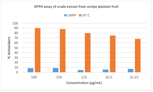
Figure 1: % DPPH Radical Scavenging Activities of Crude Extract of Musa Paradisiacal (Unripe plantain or UNRP). Values are Presented as Mean ± Standard Deviation of Triplicates. Values with High Activity Concentration are Significantly Different at P< 0.05.
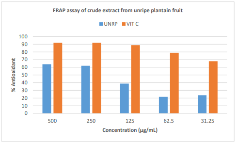
Figure 2: % Ferric Reducing Powers of Crude Extract of Musa Paradisiacal (unripe plantain or Unrp). Values are Presented as Mean ± Standard Deviation of Triplicates. Values with High Ferric Reducing Power Concentration are Significantly Different at p< 0.05.
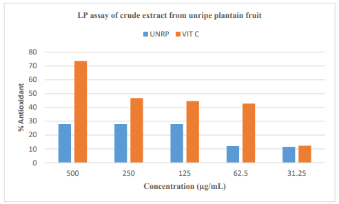
Figure 3: % Inhibitory Activities of Crude Extracts of Musa Paradisiaca (unripe plantain or unrp) on Lipid Peroxidation. Values Are Presented as Mean ± Standard Deviation of Triplicates. Values with Higher Lipid Peroxidation Activity Concentration are Significantly Different at p < 0.05.
Antioxidant Effect of DPPH 2, FRAP 2 and Lipid Peroxidation 2 on Extract Fractions
|
µg/mL |
% UH1 |
% UH2 |
% UC |
% UE |
% Vit C |
|
500 |
40.1 |
36.99 |
66.11 |
51.31 |
82.58 |
|
250 |
20.76 |
26.25 |
46.06 |
40.1 |
53.22 |
|
125 |
15.99 |
33.17 |
16.23 |
31.03 |
46.3 |
|
62.5 |
17.66 |
12.89 |
21 |
18.85 |
47.02 |
|
31.25 |
4.3 |
12.65 |
15.04 |
35.32 |
45.58 |
Table 3: Showing the % Scavenging Antioxidant Activity of Extract Fractions DPPH 2
|
µg/mL |
% UH1 |
% UH2 |
% UC |
% UE |
% Vit C |
|
500 |
67.59 |
63.31 |
71.96 |
71.54 |
95.25 |
|
250 |
50.48 |
45.24 |
61.79 |
56.84 |
95.25 |
|
125 |
36.57 |
35.71 |
35.54 |
35.99 |
93.42 |
|
62.5 |
27.87 |
17.14 |
31.44 |
35.99 |
86.37 |
|
31.25 |
8.32 |
16.92 |
29.53 |
28.8 |
77.28 |
Table 4: Showing the % Ferric Reducing Antioxidant Power (FRAP) of Extract Fractions
|
µg/mL |
% UH1 |
% UH2 |
% UC |
% UE |
% Vit C |
|
500 |
61.8 |
54.3 |
58.01 |
63 |
91.36 |
|
250 |
54.51 |
51.38 |
49.09 |
54.37 |
84.58 |
|
125 |
50.87 |
43.85 |
45.01 |
42.92 |
72.97 |
|
62.5 |
47.91 |
23.16 |
28.3 |
34.58 |
65.28 |
|
31.25 |
43.44 |
4.39 |
2.77 |
15.47 |
49.25 |
Table 5: Showing the % Inhibitory Activity of Antioxidant of Extract Fractions Lipid Peroxidation
Antidiabetic Effect of Crude Extract in Yeast Cells
Effect of crude extract of Musa paradisiaca, on glucose uptake ability by yeast cell. The crude extract of Musa paradisiaca, stirred the uptake of glucose through the moderately but not totally permeable membrane of yeast cells Figures 4, 5, and 6 respectively. The glucose uptake at an early concentration of 5mM/L and 10mM/L by the crude extract of Musa paradisiaca was agreeable to that of the known standard drug (figures 4 and 5). However, the effect of Metronidazole on glucose uptake by the yeast cell at 25mM/L glucose concentration was similar with the crude extract of Musa paradisiaca, (figure 6).
Besides, at 0.625 mg/mL the linear equations, and R2 shows that the extract were higher in dose predictability than the standard drug (Metronidazole) as shown by the equations; y = 21.416x - 15.575 and, R² = 0.9747 (97.5%) for Musa paradisiaca while y = 2.1324x + 82.881, and R² = 0.1213 (12.1%) for Metronidazole when 5 mg/ mL of crude extract of Musa paradisiaca, was used (figure 4). This suggests that increasing the concentration of crude extract of Musa paradisiaca increased the possibility of yeast cells to take up additional glucose from the surrounding; but, the standard drug though showed high glucose uptake ability, however had a very low drug-dose predictability compared to the extracts as confirmed by the very low R2 value of 12.1% beside the crude extract R2 value of 97.5%. Equally, figures 5 and 6 showed a linear upsurge in the uptake of glucose by yeast cells with a gradual upsurge in the concentration of the crude extract. Though, an inverse correlation to the molar concentration of glucose was detected, when glucose uptake by yeast cells was compared among 5mM/L, 10mM/L, and 25mM/L for the related amount of crude extract of Musa paradisiaca, (figures 4, 5, and 6).
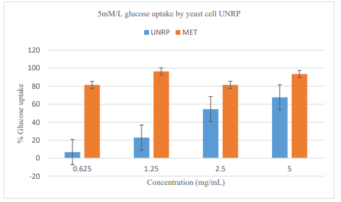
Figure 4: Glucose uptake by yeast cells at 5mM/L initial concentration of glucose in the presence of UNRP; crude extract of Musa paradisiaca (unripe plantain or UNRP); Metronidazole (MET) a synthetic diabetes drug. The error bars represent ± SE of triplicate data. The bars are significantly different at P = 0.05.
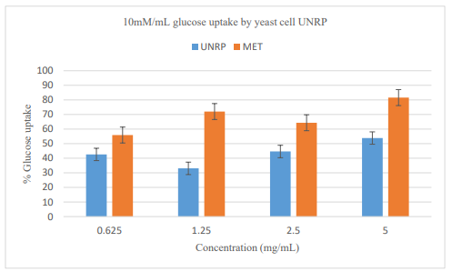
Figure 5: Glucose uptake by yeast cells at 10mM/L initial concentration of glucose in the presence of OS, BYAM, and UNRP; crude extract of Musa paradisiaca (unripe plantain or UNRP); Metronidazole (MET) The error bars represent ± SE of triplicate data. The bars are significantly different at P = 0.05.
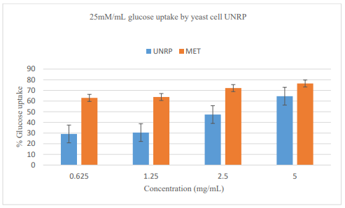
Figure 6: Glucose uptake by yeast cells at 25mM/L initial concentration of glucose in the presence of OS, BYAM, and UNRP; crude extract of) Musa paradisiaca (unripe plantain or UNRP), and Dioscorea dumetorum (bitter yam or BYAM); Metronidazole (MET). The error bars represent ± SE of triplicate data. The bars are significantly different at P = 0.05.
Antidiabetic Effect of Extract Fractions in Yeast Cell
|
µg/mL |
% UH1 |
% UH2 |
% UC |
% UE |
% MET |
|
500 |
88.06 |
47.39 |
71.32 |
65.28 |
88.18 |
|
250 |
77.97 |
40.59 |
62.39 |
53.43 |
81.93 |
|
125 |
63.11 |
37.89 |
52.54 |
51.93 |
76.5 |
|
62.5 |
56.09 |
25.5 |
49.93 |
50.26 |
54.29 |
|
31.25 |
49.63 |
15.13 |
48.39 |
47.27 |
49 |
Table 6: Showing Glucose Uptake Ability at a Concentration of 5mM/L
|
µg/mL |
% UH1 |
% UH2 |
% UC |
% UE |
% MET |
|
500 |
95.6 |
99.06 |
86.74 |
93.56 |
92.35 |
|
250 |
94.11 |
70.45 |
80.74 |
92.2 |
89.37 |
|
125 |
84.33 |
60.33 |
69.66 |
73.35 |
85.39 |
|
62.5 |
76.92 |
51.41 |
47.81 |
54.55 |
71.21 |
|
31.25 |
70.76 |
43.26 |
38.31 |
46.73 |
57.47 |
Table 7: Showing Glucose Uptake Ability at a Concentration of 10mM/L
|
µg/mL |
% UH1 |
% UH2 |
% UC |
% UE |
% MET |
|
500 |
65.04 |
61.7 |
81.8 |
68.49 |
97.43 |
|
250 |
61.88 |
52.48 |
75.53 |
66.58 |
93.67 |
|
125 |
54.27 |
47.73 |
70.91 |
62.86 |
74.09 |
|
62.5 |
51.11 |
40.17 |
65.67 |
59.59 |
69.91 |
|
31.25 |
48.55 |
38.81 |
51.28 |
49.26 |
64.86 |
Table 8: Showing Glucose Uptake Ability at a Concentration of 25mM/L
Glucose uptake ability at a glucose concentration of 5mM/L table 6 showed a decrease in the percentage of glucose uptake capacity with a decrease in extract fraction concentration. Fraction UH1 was exceptionally higher than other fractions and was comparable with the standard drug at all concentrations.
The glucose uptake ability at a concentration of 10mM/L table 7 showed a decrease in percentage glucose uptake capability with a decreased extract fraction concentration. Extract fractions UH2 (99.06 %) and UH1 (95.6 %) showed much higher glucose uptake ability than the standard drug at a higher concentration of 500µg/mL as well as UE (92.2 %) fractions were higher than the standard drug at extract fraction concentration of 250µg/mL; while extract fraction was higher compared to the standard drug in all concentrations except at extract fraction of 125µg/mL;
Discussions
Diabetes mellitus associates closely with metabolic abnormalities among which include oxidative stresses. Biochemical studies reveal increased generation of reactive oxygen species in diabetic patients' tissues and cells [39]. To combat ROS, potent body antioxidants presence is vital since it retards and prevents substance oxidation during which DPPH, FRAP (Flavonoid Radical Scavenging Antioxidant Assay System) serves as preferred method of testing potential drugs, natural products' and potential antioxidants. For instance, this modified Blois method introduced by Brand Williams et al. in 1995 serves as reference point for researchers [40]. In addition to this, some notable indicators assaying potential antidiabetic drugs include (i) potential plasma-membrane glucose uptake across muscle and adipose as well as yeast cells; (ii) glucose adsorption; (iii) inhibition of α-amylase and α-glucosidase enzymes (for in vivo studies) [41-43]. In light results obtained herein, it deductive suggesting hexane, methanol and water solvent extractions yield potent candidate for natural bioactive compounds effective in treating type 2 diabetics. Inhibiting free-radicals from locking up cell membranes which stops the flexibility of cell membrane [44, 45]. Such potent bioactive compounds identified include quercetin 3-O-glucosyl (QDG)/quercetin-3-O-glycoside (QG), oxacyclododecane 2-one imidazole, bioflavonoids, eugenol, caryophyllene, copene azulene, dodecatetraenamide, and phenethylamine [46-51]. Consequently, the presence of one the above compounds explains the mechanism behind uptake of glucose in the present study. On a general basis, muscle cells' glucose-uptakes is due to accumulation of functional glucose transporting molecules in cell membrane. Regulated leptocytes and myocytes responses to high insulin secretion in blood, hypoglycemic effect ensues [52]. Nonetheless, researchers need to explore further drug fractions in vivo trials (here the focus is only on in vitro).
Conclusions
Extract fractions proved active as drug candidates both at low and high concentrations and compare favorably well with standard drug and standard antioxidants. From the results obtained, it can be concluded that increasing extract solution concentration boosts glucose yeast cell uptake. Nutrients in extracted-fractions are naturally safe and without side effects unlike most standard drugs. Furthermore, the findings show that R² which is the metric regression-error explains the model performance used in predicting the response and the target variable values. Further research is Anticipated in encapsulating the high bioactive extract fractions separately and coding it based on its functions as diabetes-drug candidates. An animal as well as a human volunteered trial will be vital to monitor the in vivo performances of the extract fractions while following necessary research protocols.
Institutional Review Board Statement
The study was conducted in accordance with the Declaration of Helsinki, and approved by the Institutional Review Board of University of Nicosia; approval date is 15 January 2020.
Contributors
“Conceptualization, MA. and EA.; methodology, MA and EA.; software, MA and EA.; validation, KF., and CP.; formal analysis, MA.; investigation, EA, KF and CP.; resources, MA and EA.; data curation, MA and EA.; writing - original draft preparation, MA.; writing - review and editing, MA, EA; visualization, MA, EA, KF, and CP; supervision, EA, KF, and CP; project administration, EA; funding acquisition, EA, KF, and CP. All authors have read and agreed to the published version of the manuscript.”
Funding
This research received no external funding
Competing interests
None declared
References
- Adepoju, O. T., Sunday, B. E., & Folaranmi, O. A. (2012). Nutrient composition and contribution of plantain (Musa paradisiacea) products to dietary diversity of Nigerian consumers. African Journal of Biotechnology, 11(71), 13601-13605.
- World Health Organization. Global report on diabetes. Geneva. 2016; Author.
- Kwon, Y. I., Apostolidis, E., Kim, Y. C., & Shetty, K. (2007). Health benefits of traditional corn, beans, and pumpkin: in vitro studies for hyperglycemia and hypertension management. Journal of medicinal food, 10(2), 266-275.
- Odebode, F. D., Ekeleme, O. T., Ijarotimi, O. S., Malomo, S. A., Idowu, A. O., Badejo, A. A., ... & Fagbemi, T. N. (2018). Nutritional composition, antidiabetic and antilipidemic potentials of flour blends made from unripe plantain, soybean cake, and rice bran. Journal of Food Biochemistry, 42(4), e12447.
- Popkin, B. M. (2002). The shift in stages of the nutrition transition in the developing world differs from past experiences!. Public health nutrition, 5(1A), 205-214.
- Kapoor, S. K., & Anand, K. (2002). Nutritional transition: a public health challenge in developing countries. Journal of Epidemiology & Community Health, 56(11), 804-805.
- Sun, Q., Spiegelman, D., van Dam, R. M., Holmes, M. D.,Malik, V. S., Willett, W. C., & Hu, F. B. (2010). White rice, brown rice, and risk of type 2 diabetes in US men and women. Archives of internal medicine, 170(11), 961-969.
- Eleazu, C. O., & Okafor, P. N. (2012). Antioxidant effect of unripe plantain (Musa paradisiacae) on oxidative stress in alloxan-induced diabetic rabbits. International Journal of Medicine and Biomedical Research, 1(3), 232-241.
- Undie, A. S., & Akubue, P. I. (1986). Pharmacological evaluation of Dioscorea dumetorum tuber used in traditional antidiabetic therapy. Journal of ethnopharmacology, 15(2), 133-144.
- Smit, R., Neeraj, K. and Preeti, K. Traditional Medicinal Plants Used for the Treatment of Diabetes. Int. J. of Pharma 2013; 3(3):171-175.
- World Health Organization (WHO). (2002). WHO launches the first global strategy on traditional medicine: Press release WHO/38. Geneva, Switzerland.
- WHO Diet, nutrition and the prevention of chronic diseases. Report of a joint WHO/FAO Expert Consultation. 2008; Geneva. Switzerland
- Balogun, J. B., Abubakar, Z., Ibrahim, T. B., Balogun, S. U., Sadiq, I. S., Dogara, M. M., ... & Orendu, M. A. (2017). In vivo anti-trypanosomal potential of methanol root extract of Terminalia macroptera (Guill. And Perr.) in Trypanosoma brucei brucei infected Wistar rat. IOSR Journal of Pharmacy and Biological Sciences, 2(5), 12-17.
- Akpovona, A. E., Onoagbe, I. O., Eze, G. I., & Omonkhua, A.(2016). Acute and subchronic toxicity studies of ethanol extract of Terminalia macroptera stem bark in Wistar albino rats. Journal of Medicine and Biomedical Research, 15(1), 62-73.
- Mfotie Njoya, E., Weber, C., Hernandez-Cuevas, N. A., Hon,C. C., Janin, Y., Kamini, M. F., ... & Guillén, N. (2014). Bioassay-guided fractionation of extracts from Codiaeum variegatum against Entamoeba histolytica discovers compounds that modify expression of ceramide biosynthesis related genes. PLoS Neglected Tropical Diseases, 8(1), e2607.
- Toiu, A., Mocan, A., Vlase, L., Pârvu, A. E., Vodnar, D. C., Gheldiu, A. M., ... & Oniga, I. (2018). Phytochemical composition, antioxidant, antimicrobial and in vivo anti-inflammatory activity of traditionally used Romanian Ajuga laxmannii (Murray) Benth.(“Nobleman’s Beard”–Barba Împaratului). Frontiers in pharmacology, 9, 7.
- Umar, S. I., Lawal, B., Mohammed, B. A., Obiekezie, C. I., Adewuyi, A. H., Babalola, S. B., & Ariyeloye, S. D. (2019). Antioxidant and antimicrobial activities of naturally occurring flavonoids from M. heterophylla and the safety evaluation in Wistar rats. Iranian Journal of Toxicology, 13(4), 39-44.
- Chiamaka, O. S., Ndarubu, T. A., Mahmood, M. F., Adenike,R., Alfa, S., Praise, O. O., ... & Bashir, L. (2019). In vitro antioxidants, antimicrobials and biochemical response of methanol leaf extract of eucalyptus camaldulensis following sub-acute administration to rats. Saudi Journal of Biomedical Research, 4(11), 405-411.
- Madaki, F., Kabiru, A., Mann, A., Abdulkadir, A., Agadi, J., & Akinyode, A. O. (2016). Phytochemical analysis and In-vitro antitrypanosomal activity of selected medicinal plants in Niger State, Nigeria.
- Lawal, B., Shittu, O. K., Kabiru, A. Y., Jigam, A. A., Umar,M. B., Berinyuy, E. B., & Alozieuwa, B. U. (2015). Potential antimalarials from African natural products: A reviw. Journal of intercultural ethnopharmacology, 4(4), 318.
- Krishnamurthy, P., & Wadhwani, A. (2012). Antioxidant enzymes and human health. Antioxidant enzyme, 1, 3-18.
- Bagchi, K.; Puri, S. (1998) ‘Free radicals and antioxidants in health and disease: A review,’ Eastern Mediterranean Health Journal; 4(2) pp. 350-360.
- Aziz, M. A. et al. (2016) ‘The association of oxidant-antioxidant status in patients with chronic renal failure’, Renal Failure, 38(1), pp. 20–26.
- Law, B. M. H. et al. (2017) ‘Hypotheses on the potential of rice bran intake to prevent gastrointestinal cancer through the modulation of oxidative stress,’ International Journal of Molecular Sciences; 18 pp. 1-20.
- Lü, J. M. et al. (2012) ‘Chemical and molecular mechanisms of antioxidants: Experimental approaches and model systems,’ Journal of Cellular and Molecular Medicine; 14(4) pp. 840-860.
- UK Prospective Diabetes Study Group. Intensive blood-glucose control with sulphonylureas or insulin compared with conventional treatment and risk of complications in patients with type 2 diabetes (UKPDS 33) UK Prospective Diabetes Study (UKPDS) Group. Lancet. 1998; 352:837-853.
- Stratton IM, Adler AI, Neil HA. Association of glycaemia with macrovascular and microvascular complications of type 2 diabetes (UKPDS 35): prospective observational study. BMJ. 2000; 321:405-412.
- Centers for Disease Control and Prevention. National diabetes fact sheet, 2011. www.cdc.gov/diabetes/pubs/pdf/ndfs_2011. pdf. Accessed January 15, 2015
- Akinyemi, K. O., Oladapo, O., Okwara, C. E., Ibe, C. C., & Fasure, K. A. (2005). Screening of crude extracts of six medicinal plants used in South-West Nigerian unorthodox medicine for anti-methicillin resistant Staphylococcus aureus activity. BMC complementary and alternative medicine, 5, 1-7.
- Oyaizu, M. (1986). Studies on products of browning reaction antioxidative activities of products of browning reaction prepared from glucosamine. The Japanese journal of nutrition and dietetics, 44(6), 307-315.
- Halliwell B. How to characterize an antioxidant: an update In: Biochemical Society Symposia. 1995 61:73-101). Portland Press Limited.
- Cirillo VP. Yeast Cell Membrane. 1962;485–91.
- Kusuma IW, Arung ET, Kim, YU. Antimicrobial and antioxidant properties of medicinal plants used by the Bentian tribe from Indonesia. Food Sci. Hum. Wellness. 2014; 3(3-4):191-196.
- Mileva M, Kusovaski V, Krastev D, Dobreva A, Galabov A. Chemical composition, in vitro antiradical and antimicrobial activities of Bulgarian Rosa alba L. essential oil against some oral pathogens. Int J Curr Microbiol App Sci. 2014;3(7):11–20.
- Li, W., Hosseinian, F. S., Tsopmo, A., Friel, J. K., & Beta, T. (2009). Evaluation of antioxidant capacity and aroma quality of breast milk. Nutrition, 25(1), 105-114.
- Gruz, J., Ayaz, F. A., Torun, H., & Strnad, M. (2011). Phenolic acid content and radical scavenging activity of extracts from medlar (Mespilus germanica L.) fruit at different stages of ripening. Food Chemistry, 124(1), 271-277.
- Amari, N. O., Bouzouina, M., Berkani, A., & Lotmani, B. (2014). Phytochemical screening and antioxidant capacity of the aerial parts of Thymelaea hirsuta L. Asian Pacific journal of tropical disease, 4(2), 104-109.
- Halliwell, B. (1989). Protection against tissue damage in vivo by desferrioxamine: what is its mechanism of action?. Free Radical Biology and Medicine, 7(6), 645-651.
- USUKI, R., ENDOH, Y., & KANEDA, T. (1981). A simple and sensitive evaluation method of antioxidant activity by the measurement of ultraweak chemiluminescence. Nippon Shokuhin Kogyo Gakkaishi, 28(11), 583-587.
- Atlas, D. (2015). International diabetes federation. IDF Diabetes Atlas, 7th edn. Brussels, Belgium: International Diabetes Federation, 33(2).
- Lebeau, J., Furman, C., Bernier, J. L., Duriez, P., Teissier, E., & Cotelle, N. (2000). Antioxidant properties of di-tert-butylhydroxylated flavonoids. Free Radical Biology and Medicine, 29(9), 900-912.
- Marinova G, Batchvarov V. Methods DPPH. Bulg J Agric Sci. 2011;17(1):11–24.
- Bhutkar, M., & Bhise, S. (2013). In vitro hypoglycemic effects of Albizzia lebbeck and Mucuna pruriens. Asian Pacific journal of tropical biomedicine, 3(11), 866-870.
- Gulati, V., Gulati, P., Harding, I. H., & Palombo, E. A. (2015). Exploring the anti-diabetic potential of Australian Aboriginal and Indian Ayurvedic plant extracts using cell-based assays. BMC complementary and alternative medicine, 15, 1-11.
- Rajeswari, R., & Sriidevi, M. (2014). Study of in vitro glucose uptake activity of isolated compounds from hydro alcoholic leaf extract of Cardiospermum halicacabum linn. Int J Pharm Pharm Sci, 6(11), 181-185.
- Srividhya, M., Hridya, H., Shanthi, V., & Ramanathan, K.(2017). Bioactive Amento flavone isolated from Cassia fistula L. leaves exhibits therapeutic efficacy. 3 Biotech, 7, 1-5.
- Gray, A. M., & Flatt, P. R. (1997). Nature's own pharmacy: the diabetes perspective. Proceedings of the Nutrition Society 56(1B), 507-517.
- Jothy, S. L., Zakaria, Z., Chen, Y., Lau, Y. L., Latha, L. Y., Shin, L. N., & Sasidharan, S. (2011). Bioassay-directed isolation of active compounds with antiyeast activity from a Cassia fistula seed extract. Molecules, 16(9), 7583-7592.
- Arapitsas, P. (2008). Identification and quantification of polyphenolic compounds from okra seeds and skins. Food Chemistry, 110(4), 1041-1045.
- Shui, G., & Leong, L. P. (2004). Analysis of polyphenolic antioxidants in star fruit using liquid chromatography and mass spectrometry. Journal of chromatography A, 1022(1-2), 67-75.
- Atawodi, S. E., Atawodi, J. C., Idakwo, G. A., Pfundstein, B., Haubner, R., Wurtele, G., ... & Owen, R. W. (2009). Polyphenol composition and antioxidant potential of Hibiscus esculentus L. fruit cultivated in Nigeria. Journal of medicinal food, 12(6), 1316-1320.
- Rajagopal, P. L., Premaletha, K., Kiron, S. S., & Sreejith, K. R. (2013). PHYTOCHEMICAL AND PHARMACOLOGICAL REVIEW ON CASSIA FISTULA LINN.-" THE GOLDEN SHOWER". International Journal of Pharmaceutical,Chemical & Biological Sciences, 3(3).
- Kadhim, M. J., Mohammed, G. J., & Hameed, I. H. (2016). In vitro antibacterial, antifungal and phytochemical analysis of methanolic extract of fruit Cassia fistula. Oriental Journal of Chemistry, 32(3), 1329.
- Rehman, G., Hamayun, M., Iqbal, A., Islam, S. U., Arshad, S., Zaman, K., ... & Lee, I. (2018). In vitro antidiabetic effects and antioxidant potential of Cassia nemophila pods. BioMed research international, 2018.
- Aziz, M. A., Majeed, G. H., Diab, K. S., & Al-Tamimi, R.J. (2016). The association of oxidant–antioxidant status in patients with chronic renal failure. Renal failure, 38(1), 20-26.56.Law, B. M., Waye, M. M., So, W. K., & Chair, S. Y. (2017).
- Hypotheses on the potential of rice bran intake to prevent gastrointestinal cancer through the modulation of oxidative stress. International Journal of Molecular Sciences, 18(7), 1352.