

Advances in Bioengineering and Biomedical Science Research(ABBSR)
ISSN: 2640-4133 | DOI: 10.33140/ABBSR

Impact Factor: 1.7

Review Article - (2023) Volume 6, Issue 6
Fetal Electrocardiogram: St Waveform Analysis in Intrapartum Surveillance (Analisi Del Tratto St Dell elettrocardiografia Fetale In Travaglio Di Parto)
Received Date: Jun 15, 2023 / Accepted Date: Jun 23, 2023 / Published Date: Jun 23, 2023
Copyright: ©Â©2023 S Felis, et al. This is an open-access article distributed under the terms of the Creative Commons Attribution License, which permits unrestricted use, distribution, and reproduction in any medium, provided the original author and source are credited.
Citation: Felis, S., Cremonini, F., Primizia, E. (2023). Fetal Electrocardiogram: St Waveform Analysis in Intrapartum Surveillance (Analisi Del Tratto St Dell
Abstract
ST-analysis of the fetal electrocardiogram (ECG) (STAN (®)) combined with cardiotocography (CTG) for intrapartum fetal monitoring has been developed following many years of animal research. Changes in the ST-segment of the fetal ECG correlated with fetal hypoxia occurring during labor. In 1993 the first randomized controlled trial (RCT), com- paring CTG with CTG + ST-analysis was published. STAN (®) was introduced for daily practice in 2000. To date, six RCTs have been performed, out of which five have been published. Furthermore, there are six published meta-analyses. The meta-analyses showed that CTG + ST-analysis reduced the risks of vaginal operative delivery by about 10% and fetal blood sampling by 40%. There are conflicting results regarding the effect on metabolic acidosis, much because of controveries about which RCTs should be included in a meta-analysis, and because of differences in methodology, exe- cution and quality of the meta-analyses. Several cohort studies have been published, some showing significant decrease of metabolic acidosis after the introduction of ST-analysis. In this review, we discuss not only the scientific evidence from the RCTs and meta-analyses, but also the limitations of these studies. In conclusion, ST-analysis is effective in reducing operative vaginal deliveries and fetal blood sampling but the effect on neonatal metabolic acidosis is still under debate. Further research is needed to determine the place of ST-analysis in the labor ward for daily practice.
Keywords
ST-Analysis; STAN; Cardiotocography; Fetal ECG; Intrapartum Fetal Monitoring.
Introduction
The cornerstone of intrapartum fetal well-being monitoring is cardiotocography (CTG), whose diagnostic accuracy has always been intensely debated in the scientific community. In fact, it is well known that there is a wide heterogeneity between insti-tutions and different countries as regards the interpretation of cardiotocography, just as it has been demonstrated that the use of this technology leads to an increase in operations in the delivery room [1]. This last aspect has prompted the scientific community to question how to define non-reassuring CTG patterns in a more specific way. Some countries have responded by recommending intermittent auscultation in labor at low obstetric risk and thus reserving continuous cardiotocography for labor at high risk [2,3]. Similarly, to this trend, an effort has arisen over time to produce guidelines that make the interpretation of cardiotoco¬graphic monitoring easier. However, this effort was immediately limited by the existence of a wide variability in the interpretation of the tracings among different operators, even among cardioto¬cography experts [4].
Therefore, in an attempt to improve the diagnostic accuracy of cardiotocography, some ancillary methods have been explored.
Still today, some northern European centers use fetal scalp sam¬pling to evaluate fetal oxygenation through pH or lactate in theevent of a non-reassuring CTG tracing, in such a way as to limit the rate of unnecessary operative deliveries and caesarean sections [5,6]. The utility of fetal scalp harvesting has recently been questioned for various reasons [7]. First, only a few studies have demonstrated that scapula sampling correlates with perinatal hypoxia; secondly, it is not clear whether this method actually leads to a significant reduction in operating parts; finally, serious complications, albeit rare, have been attributed to the method, such as fetal haemorrhage, scalp abscess and CSF leakage [8-10]. These reasons have led some countries to abandon the use of this procedure.
A second ancillary method proposed is fetal scalp stimulation, which has recently been the subject of a Cochrane review. The latter included two randomized clinical trials for a total of 377 singleton and full-term pregnant women, failing to obtain defin¬itive conclusions regarding the efficacy of this procedure [11].
The third ancillary method to CTG is represented by STAN, the analysis of the ST segment of fetal electrocardiography. With 6 randomized clinical trials, 10 meta-analyses, and over 20 pub¬lished observational studies, the latter is one of the most studied and discussed, and therefore the most controversial, techniques for monitoring fetal well-being [12].
What is STAN
STAN combines standard CTG monitoring with simultaneous assessment of fetal electrocardiography (ECG). The ECG represents the electrical currents that are generated in the myocardium. The first wave, the P wave, corresponds to the contraction of the atria. Next comes the phase of contrac¬tion of the ventricles, which corresponds to the QRS complex. Finally, the last component is the T wave, which corresponds to the regeneration of membrane potentials, precisely because the heart prepares for the next beat. The QRS complex is robust and is ideal for recording heart rate, which can be obtained by taking the R-R interval into consideration. A cardiotocograph only takes into consideration this single part of the fetal ECG. The STAN system, on the other hand, associate’s changes in the ST interval with the R-R interval measurement. The ratio of the height of the T wave to the amplitude of the QRS complex (T/ QRS ratio) provides an accurate measure of the changes that can affect the T wave.
Graphically, a horizontal or above-baseline ST segment and a T wave with stable, non-increasing amplitude are characteristic of a normal ST segment. This indicates a positive energy balance, typical of an aerobic myocardial metabolism.
When subjected to hypoxia, myocardial cells exploit anaerobic metabolism to produce energy. Cardiac hypoxia and ischemia can lead to ST segment depression and T wave inversion, similar to what occurs in ischemic myocardial disease in adults [13,14].
For the purpose of the ST analysis, a special electrode with tips is placed on the fetal scalp spiral. The analysis comes from the automatic evaluation of 30 ECG complexes. From this set the calculation of the T/QRS ratio is performed; ST segment analy¬sis is also performed with consequent identification of biphasic ST. If, for example, the heart rate is 120 bpm and the signal quality is good, the evaluation of 4 ST segments per minute is performed [13,14].
Types of STAN Events
The fetus usually shows a stable T/QRS ratio on the ECG during labor. Not usually there are marked ST elevations and no bipha¬sic ST either. Under these circumstances the event diary it does not report any ST events. The absence of significant ST events indicates that the fetus has the situation under control, and its en¬ergy balance within the myocardium is positive. ST analysis of¬fers us therefore the possibility of recording a situation in which the fetus is defending itself from hypoxia.
When significant ST segment changes occur, an “ST event” alarm appears on the screen (marked by a small black box indi¬cating the moment of its appearance on the CTG trace). In the lateral space, the details of the ST event appear, i.e. type and entity.
There are three Types of ST Events
1. Episodic increase in T/QRS: this is given by the increase in the T/QRS ratio (which in turn indicates an increase in the am-plitude of the T wave) lasting less than 10 minutes. It is possible to notice that the crosses at the base of the screen first tend to go up and then to go down. If the increase is greater than 0.11 it is identified as significant and recorded as an ST event. Physiolog¬ically, an episodic increase in T/QRS corresponds to short-term hypoxia, during which the fetus is forced to use anaerobic me¬tabolism to maintain cardiac function
Evento ST bifasico:
esso è caratterizzato da una discesa del tratto ST. A differenza degli aumenti del T/QRS, non viene noti-ficata l’entità della discesa del tratto ST. Al contrario, la depres-sione viene classificata in 3 gradi, in base al rapporto della curva con la linea di base. Avremo pertanto: a) il tipo 1, in cui il tratto ST tende a scendere ma si mantiene al di sopra della linea di base; b) il tipo 2 in cui il tratto ST tende a scendere e attraversa la linea di base ma non è mai completamente al di sotto di questa; c) il tipo 3, in cui il tratto ST è situato completamente al di sotto della linea di base (15). Gli eventi ST bifasici generalmente indicano una situazione in cui l’ipossia si sta verificando ma il feto non può o non ha ancora avuto il tempo di rispondere con un metabolismo anaerobico.
Biphasic ST Event:
it is characterized by a descent of the ST segment. Unlike T/QRS elevations, there is no notification of the extent of ST-segment descent. Conversely, depression is classified into 3 grades, based on the relationship of the curve to the baseline. We will therefore have: a) type 1, in which the ST segment tends to descend but remains above the baseline; b) type 2 in which the ST segment tends to descend and crosses the baseline but is never completely below it; c) type 3, in which the ST segment is completely below the baseline (15). Biphasic ST events generally indicate a situation in which hy¬poxia is occurring but the fetus cannot or has not yet had time to respond with anaerobic metabolism.
How to Interpret STAN Events?
STAN technology is ancillary to cardiotocography, and as such depends on it. Therefore, when using this tool, the first step con¬sists in interpreting the current CTG tracing according to the guidelines adopted by the technology itself (see table 1) (15). The subsequent clinical behavior depends on the category of the CTG trace.
If the CTG trace is normal, any STAN event should be ignored, as this represents the result of an adrenal surge in a healthy fetus. Consequently, no action should be taken in this condition.
Even in the case of a preterminal CTG tracing, STAN events should not be considered, as the severity of the situation often dictates immediate delivery.
If, on the other hand, the CTG trace is of an intermediate cate¬gory, then it is advisable to evaluate the type and extent of the STAN event. Intervention is recommended when the STAN event exceeds the threshold indicated in the guidelines (Table 2). The rationale behind this approach is the following: the more worrying the CTG monitoring is, the lower will be the entity of the STAN event necessary to determine the indication for inter¬vention (15).
In the first stage of labor, the intervention consists in the use of intrauterine resuscitation maneuvers with the aim of removing the causes that could justify fetal suffering. If the measures taken do not lead to an improvement in CTG monitoring and STAN events persist, then delivery should take place within a reason-able time (and without resorting to fetal scab sampling).
During the second stage of labor a STAN event should require operative delivery unless spontaneous birth is imminent.
Scientific Evidence on STAN Technology
The first randomized clinical trial comparing ST analysis plus CTG with CTG alone was published in 1993 in Plymouth, UK (16). This study included 2434 women with high-risk labor, in whom continuous CTG monitoring was indicated. The prima-ry outcome was the operation rate for intrapartum fetal distress. The total operation rate for fetal distress was 5% (61/1219) in the CTG plus ST arm, i.e. significantly reduced compared to that observed in the CTG only arm, which was 9.1% (111/1215). In contrast, the rate of metabolic acidosis was not significantly different between the two groups. However, what distinguished this study was the fact that the technique employed was differ¬ent from today, i.e. the identification of ST-segment changes did not happen automatically. Consequently, the technology did not include automatic alarms, but a careful eye on the part of the clinician. Therefore, this trial provided the opportunity and the stimulus to further develop the STAN technology.
Between 2000 and 2010, 4 more randomized clinical trials were performed in Europe (Sweden, Finland, France and the Neth¬erlands) with a later version of STAN. As in the previous trial, patients monitored with CTG plus STAN were compared with patients monitored with CTG alone.
Between December 1998 and June 2000, the Swedish trial was conducted which included pregnant women with gestational age > 36 weeks, active labour, fetus in cephalic presentation and indication for continuous CTG monitoring (17). 4966 patients were enrolled. In this case the primary outcome was the rate of metabolic acidosis (defined by pH < 7.05 and base deficit > 12 mmol/L), which was 0.7% (15 of 2159) in the CTG plus STAN arm and 2 % (31 of 2079) in the CTG only arm. The operation rate for fetal distress was 8% (193 of 2519) in the CTG plus STAN arm and 9% (227 of 2447) in the CTG only arm. A sub-sequent analysis performed with an intention to treat approach confirmed the benefit obtained from monitoring with the STAN technique (18).
Two more RCTs were performed between 2003 and 2006, one in Finland in 2003 and the second in France between 2004 and 2006. The Finnish trial enrolled women with gestational age > 36 weeks, active labor, cephalic fetus and underwent amniorexi, while women in the second stage of labor were deliberately ex-cluded. The purpose of the Finnish trial was to test whether the STAN technique could be able to modify the rate of metabolic acidosis (defined by a pH < 7.10), the operative rate and the need for sampling on the fetal scalp (19). The prevalence of metabolic acidosis was 0.7% in the CTG arm, furthermore the population included was found to be at lower risk than that enrolled in the Swedish trial. The French trial instead included women with singleton pregnancy, gestational age > 36 weeks, cephalic fetus and suspicious or abnormal CTG monitoring (20). The objective of the trial was to verify whether STAN technology was able to reduce the operation rate for non-reassuring fetal status. 799 patients were included, and the prevalence of metabolic acidosis was 1.3% in the CTG arm alone.
Neither the Finnish nor the French trials were able to demon-strate a significant difference between the groups in terms of metabolic acidosis at birth and functioning. However, both stud¬ies demonstrated a significant reduction in the use of fetal scalp sampling in the CTG plus STAN arm.
Finally, between 2006 and 2008, the Dutch trial was conducted which considered the difference in the rate of metabolic acido¬sis between the two groups as the main outcome. The women enrolled were adults, with single pregnancy at high obstetric risk, cephalic fetus, active labor and gestational age of at least 36 weeks (21). The prevalence of metabolic acidosis was 0.1% in the CTG arm (30/2840), not significantly different from the prevalence in the CTG plus STAN arm (0.7%, 20/2827).
From this overview of the first 5 randomized clinical trials, it is clear that there is no uniformity between the results. Only the Swedish trial demonstrated a significant reduction in the rate of metabolic acidosis in the CTG plus STAN arm compared to the CTG alone arm. However, it is also true that both the Finnish and the French trials were not developed with the aim of identifying whether there were significant differences between the groups regarding this outcome. Operation rates for nonreassuring fetal status or suspected fetal distress were significantly different in the STAN plus CTG arm in the English and Swedish trials only.
In 2015, the last randomized clinical trial was published involving 26 centers in the United States including 11108 women with singleton at term pregnancy (>36 weeks of gestation) and ce¬phalic fetus (22). Unlike previous trials, this one also included patients with low obstetric-risk pregnancies. The primary out¬come of the study was composite, and included: intrapartum fetal death, neonatal death, Apgar score < 3 to 5 minutes, neonatal seizures, metabolic acidosis, intubation for ventilation at deliv¬ery, and neonatal encephalopathy. Well, no significant difference was found between the two groups in relation to this compos¬ite outcome nor in relation to the operation rate in the delivery room. However, it must be said that this trial was characterized by important differences in methodology compared to previous trials (12):
1. The trial was performed in 26 centers taking over 3.5 years (41 months). The very low inclusion rate in some centers sug¬gests a lack of adequate experience with STAN technology;
2. Recruitment was limited to weekdays from 8 to 14, and the population was predominantly made up of patients at low risk of fetal hypoxia. With 43,376 patients evaluated for eligibility and as many as 32,268 patients excluded because they were deemed ineligible, selection bias represents a real possibility.
3. The composite outcome chosen as the primary outcome of the study turned out to be surprisingly low in both arms of the study: 0.94% in the CTG + STAN arm and 0.72% in the CTG arm, compared to the expected rate of 1.75%. This suggests the presence of the Hawthorne effect.
4. The STAN guidelines used in Europe were simplified in the United States, lowering the cut-off for interventionism. In detail, the American guidelines recommended a shorter wait (60 in-stead of 90 minutes) and smaller entities of the ST events before intervening. Furthermore, the guidelines also contained the suggestion not to rely only on STAN events to intervene, but also on clinical judgment. In case of cardiotocographic or clinical suspicion of fetal distress, the clinician could ignore the STAN and intervene as he thought best. It is therefore understandable how these recommendations could have had important consequenc- es. First, it is likely that these suggestions may have led to an increase in the rate of unnecessary interventionism. Secondly, a study structured in this way could not really demonstrate wheth¬er the addition of ST analysis of the fetal ECG to cardiotocogra¬phy could actually reduce the risk of fetal compromise.
In conclusion, the differences between the trials in terms of study protocols, inclusion criteria, enrollment rates, guidelines, use of fetal scalp harvesting and definition of critical outcomes make the interpretation of the results very difficult. The most recent meta-analysis using adjusted data from each clinical trial demonstrated that STAN technology is capable of reducing scalp puncture rate by 41%, operation rate by 8%, and metabolic acidosis rate by 8%. 36%. Similar benefits have not been de¬scribed for other intrapartum fetal monitoring techniques (12).

Figure 1: Schematic representation of a normal ECG complex. The P wave represents atrial contraction, the QRS complex represents contraction of the ventricles, and the T wave indicates repolarization of the ventricles.
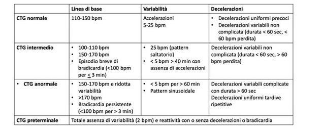
Table 1: Cardiotocography classification system to be used with STAN technology. The goal is to increase the specificity of the interpretative ability of cardiotocography, in situations in which the fetal state is considered not reassuring.

Table 2: ST analysis and situations where intervention* is required
*The intervention could include delivery or maternal-fetal re-suscitation by removing the possible causes of fetal distress (for example uterine hyperstimulation by oxytocin infusion, mater¬nal hypotension, etc.) **Duration between biphasic ST events should be related to CTG pattern and clinical situation.
References
- Clark, S. L., Hamilton, E. F., Garite, T. J., Timmins, A., Warrick, P. A., & Smith, S. (2017). The limits of electronic fetal heart rate monitoring in the prevention of neonatal metabolic acidemia. American journal of obstetrics and gynecology, 216(2), 163-e1.
- National Institute for Health and Clinical Excellence. Intrapartum care for healthy women and babies. Clinical guideline [CG190].
- World Health Organization. WHO recommendations: intrapartum care for a positive childbirth experience. Geneva: World Health Organization; 2018.
- Sabiani, L., Le Dû, R., Loundou, A., d’Ercole, C., Bretelle, F., Boubli, L., & Carcopino, X. (2015). Intra-and interob-server agreement among obstetric experts in court regarding the review of abnormal fetal heart rate tracings and obstetrical management. American journal of obstetrics and gynecology, 213(6), 856-e1.
- Jørgensen, J. S., & Weber, T. (2014). Fetal scalp blood sampling in labor–a review. Acta obstetricia et gynecologica Scandinavica, 93(6), 548-555.
- East, C. E., Leader, L. R., Sheehan, P., Henshall, N. E., Colditz, P. B., & Lau, R. (2015). Intrapartum fetal scalp lactate sampling for fetal assessment in the presence of a non-reassuring fetal heart rate trace. Cochrane Database of Systematic Reviews, (5).
- Chandraharan, E., & Wiberg, N. (2014). Fetal scalp blood sampling during labor: an appraisal of the physiological basis and scientific evidence. Acta Obstetricia et Gynecologica Scandinavica, 93(6), 544-547.
- Holzmann, M., Wretler, S., Cnattingius, S., & Nordström,L. (2015). Neonatal outcome and delivery mode in labors with repetitive fetal scalp blood sampling. European Journal of Obstetrics & Gynecology and Reproductive Biology, 184, 97-102.
- Sabir, H., Stannigel, H., Schwarz, A., & Hoehn, T. (2010). Perinatal hemorrhagic shock after fetal scalp blood sampling. Obstetrics & Gynecology, 115(2 Part 2), 419-420.
- Schaap, T. P., Moormann, K. A., Becker, J. H., Westerhuis,M. E., Evers, A., Brouwers, H. A., ... & Kwee, A. (2011). Cerebrospinal fluid leakage, an uncommon complication of fetal blood sampling: a case report and review of the literature. Obstetrical & gynecological survey, 66(1), 42-46.
- Murphy, D. J., Devane, D., Molloy, E., & Shahabuddin, Y. (2023). Fetal scalp stimulation for assessing fetal well-being during labour. Cochrane Database of Systematic Reviews, (1).
- Amer-Wåhlin, I., Ugwumadu, A., Yli, B. M., Kwee, A., Timonen, S., Cole, V., ... & Vayssiere, C. Study group of intrapartum fetal monitoring (European association of perinatal medicine).: fetal electrocardiography ST-segment analysis for intrapartum monitoring: a critical appraisal of conflicting evidence and a way forward. Am. J. Obstet. Gy-necol, 221(6), 577-601.
- Amer-Wåhlin, I., & Maršál, K. (2011, February). ST analysis of fetal electrocardiography in labor. In Seminars in Fetal and Neonatal Medicine (Vol. 16, No. 1, pp. 29-35). WB Saunders.
- Amer-Wahlin, I., & Kwee, A. (2016). Combined cardiotocographic and ST event analysis: a review. Best Practice & Research Clinical Obstetrics & Gynaecology, 30, 48-61.
- Sacco, A., Muglu, J., Navaratnarajah, R., & Hogg, M. (2015). ST analysis for intrapartum fetal monitoring. The Obstetrician & Gynaecologist, 17(1), 5-12.
- Westgate, J., Harris, M., Curnow, J. S., & Greene, K. R. (1993). Plymouth randomized trial of cardiotocogram only versus ST waveform plus cardiotocogram for intrapartum monitoring in 2400 cases. American journal of obstetrics and gynecology, 169(5), 1151-1160.
- Amer-Wåhlin, I., Hellsten, C., Norén, H., Hagberg, H., Herbst, A., Kjellmer, I., ... & Maršál, K. (2001). Cardiotocography only versus cardiotocography plus ST analysis of fetal electrocardiogram for intrapartum fetal monitoring: a Swedish randomised controlled trial. The Lancet, 358(9281), 534-538.
- AMER-WÅHLIN, I. S. I. S., Kjellmer, I., Maršál, K., Olofs-son, P., & Rosen, K. G. (2011). Swedish randomized controlled trial of cardiotocography only versus cardiotocography plus ST analysis of fetal electrocardiogram revisited: analysis of data according to standard versus modified in-tention-to-treat principle. Acta obstetricia et gynecologica Scandinavica, 90(9), 990-996.
- Ojala, K., Vääräsmäki, M., Mäkikallio, K., Valkama, M., & Tekay, A. (2006). A comparison of intrapartum automated fetal electrocardiography and conventional cardiotocogra-phy—a randomised controlled study. BJOG: an international journal of obstetrics & gynaecology, 113(4), 419-423.
- Vayssière, C., David, E., Meyer, N., Haberstich, R., Seba-houn, V., Roth, E., ... & Langer, B. (2007). A French randomized controlled trial of ST-segment analysis in a population with abnormal cardiotocograms during labor. American journal of obstetrics and gynecology, 197(3), 299-e1.
- Westerhuis, M. E., Visser, G. H., Moons, K. G., Van Beek,E., Benders, M. J., Bijvoet, S. M., ... & Kwee, A. (2010). Cardiotocography plus ST analysis of fetal electrocardiogram compared with cardiotocography only for intrapartum monitoring: a randomized controlled trial. Obstetrics & Gynecology, 115(6), 1173-1180.
- Belfort, M. A., Saade, G. R., Thom, E., Blackwell, S. C.,Reddy, U. M., Thorp Jr, J. M., ... & VanDorsten, J. P. (2015). A randomized trial of intrapartum fetal ECG ST-segment analysis. New England Journal of Medicine, 373(7), 632-641.