

Advances in Bioengineering and Biomedical Science Research(ABBSR)
ISSN: 2640-4133 | DOI: 10.33140/ABBSR

Impact Factor: 1.7

Research Article - (2023) Volume 6, Issue 12
Efficacy of the ExVent Accessory for the O2Vent Optima Oral Appliance in the Treatment of Obstructive Sleep Apnea
2Windsor Sleep Disorders Clinic, Windsor, Canada
Received Date: Nov 20, 2023 / Accepted Date: Dec 12, 2023 / Published Date: Dec 15, 2023
Copyright: ©Â©2023 Sat Sharma, et al. This is an open-access article distributed under the terms of the Creative Commons Attribution License, which permits unrestricted use, distribution, and reproduction in any medium, provided the original author and source are credited.
Citation: Sharma, S., Conflitti, A., Reiter, H., Rajkumar, W., Simeunovic, B. (2023). Efficacy of the ExVent Accessory for the O2Vent Optima Oral Appliance in the Treatment of Obstructive Sleep Apnea. Adv Bioeng Biomed Sci Res, 6(12), 131-137.
Abstract
Study Objectives: An optional accessory for the O2Vent Optima mandibular advancement device, the ExVent, provides oral expiratory positive airway pressure (EPAP). Similar to nasal EPAP, oral EPAP results in passive airway dilatation, reducing flow limitation. The present study was conducted to evaluate the safety and efficacy of the ExVent accessory for the treatment of mild to moderate obstructive sleep apnea (OSA).
Methods: This prospective, multicenter, open-label, single-arm study enrolled participants diagnosed with mild to moderate OSA (defined as apnea–hypopnea index [AHI] 5–29). During the Home Use Phase, study participants logged their hours of O2Vent Optima + ExVent usage at home over 3 months. Participants were then evaluated by in-laboratory polysomnogram while using the O2Vent Optima + ExVent. The primary effectiveness measure was a change in AHI relative to baseline while using the O2Vent Optima + ExVent.
Results: Treatment with the O2Vent Optima + ExVent improved AHI by 62% on average, from 15.3 (interquartile range [IQR]: 11.5–19.5) to 5.8 (IQR: 3.9–6.8; p<0.001). Mean oxygen saturation improved from 93.5%±1.2% to 94.6%±1.34% (p=0.006). The lowest oxygen saturation increased from 85% (IQR: 83%–87%) to 90% (IQR: 89%–95; p<0.001). The overall treatment success rate (AHI<5) was 66%, and the treatment response rate (>50% reduction in AHI) was 77%. No excessive adverse events or device malfunction events were reported.
Conclusions: Use of the O2Vent Optima with the ExVent oral EPAP accessory successfully reduced AHI in mild to moderate OSA with no significant adverse effects.
Keywords
Obstructive Sleep Apnea, Mandibular Advancement Device, MAD, ExVent, Oral Expiratory Positive Airway Pressure
Abbreviation List
AHI – Apnea hypopnea index
CPAP- Continuous positive airway pressure
EPAP - Expiratory positive airway pressure
EEG – Electroencephalogram
EOG – Electrooculogram
EMG - Electromyogram Hypos – Hypopneas
IQR – Inter quartile range
MAD – Mandibular advancement device
NREM – non rapid eye movement
REM – Rapid eye movement
OSA – Obstructive sleep apnea
PSG – Polysomnographic sleep study
SpO2 – Percutaneous oxygen saturation.
Brief Summary
Despite being better tolerated by patients with OSA, Mandibular advancement devices (MAD) therapy remains less than optimal for greater than 50% of patients (residual apnea–hypopnea index [AHI]>5). Therefore, the development of novel and effective therapies for OSA remains a priority.
The study findings provide novel insight into the potential benefit of oral EPAP accessory, a simple, cheap, and flexible addition to MAD therapy among people with mild to moderate OSA.
Introduction
Obstructive sleep apnea (OSA) is a common chronic sleep- related breathing disorder in which intermittent hypoxia and sleep fragmentation result in the development of symptoms that include excessive daytime sleepiness, impaired concentration, and fatigue [1-3]. Untreated OSA is associated with myriad long¬term adverse cardiovascular, metabolic, and neurocognitive7 health outcomes and comorbidities, which impair quality of life and safety, resulting in substantial economic effects [4-10].
Current first-line treatment for OSA is continuous positive airway pressure (CPAP), which is highly effective but not well tolerated [11]. Greater than 50% of patients with OSA on CPAP therapy report the use of CPAP devices for less than half the night or not at all, resulting in the failure to achieve the desired clinical benefits [12-14]. Mandibular advancement devices (MADs) are better tolerated than CPAP therapy [15]. MADs protrude the mandible anteriorly to enlarge the upper airway volume and reduce pharyngeal collapsibility during sleep [16,17]. MAD therapy often yields significant reductions in OSA severity, accompanied by improvements in daytime symptoms and quality of life [15,18, 18-20]. Despite being better tolerated by patients with OSA, MAD therapy remains less than optimal for greater than 50% of patients (residual apnea–hypopnea index [AHI]>5) [21]. Because patients with OSA who did not respond to MAD therapy were also intolerant to CPAP therapy, treatment failure is associated with considerable health, safety, and financial costs [10,13,22]. Therefore, the development of novel and effective therapies for OSA remains a priority.
The ExVent is an optional accessory that can be inserted into the O2Vent Optima MAD to provide upper airway support via oral expiratory positive airway pressure (EPAP) using passive airway dilation to reduce flow limitation, similar to the mechanism used by nasal EPAP devices in commercial distribution as stand-alone therapies for OSA [23]. Oral EPAP provided by the ExVent accessory is designed to augment the MAD therapy provided by the O2Vent Optima for patients with OSA.
The present study was conducted to evaluate the safety and efficacy of using the ExVent accessory with the O2Vent Optima MAD in the treatment of mild to moderate OSA. The primary effectiveness measure was the difference in AHI between baseline and treatment with the O2Vent Optima + ExVent. The outcome measure was the final AHI value. The frequency and type of adverse events were summarized descriptively.
Methods
Device Overview
The ExVent is an oval-shaped, passive, flapper-type valve that can be inserted into the extended anterior airway inlet of the O2Vent Optima (Fig. 1). When the patient is breathing through the airway, the valve fully opens during inspiration (Fig. 2a) and closes upon expiration, with airflow directed through “holes” in the flapper valve, resulting in increased EPAP (Fig. 2b). The ExVent is secured to the O2Vent Optima by a retention clip that allows for the easy removal of the ExVent accessory if desired. The ExVent is a single-patient, multiple-use device.

Figure 1: The ExVent is inserted into the O2Vent Optima Anterior Airway Opening.
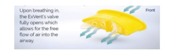
Figure 2a: During inspiration, the ExVent valve is open. 
Figure 2b: During expiration, the ExVent closes to create EPAP.
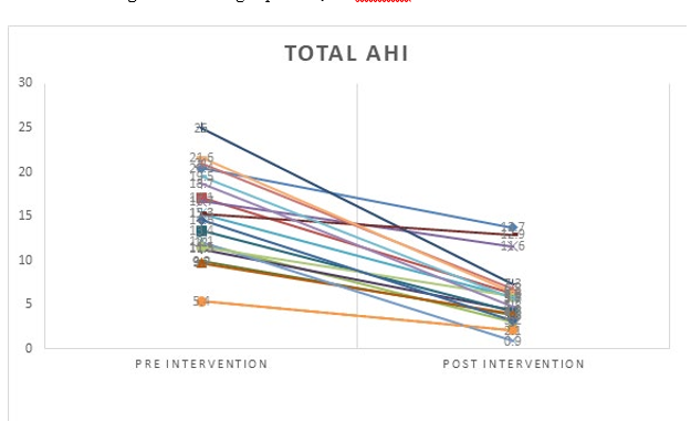
Figure 3: Total AHI for individual participant pre and post intervention.
Study Design
A prospective, open-label, single-arm study was conducted at four North American sites. Each participant signed an informed consent form approved by a central Institutional Review Board, WCG IRB Connexus, IRB Tracking ID: 20216980. All individuals who were diagnosed with mild to moderate OSA (defined as AHI 5–29) during a polysomnogram (PSG) study conducted by the sleep lab at the investigational site during the previous 6 months were asked to participate in this study. Inclusion criteria included current and adherent use of the O2Vent Optima. Adherence was defined as self-reported device use for at least 4 hours each night for at least 5 nights each week. Exclusion criteria included age younger than 22 years; oral cavity infection; any concomitant sleep disorder, including insomnia or central apnea; history of any prior surgical treatments for OSA, including radiofrequency ablation or palatal stent devices; and current enrollment in any other research project. Participants who met both inclusion and exclusion criteria and agreed to participate were asked to sign informed consent.
Screening Phase
After patients signed informed consent, routine dental and clinical evaluations were performed. The sleep dentist optimized the fit of the O2Vent Optima and made necessary adjustments according to standard practice. Demographic data were collected for all participants, including age, sex, height (in inches), weight (in pounds), prior OSA treatments, severe nasal allergies, sinusitis or nasal blockages, all current prescription medications or other agents that may affect sleep or PSG results (listing and dosage), and cumulative duration of O2Vent Optima usage (estimate of total months and years)
Home Use Phase
Participants who met all inclusion and exclusion criteria and signed informed consent were enrolled in the study. Enrolled participants received an ExVent device and were provided with instructions on device insertion and removal. Participants were asked to use the O2Vent Optima + ExVent every night for a 12-week period and to record hours of use. Adherence was assessed by phone at Weeks 1, 3, 6, and 8. Successful completion of the Home Use Phase was defined as reported device use for at least 4 hours each night for at least 5 nights each week. All participants who successfully completed the Home Use Phase proceeded to the PSG study, during which participants used the O2Vent Optima + ExVent device during an in-laboratory PSG study.
PSG Procedures
Participants were instrumented with electroencephalograms (EEGs; frontal, central, and occipital); right and left electrooculograms (EOGs); surface submentalis electromyograms (EMG); a nasal cannula (Pro-Tech Pro-Flow Nasal Cannula, Philips Respironics, Murrysville, PA) and pressure transducer for nasal airflow; a piezoelectric vibration sensor for snoring; thoracic and abdominal respiratory effort belts; body position sensor; 3-lead electrocardiogram; leg EMGs; and a pulse oximeter. The following data were collected at baseline and after the Home Use Phase: date of sleep night, time at lights off (24-hour clock), time at lights on (24-hour clock), total sleep time (minutes), sleep night notes (i.e., in-room adjustments, equipment technical issues), overall AHI, supine AHI, rapid eye movement (REM) AHI, non–rapid eye movement (NREM) AHI, mean sleep oxygen saturation (SpO2), nadir SpO2, arousal index, fraction of hypopneas, and sleep efficiency.
The sleep records were scored by the certified scorers blinded to the study protocol and no knowledge of the therapy utilized or whether the records belonged to a study participant. Apnea was scored when there was a drop in the peak signal excursion by ≥ 90% of pre-event baseline using an oronasal thermal sensor for ≥ 10 seconds. Hypopnea was scored when the peak signal excursions drop by ≥ 30% of pre-event baseline using nasal pressure for ≥ 10 seconds in association with either ≥ 3% arterial oxygen desaturation or an arousal.
Statistical Considerations
Sample Size Calculation
The minimum sample size was determined based on data from a comparable group of patients, which had a mean change in AHI of −8.37 relative to baseline, with a standard deviation of 7.73 for paired differences. Setting alpha at 0.05 and power at 0.80, we determined that nine participants would be necessary to observe a significant effect of treatment. Because of the imperfect predictability of historical data, we decided to enroll 18 evaluable patients to ensure adequate power.
Primary Endpoint Analysis
The primary endpoint in this study was a significant reduction in AHI relative to baseline following treatment with the O2Vent Optima + ExVent.
Safety Analysis
The type and frequency of adverse events are summarized descriptively. Serious adverse events and unanticipated adverse device effects were individually collected and reported. Adverse events which might occur or have been known to occur with the use of MADs include but are not limited to tooth movement, discomfort, pain, or changes in dental occlusion; loss of dental restorations; dental soreness; pain or soreness to the temporomandibular joint; excessive salivation; cheek or tongue pain; jaw discomfort, pain, or jaw set; gingivitis; dry mouth; and difficulty sleeping. Because the ExVent accessory is an investigational device, additional risks and discomforts may be associated with device use that are not yet known.
Statistical Analysis
All data are summarized descriptively. Categorical variables are summarized as frequency and percentage, and continuous variables are summarized as the number, mean, median, standard deviation, and range. For continuous outcomes, normality was assessed using the Shapiro–Wilk test. Normality was assessed for each pre-intervention and post-intervention observation of each outcome. When the normality assumption was met for both observations, paired-sample t-tests were used to test for significant changes across time. Means and standard deviations are reported and were interpreted for the t-test analyses. When either or both sets of observations violated normality, non-parametric Wilcoxon signed-rank tests were used to test for significant changes across time. Medians and interquartile ranges (IQRs) are reported and were interpreted for non-parametric analyses. The descriptive statistics for all analyses are presented in tabular format. All analyses were performed using SPSS Version 29 (Armonk, NY: IBM Corp.), and significance was assumed at an alpha value of 0.05.
Results
Patients with mild to moderate OSA were treated with the O2Vent Optima + ExVent for 3 months at four different sites in North America. Out of 22 participants enrolled, 18 completed the home use phase. Participant characteristics and anthropometric data are shown in Table 1. During the trial, no excessive adverse events or device malfunction events were reported.
|
Age (years) |
52.2±14.1 |
|
Sex (M/F) |
11/7 |
|
Body mass index (kg/m2) |
30.2±4.2 |
|
Epworth Sleepiness Scale (0–24 point scale) |
9±3 |
|
MAD advancement level |
61±11 |
MAD, mandibular advancement device; M, male; F, female
Table 1. Participant Characteristics
|
Variable |
Pre-intervention |
Post-intervention |
p-value |
|
Total Sleep Time (mins)* |
331.86 (51.26) |
332.71 (43.98) |
0.94 |
|
Supine Sleep (mins)* |
170.26 (102.99) |
163.35 (97.24) |
0.73 |
|
Side Sleep (mins)* |
188.23 (119.92) |
184.66 (100.95) |
0.90 |
|
NREM AHI* |
12.23 (4.75) |
5.14 (2.76) |
<0.001*** |
|
Max Length of Hypos (sec)* |
45.48 (10.79) |
35.28 (12.84) |
0.014*** |
|
Mean SpO2* |
93.5 (1.20) |
94.6 (1.34) |
0.006*** |
|
Sleep EFFE** |
83.3 (75.9–91.9) |
88.5 (80.0–91.8) |
0.27 |
|
Prone Sleep (mins)** |
0.0 (0.0–0.0) |
0.0 (0.0–0.0) |
0.66 |
|
AHI** |
15.3 (11.5–19.5) |
5.8 (3.9–6.8) |
<0.001*** |
|
REM AHI** |
22.5 (18.8–31.5) |
6.9 (4.9–11.7) |
0.001*** |
|
Supine AHI** |
22.6 (8.7–28.3) |
6.9 (3.9–12.6) |
0.007*** |
|
Total Hypos** |
56.0 (36.0–90.0) |
23.0 (19.0–26.0) |
<0.001*** |
|
Total Apneas and Hypos** |
89.0 (56.0–119.0) |
32.5 (23.0–41.0) |
<0.001*** |
|
Fractional Hypops/Hypos** |
93.9 (86.7–97.0) |
89.2 (79.1–95.0) |
0.28 |
|
Nadir SpO2** |
85.0 (83.0–87.0) |
90.0 (89.0–92.0) |
<0.001*** |
|
Arousal Index (per hour) ** |
19.8 (17.5–21.9) |
7.2 (4.1–8.6) |
<0.001*** |
AHI, apnea–hypopnea index; REM, rapid eye movement; EFFE, efficiency; SpO2, oxygen saturation; hypos, hypopneas Note: * Values are mean (standard deviation), ** values are median (interquartile range), ***p<0.05
Table 2. Within-Subjects Comparisons
Outcomes that violated the normality assumption were evaluated using the non-parametric Wilcoxon signed-rank test. Relative to baseline, significant decreases in overall AHI (p<0.001), REM AHI (p=0.001), supine AHI (p=0.007), total hypopneas (p<0.001), total apneas and hypopneas (p<0.001), and arousal index (p<0.001) were observed after treatment. Relative to baseline, a significant increase in nadir SpO2 (p<0.001) was observed after treatment. No significant differences were detected between pre-intervention and post-intervention measures of sleep efficiency (p=0.027), prone sleep (p=0.066), or the ratio of fractional hypopneas to total hypopneas (p=0.28). Descriptive statistics for non-parametric comparisons are presented in Table 2.
Discussion
This study is one of the first to evaluate the efficacy of an external EPAP device, the ExVent accessory attached to the O2Vent Optima. The primary findings of this study indicate that the addition of an oral EPAP valve to a novel MAD was effective for the management of OSA. Treatment with the O2Vent Optima combined with the ExVent accessory reduced AHI from 15.3 (IQR: 11.5–19.5) at baseline to 5.8 (IQR: 3.9–6.8; p<0.001) post-intervention, an average reduction of 62%. AHI improvements were observed during NREM sleep, REM sleep, and supine sleep. The length of hypopneas also decreased significantly, from 45.48±10.79 seconds at baseline to 35.28±12.84 seconds after intervention (p=0.014). The mean SpO2 improved from 93.5%±1.2% at baseline to 94.6%±1.34% (p=0.006) after the intervention, and the nadir SpO2 increased from 85% (IQR: 83%–8%7) at baseline to 90% (IQR: 89%–92%; p<0.001) after the intervention. A significant improvement in arousal index was also observed, from 19.8 (IQR: 17.5–21.9) at baseline to 7.2 (IQR: 4.1–8.6; p<0.001) after the intervention. During the trial, patients using the O2Vent Optima + ExVent reported no excessive adverse events or device malfunctions
On average, MAD therapy reduces OSA severity by approximately 50%, and similar results were reported for the O2Vent Optima [18-21, 24]. In a multicenter analysis, Sutherland and colleagues found that 37% of individuals diagnosed with OSA achieved complete treatment success (defined as AHI<5) following MAD therapy, with almost 65% reporting a 50% or greater reduction in AHI [21]. However, a substantial proportion of individuals diagnosed with OSA and prescribed MAD therapy experience incomplete treatment, many of whom have already failed CPAP therapy [15-17, 20]. Identifying and developing novel therapies able to mitigate the risks of adverse health outcomes due to untreated OSA remain urgent needs. The simple addition of the ExVent accessory to provide oral EPAP in our study was associated with an overall treatment success rate (defined as AHI<5) of 66% and a response rate (defined as a >50% reduction in AHI) of 77%.
Potential Mechanisms of Action
Several potential mechanisms of action may explain the reduction in OSA severity observed in response to MAD therapy combined with an EPAP valve. Magnetic resonance imaging studies revealed that MAD therapy primarily prevents upper airway collapse by increasing the velopharyngeal segment volume or protruding the tongue [17,25]. However, upper airway collapse occurs at multiple levels in 68% of individuals with OSA,26 which could render MAD therapy ineffective for certain individuals and may partially account for the reduced efficacy of MAD therapy among patients with severe OSA [26,27]. Vroegop et al. detected multilevel collapse of the velopharyngeal and hypopharyngeal segments in greater than 20% of participants with OSA. Nasal EPAP devices increase the end-expiratory lung volume and may, therefore, dilate different segments of the upper airway, especially the hypopharyngeal segment [26,28]. Accordingly, combining MAD therapy with EPAP therapy may represent a more effective solution for preventing multilevel upper airway collapse than MAD therapy alone.
The upper airway narrows progressively toward the end of expiration when preceding an apnea event, leaving the upper airway vulnerable to collapse during subsequent inspiration when negative pressure forces are generated [28]. Supplementing MAD therapy with an EPAP valve may address these vulnerabilities. Nasal EPAP increases end-expiratory lung volume, which increases the longitudinal tension or “caudal traction” on the upper airway, reducing airway collapsibility during this critical phase [28,29].
High nasal resistance is a recognized risk factor for OSA, and several studies have shown that high nasal resistance contributes to increased OSA severity [30-33]. Additionally, patients with OSA and high nasal resistance tend to be intolerant of CPAP and other oral appliance therapies [13,15,21]. The O2Vent Optima, a novel oral appliance with a built-in oral airway that allows for oral breathing without mouth opening and consequent mandible retraction, has demonstrated efficacy in patients with OSA and nasal obstruction [34,35]. The ExVent accessory provides oral EPAP, which may augment the therapeutic benefits of the O2Vent Optima. In vitro proprietary studies of ExVent demonstrated that the medium 0.38mm ExVent Flapper Valves generated exhalation resistance of 4.2 to 5.9 cmH2O/L/Min. at a flow rate of 20 to 30 L/Min.
Our study has significant limitations. The comparison was pretreatment versus treatment with both the O2Vent Optima and EPAP add on. Therefore, it is not possible to know what the contribution of the EPAP add on was on AHI reduction. Our study did not measure whether the ExVent increased either resistance or end expiratory pressure (EPAP). Given that the nasal airway was open, it is possible that the ExVent causes preferential nasal exhalation thereby nominally or not at all altering EPAP. Therefore, a randomized study is necessary to assess whether ExVent improves outcome beyond the O2Vent Optima.
Conclusion
The present clinical trial confirmed that the addition of an oral EPAP accessory, the ExVent, to the O2Vent Optima MAD resulted in treatment success in patients with mild to moderate OSA. Combined treatment was well tolerated with no excessive adverse effects. Although further randomized clinical studies remain necessary to assess the efficacy of the ExVent + O2Vent Optima compared with the O2Vent Optima alone, the current findings provide novel insight into the potential benefit of simple, cheap, and flexible addition to MAD therapy among people with mild to moderate OSA.
References
- Adult Obstructive Sleep Apnea Task Force of the American Academy of Sleep Medicine. (2009). Clinical guideline for the evaluation, management and long-term care of obstructive sleep apnea in adults. Journal of clinical sleep medicine, 5(3), 263-276.
- Peppard, P. E., Young, T., Barnet, J. H., Palta, M., Hagen,E. W., & Hla, K. M. (2013). Increased prevalence of sleep-disordered breathing in adults. American journal of epidemiology, 177(9), 1006-1014.
- Young, T., Palta, M., Dempsey, J., Skatrud, J., Weber, S., & Badr, S. (1993). The occurrence of sleep-disordered breathing among middle-aged adults. New England journal of medicine, 328(17), 1230-1235.
- Hla, K. M., Young, T., Hagen, E. W., Stein, J. H., Finn, L. A., Nieto, F. J., & Peppard, P. E. (2015). Coronary heart disease incidence in sleep disordered breathing: the Wisconsin Sleep Cohort Study. Sleep, 38(5), 677-684.
- Redline, S., Yenokyan, G., Gottlieb, D. J., Shahar, E., O'Connor, G. T., Resnick, H. E., ... & Punjabi, N. M. (2010). Obstructive sleep apnea–hypopnea and incident stroke: the sleep heart health study. American journal of respiratory and critical care medicine, 182(2), 269-277.
- Aurora, R. N., & Punjabi, N. M. (2013). Obstructive sleep apnoea and type 2 diabetes mellitus: a bidirectional association. The Lancet Respiratory Medicine, 1(4), 329-338.
- Osorio, R. S., Gumb, T., Pirraglia, E., Varga, A. W., Lu, S. E., Lim, J., ... & Alzheimer's Disease Neuroimaging Initiative. (2015). Sleep-disordered breathing advances cognitive decline in the elderly. Neurology, 84(19), 1964-1971.
- Lacasse, Y., Godbout, C., & Series, F. (2002). Health-related quality of life in obstructive sleep apnoea. European Respiratory Journal, 19(3), 499-503.
- Ellen, R., Marshall, S. C., Palayew, M., Molnar, F. J., Wilson, K. G., & Man-Son-Hing, M. (2006). Systematic review of motor vehicle crash risk in persons with sleep apnea. J Clin Sleep Med, 2(2), 193-200.
- Hillman, D., Mitchell, S., Streatfeild, J., Burns, C., Bruck, D., & Pezzullo, L. (2018). The economic cost of inadequate sleep. Sleep, 41(8), zsy083.
- Sullivan, C., Berthon-Jones, M., Issa, F., & Eves, L. (1981). Reversal of obstructive sleep apnoea by continuous positive airway pressure applied through the nares. The Lancet, 317(8225), 862-865.
- McEvoy, R. D., Antic, N. A., Heeley, E., Luo, Y., Ou, Q., Zhang, X., ... & Anderson, C. S. (2016). CPAP for prevention of cardiovascular events in obstructive sleep apnea. New England Journal of Medicine, 375(10), 919-931.
- Weaver, T. E., & Grunstein, R. R. (2008). Adherence to continuous positive airway pressure therapy: the challenge to effective treatment. Proceedings of the American Thoracic Society, 5(2), 173-178.
- Weaver, T. E., Maislin, G., Dinges, D. F., Bloxham, T.,George, C. F., Greenberg, H., ... & Pack, A. I. (2007). Relationship between hours of CPAP use and achieving normal levels of sleepiness and daily functioning. Sleep, 30(6), 711-719.
- Phillips, C. L., Grunstein, R. R., Darendeliler, M. A., Mihailidou, A. S., Srinivasan, V. K., Yee, B. J., ... & Cistulli,P. A. (2013). Health outcomes of continuous positive airway pressure versus oral appliance treatment for obstructive sleep apnea: a randomized controlled trial. American journal of respiratory and critical care medicine, 187(8), 879-887.
- Chan, A. S., Sutherland, K., Schwab, R. J., Zeng, B.,Petocz, P., Lee, R. W., ... & Cistulli, P. A. (2010). The effect of mandibular advancement on upper airway structure in obstructive sleep apnoea. Thorax, 65(8), 726-732.
- Ng, A. T., Gotsopoulos, H., Qian, J., & Cistulli, P. A. (2003). Effect of oral appliance therapy on upper airway collapsibility in obstructive sleep apnea. American journal of respiratory and critical care medicine, 168(2), 238-241.
- Blanco, J., Zamarron, C., Abeleira Pazos, M. T., Lamela, C., & Suarez Quintanilla, D. (2005). Prospective evaluation of an oral appliance in the treatment of obstructive sleep apnea syndrome. Sleep and Breathing, 9, 20-25.
- Galic, T., Bozic, J., Pecotic, R., Ivkovic, N., Valic, M., & Dogas, Z. (2016). Improvement of cognitive and psychomotor performance in patients with mild to moderate obstructive sleep apnea treated with mandibular advancement device: a prospective 1-year study. Journal of Clinical Sleep Medicine, 12(2), 177-186.
- Petri, N., Svanholt, P., Solow, B., Wildschiødtz, G., & Winkel, P. (2008). Mandibular advancement appliance for obstructive sleep apnoea: results of a randomised placebo controlled trial using parallel group design. Journal of sleep research, 17(2), 221-229.
- Sutherland, K., Vanderveken, O. M., Tsuda, H., Marklund, M., Gagnadoux, F., Kushida, C. A., & Cistulli, P. A. (2014). Oral appliance treatment for obstructive sleep apnea: an update. Journal of Clinical Sleep Medicine, 10(2), 215-227.
- Young, T., Skatrud, J., & Peppard, P. E. (2004). Risk factors for obstructive sleep apnea in adults. Jama, 291(16), 2013-2016.
- Berry RB, Kryger MH, Massie CA. (2011). A novel nasal expiratory positive airway pressure (EPAP) device for the treatment of obstructive sleep apnea: a randomized controlled trial. Sleep, 34(4), 479-485.
- Tong, B. K., Tran, C., Ricciardiello, A., Chiang, A., Donegan, M., Murray, N., ... & Eckert, D. J. (2020). Efficacy of a novel oral appliance and the role of posture on nasal resistance in obstructive sleep apnea. Journal of Clinical Sleep Medicine, 16(4), 483-492.
- Vroegop AV, Vanderveken OM, Boudewyns AN, et al. (2014). Drug-induced sleep endoscopy in sleep-disordered breathing: report on 1,249 cases. Laryngoscope, 124(3),797-802.
- Morrell, M. J., Arabi, Y., Zahn, B., & Badr, M. S. (1998). Progressive retropalatal narrowing preceding obstructive apnea. American journal of respiratory and critical care medicine, 158(6), 1974-1981.
- Sutherland, K., Takaya, H., Qian, J., Petocz, P., Ng, A. T., & Cistulli, P. A. (2015). Oral appliance treatment response and polysomnographic phenotypes of obstructive sleep apnea. Journal of Clinical Sleep Medicine, 11(8), 861-868.
- Braga, C. W., Chen, Q., Burschtin, O. E., Rapoport, D. M., & Ayappa, I. (2011). Changes in lung volume and upper airway using MRI during application of nasal expiratory positive airway pressure in patients with sleep-disordered breathing. Journal of Applied Physiology, 111(5), 1400-1409.
- Heinzer, R., White, D. P., Malhotra, A., Lo, Y. L., Dover, L., Stevenson, K. E., & Jordan, A. S. (2008). Effect of expiratory positive airway pressure on sleep disordered breathing. Sleep, 31(3), 429-432.
- Zwillich, C. W., Pickett, C., Hanson, F. N., & Weil, J. V. (1981). Disturbed sleep and prolonged apnea during nasal obstruction in normal men. American Review of Respiratory Disease, 124(2), 158-160.
- Suratt, P. M., Turner, B. L., & Wilhoit, S. C. (1986). Effect of intranasal obstruction on breathing during sleep. Chest, 90(3), 324-329.
- Taasan, V., Wynne, J. W., Cassisi, N., & Block, A. J. (1981). The Effect of nasal packing on sleep-disordered breathing and nocturnal oxygen desaturation. The Laryngoscope, 91(7), 1163-1172.
- Zeng, B., Ng, A. T., Qian, J., Petocz, P., Darendeliler, M. A., & Cistulli, P. A. (2008). Influence of nasal resistance on oral appliance treatment outcome in obstructive sleep apnea. Sleep, 31(4), 543-547.
- Lavery, D., Szollosi, I., Czyniewski, S., Beer, F., McCloy, K., & Hart, C. (2017). Safety and efficacy of a novel oral appliance in the treatment of obstructive sleep apnea. J Dent Sleep Med, 4(3), 57-63.
- Sugiura, T., Noda, A., Nakata, S., Yasuda, Y., Soga, T., Miyata, S., ... & Koike, Y. (2006). Influence of nasal resistance on initial acceptance of continuous positive airway pressure in treatment for obstructive sleep apnea syndrome. Respiration, 74(1), 56-60.
- Summary Report - Pressure Flow Characterisation of ExVent vs External Regular vs Provent vs Theravent. Oventus Testing Report: 086 Author: J Choi. Oventus Proprietary Documents.