

Advances in Bioengineering and Biomedical Science Research(ABBSR)
ISSN: 2640-4133 | DOI: 10.33140/ABBSR

Impact Factor: 1.7

Research Article - (2021) Volume 4, Issue 4
Comprehensive Genetic Features of Serous Ovarian Tumor Patients Revealed PIK3CA Mutation and Chromosome Instability as Prognostic Biomarkers
2Nanjing Geneseeq Technology Inc, Nanjing, China
3Translational Medicine Research Institute, Geneseeq Technology Inc, Toronto, Ontario, China
4School of Public Health, Nanjing Medical University, Nanjing, China
Received Date: Nov 03, 2021 / Accepted Date: Nov 10, 2021 / Published Date: Nov 15, 2021
Copyright: ©Copyright: ©2021 Zheng Hong, et al. This is an open-access article distributed under the terms of the Creative Commons Attribution License, which permits unrestricted use, distribution, and reproduction in any medium, provided the original author and source are credited.
Citation: Jinjing Wang, Cunyi Gao, Junjun chen, Xue Li, Sisi Liu, Xue Wu, Yang Shao, Na Tan. Hong Zheng (2021) Comprehensive Genetic Features of Serous Ovarian Tumor Patients Revealed PIK3CA Mutation and Chromosome Instability as Prognostic Biomarkers. Adv Bioeng Biomed Sci Res 4(4): 100-107.
Abstract
Background: Ovarian cancer is the seventh most common cancer in women worldwide among which the most frequently occurred histological type is serous ovarian cancer (SOC). Since efficacious treatments for SOC have not advanced beyond platinum-based combination chemotherapy and more than 75% of high-grade SOC will relapse after first-line therapy, it is urgent to observe the genomic abnormalities and identify novel therapeutic targets and prognosis biomarkers.
Methods: In order to comprehensively identify molecular features of serous ovarian cancer, we performed targeted sequencing with 425 cancer-related genes on four serous ovarian tumor (SOT) cohorts, classified as ovarian serous adenoma (OSA), ovarian serous borderline tumor (OSBT), low-grade serous cancer (LGSC) and high-grade serous cancer (HGSC). The association between genetic alterations and patients’ overall survival (OS) was analyzed.
Results: Genomic profiling revealed distinct molecular features among these four cohorts. The frequency of genetic alterations in OSA was relatively low, and in OSBT cohort, the predominantly mutated genes, BRAF and KRAS, were identified at prevalence of 52.6% (10/19) and 36.8% (7/19) respectively with two patients harbored both these two mutations. In LGSC cohort, alterations of KRAS still occupied the highest percentage of patients which was up to 50.0% (5/10) while BRAF was not common (1/10, 10.0%). The most frequently mutated gene was TP53 in HGSC (46/47, 97.9%), whereas BRAF or KRAS mutation was rare. Meanwhile, a higher prevalence of gene copy gains in PTK2 (12/47, 25.5%), MYC (9/47, 19.1%), MDM4 (5/47, 10.6%) and ZNF217 (5/47, 10.6%) were identified only in HGSC group which indicated cancer progression promoted by chromosomal instability in this group. The median tumor mutational burden (TMB) and chromosome instability score (CIS) in cases with LGSC and HGSC higher than that in OSBT. Additionally, analysis of DNA damage repair (DDR) relevant genes showed most altered genes enriched in homologous recombination (HR) pathway in HGSC. Finally, we correlated genomic profiles with overall survival (OS) and found that PIK3CA wildtype or chromosome instability score (CIS) low patients had significantly longer OS in HGSC. Conclusion: In this study, we revealed the comprehensive genomic profiling among four SOT cohorts. Additionally, we correlated PIK3CA status and first associated chromosome instability with clinical outcomes of patients and found them to be useful clinical biomarkers in HGSC prognosis
Keywords
Serous Ovarian Tumor (SOT), High-Grade Serous Cancer (HGSC), Low-Grade Serous Cancer (LGSC), PIK3CA, Chromosome Instability
Introduction
Ovarian cancer is the seventh most common cancer in women both in China and worldwide among which the most frequent¬ly occurred histological type is serous ovarian cancer [1]. Sev¬eral studies further classified serous ovarian carcinoma into low-grade serous cancer (LGSC) and high-grade serous cancer (HGSC) according to a two-tiered grading system [2, 3]. On the basis of the International Federation of Gynecology and Obstet¬rics (FIGO) grading system, serous ovarian cancer could also be assorted into grade I, II, III, and IV [4].
The 5-year survival of HGSC is about 25% which is attributable to the lack of successful treatment strategies beyond combina¬tion chemotherapy, and more than 75% of HGSC will relapse after first-line therapy [5, 6]. Therefore, it is urgent to observe the genomic abnormalities and identify therapeutic targets on clinically annotated SOC patients.
In order to deeply study the difference of the genomic alterations among serous ovarian tumor cohorts and identify reliable pre-dictive biomarkers, comprehensive genomic mutation profiling analysis with 425 cancer-related genes was performed in ovar-ian serous adenoma (OSA), ovarian serous borderline tumor (OSBT), LGSC, and HGSC cohorts and revealed distinct ge-nomic features among these four groups. The genomic alteration occurs scarcely in OSA. KRAS and BRAF mutations frequently occurred in OSBT cohort while only KRAS was still so common in LGSC group. Different gene alteration profiling was observed in HGSC cohorts as TP53 mutation was universal presence but KRAS and BRAF alterations were rare. Most of HGSC patients harbored one or more genetic alterations related with DNA dam-age repair (DDR) pathway. In addition, we found PIK3CA wild-type or chromosome instability score (CIS) low cases had longer overall survival (OS) in HGSC patients.
Materials and Methods
Tissue Collection
Tumor and matched normal tissue biopsies were collected from 86 patients in Zunyi Medical University between 2013 and 2019, fixed in 10% neutral buffered formalin and embedded in paraffin. These specimens were stained with hematoxylin and eosin (HE), evaluated by a pathologist and further classified as OSA (n=10), OSBT (n=19), LGSC (n=10) and HGSC (n=47) according to the newest edition of WHO classification. The study was approved by the Ethical Review Board of the Zunyi Medical University. DNA extraction, Library Preparation and Targeted Sequencing Genomic DNA were extracted with QIAamp DNA FFPE Tis¬sue Kit (Qiagen, Hilden, Germany) from FFPE tissues de-par-affinized with xylene, according to manufacturer’s instructions.
Then DNA concentrations were determined using a Qubit DNA HS Assay Kit with Qubit 3.0 fluorometer (Life Technologies). For each sample, 1000 ng of genomic DNA was sheared into 350 bp fragments using Covaris M220 instrument (Covaris) and processed into library construction with KAPA HyperPrep Library Preparation Kit (KAPA Biosystems, Wilmington, MA) according to the manufacturer’s protocol. Libraries with differ- ent indexes were pooled and enriched with probes targeted 425 cancer-related genes with a customized xGen Lockdown panel (IDT). Prior to sequencing, the captured libraries were exam¬ined for quality and quantity using the KAPA Library Quantifi¬cation Kit (KAPA Biosystems) by qRT-PCR (CFX384 real time system, Bio-Rad Laboratories). The final libraries were then sequenced on a HiSeq 4000 platform (Illumina) to a mean cov¬erage of 1000x following the manufacturer’s instructions. Bioinformatics Analysis Quality control for fastq data and subsequent removal of low quality (quality reading below 15) adapters was performed with the trimmomatic software, which is a flexible trimer for Illumina sequence data [7]. Paired-end sequencing reads were then aligned to the reference human genome (build hg19) us¬ing the Burrows-Wheeler Aligner (BWA) with the parameters and further processed to PCR deduplication using the Picard suite (http://picard.sourceforge.net/) [8]. The Genome Analysis Toolkit (GATK) was used to base quality score recalibration of local realignment around indels [9]. In order to identify the so¬matic single nucleotide variants (SNVs), MuTect software was applied to tumor and paired normal BAM files [10]. The small insertions and deletions were detected with SCALPEL (http:// scalpel.sourceforge.net/). For the copy number variation (CNV) pipeline, a ≥1.6-fold change in DNA copy number was set as the cutoff for amplification, while a ≤ 0.6-fold change was the cutoff for deletion. Tumor mutational burden (TMB) and chromosome instability score (CIS) was calculated as previously described [11].
Statistical Analysis
The Kaplan-Meier method was used to calculate survival rates, and the log-rank test was used to analyzed difference between cohorts. A significant threshold was set at P-value < 0.05.
Result
Demographic and Clinical Characteristics of Patients Diagnosed With SOT A total of 86 patients were classified into four SOT cohorts termed as OSA (Figure 1A, n=10), OSBT (Figure 1B, n=19), LGSC (Figure 1C, n=10) and HGSC (Figure 1D, n=47) accord¬ing to pathological features (Figure 1, Table 1), and the median age of patients in these four groups was 62 years (range 48-88), 38 years (range 20-74), 41 years (range 19-66) and 53 years (range 40-70), respectively (Table 1). Most patients in OSBT cohort were diagnosed at relatively early stage (stage I-II, 18/19, 94.7%), whereas for HGSC cases, most patients were diagnosed at stage III-IV (37/47, 77.7%). In our cohorts, all of OSA pa¬tients only received surgery. In 19 OSBT cases, the majority of patients (12/19, 63.2%) only received surgery and additional pa¬tients (6/19, 31.6%) except one case with unknown treatment status, received adjuvant chemotherapy after surgery. In LGSC cohort, all patients received adjuvant chemotherapy after sur¬gery. 37.7% patients (13/47) underwent neoadjuvant chemother¬apy before surgery, 34.0% (16/47) suffered adjuvant chemother-apy after surgery and 38.3% (18/47) only received chemotherapy in HGSC patients.

Figure 1: Pathological features of four serous ovarian tumor (SOT) cohorts. (A) ovarian serous adenoma (OSA), cyst wall lining monolayer cubic or low columnar ciliated epithelial cells and cell without atypia can be seen. (B) ovarian serous borderline tumor (OBST), tumor cells form gradually branched nipples, epithelium appear stratified and budding, single cells or cell clusters can be seen in the glandular cavity, spike-like cells are visible and rare mitotic. (C) low-grade serous carcinoma (LGSC), the invasive micropapillary pattern, consistent cell size, higher nuclear plasma ratio and obvious nuclear atypia were observed in this cohort. (D) high-grade serous carcinoma (HGSC), which display typical morphology of papillary and/or solid areas, hyperplasia of fibrous tissue around tumor, locally visible necrosis and highly atypical nuclei.
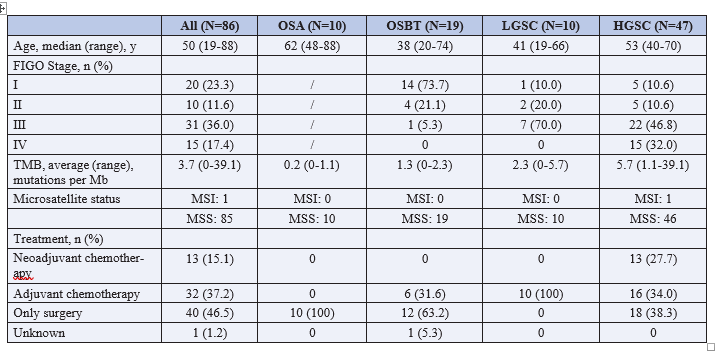
Note: TMB, tumor mutational burden; OSA, ovarian serous adenoma; OSBT, ovarian serous borderline tumor; LGSC, low-grade serous carcinoma; HGSC, high-grade serous carcinoma. /: not applicable.
The Genomic Features of Chinese SOT Cohorts
Genomic profiling revealed distinct molecular features among these four cohorts (Figure 2A). As expected, the frequency of genetic alterations in OSA was relatively low with only mis-sense mutations in AKT1 and KMT2A were observed (Figure 2A). For OSBT cohort, the predominantly mutated genes, BRAF and KRAS, were identified at prevalence of 52.6% (10/19) and 36.8% (7/19) respectively with two patients harbored both BRAF and KRAS mutations. In LGSC group, alterations of KRAS still occupied the highest percentage of patients which was up to 50.0% (5/10) while BRAF was not common (1/10, 10.0%). In HGSC patients, different genetic alteration profile was ob¬served as the most frequently mutated gene was TP53 (46/47, 97.6%), whereas the mutation of BRAF or KRAS mutation was rare. Several genomic alterations including PTK2 (2/10), MYC (2/10), NF1 (2/10) and ARID1A (2/10) were detected in LGSC. Interestingly, a higher prevalence of gene copy gains in PTK2 (12/47, 25.5%), MYC (9/47, 19.1%), MDM4 (5/47, 10.6%) and ZNF217 (5/47, 10.6%) were identified in HGSC group which indicated cancer progression promoted by chromosomal insta-bility in this group.
Then, we further analyzed MSI, TMB and CIN which were correlated with instability at the genome-wide level in OSBT, LGSC and HGSC groups. Within 86 patients, only one case was tested to be microsatellite instability-high (MSI-H) while the other 85 cases remained to be microsatellite stability (MSS) (Table 1). The average tumor mutational burden (TMB) in cas-es with LGSC and HGSC were 2.3 and 5.7 mutations per Mb, higher than that in OSBT (1.6) (Table 1) which indicated an in-creasement of gene mutations during tumor progression (Figure 2B). The result of CIS was consistent with TMB as the levels of CIS in LGSC and HGSC were significantly higher than that in OSTB. Although CIS values in HGSC were high, no significant difference was observed in LGSC and HGSC cohorts (Figure 2C).
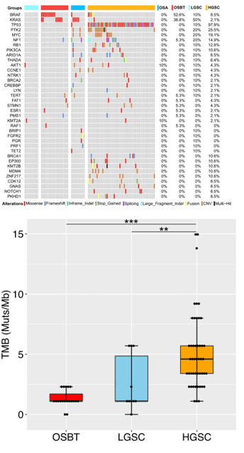
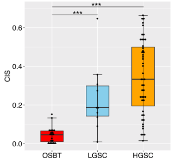
Figure 2: The molecular feature of serous ovarian tumor co-horts. (A) Co-mutation plot of the most frequently altered genes identified by next-generation sequencing of OSA, OSBT, LGSC and HGSC cohorts. Box plots comparing genomic features of TMB (B) and CIS (C) among three serous ovarian tumor cohorts apart from OSA group. The top and bottom of the boxes are the lower and upper quartiles, the middle line in the box is median. Wilcoxon’s rank-sum test was used for inter-group comparison and P value calculation with two-side. * indicates a significant threshold of P-value <0.05. ** and*** represent the P-value were <0.01 and <0.001, respectively.
Additionally, germline mutation in these four groups was ana-lyzed. The frequency of germline mutations was relatively low in both OSA and OSBT cohorts, whereas higher in LGSC (70%, 7/10) and HGSC (36%, 17/47) cohorts (Figure 3A). For LGSC cohort, BRCA2 mutations were detected, accompanied with DYPD and APC mutations. The most frequent germline muta¬tion we detected in HGSC cohort was BRCA1 (21.3%, 10/47), followed by BRCA2 (6.4%, 3/47) and WRN (4.26%, 2/47)
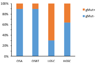

Figure 3: Analysis of germline mutation in serous ovarian tumor (SOT). (A) Frequency of germline mutation in SOT. (B) Germ¬line mutation profiling of serous
Comprehensive Molecular Profiles Revealed Many Muta¬tions Associated with DNA Damage Repair (DDR) System
Since the deficiency of DNA damage repair (DDR) system sig¬nificantly affect genomic stability and finally lead to occurrence of cancer in multiple cancer types, we further studied the genetic alterations related to DDR signaling. As expected, low frequen¬cy of DDR-relevant gene mutation was observed both in OSA (0%, 0/10) and OSBT (10.5%, 2/19) cohorts (Figure 4A). On the contrary, 10.0% (1/10) of LGSC patients and 55.3% (26/47) of HGSC patients harbored at least one gene alteration associated with DDR signaling and enriched in homologous recombination (HR) pathway (Figure 4A). Further analysis in 26 HGSC pa¬tients with DDR-relevant mutations showed that 61.5% (16/26) cases had somatic mutations among which the most frequent¬ly mutated were BRCA1 (17%), followed by ATM (10%) and PARP1 (10%). Additionally, in the other 53.8% (14/26) cases with germline mutation, BRCA1 (67%) and BRCA2 (20%) were the most frequently altered (Figure 4B, right panel).
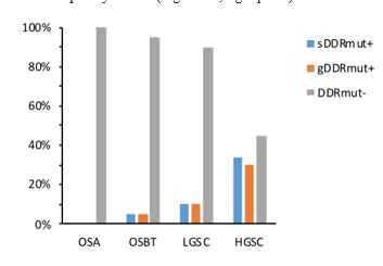
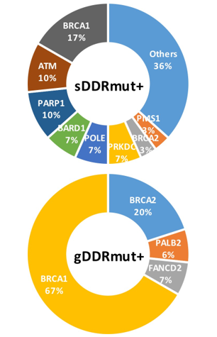
Figure 4: Mutation analysis of gene included in 425 Panel as¬sociated with DNA damage repair (DDR) pathway in serous ovarian tumor. (A) Mutation frequency of gene associated with DDR pathway in four serous ovarian tumor cohorts. High fre¬quency of somatic mutation gene (B, left panel) and germline mutation gene (B, right panel) associated with DDR pathway in HGSC cohort. sDDRmut+, gDDRmut+ represents somatic mu¬tation and germline mutation associated with DNA damage re¬pair pathway, respectively. Whereas DDRmut- indicates without mutated events associated with DDR pathway. The total number of somatic alteration and germline alteration incidents were 30 and 16, respectively.
PIK3CA Wildtype and CIS Low are related with Longer Overall Survival in HGSC
Genetic alterations could be potential predictors of prognosis in ovarian cancer therapy. Therefore, to deeply evaluate the relationship between molecular profiling and clinical outcomes, andidentify biomarkers correlated with longer overall survival (OS), we conducted survival analysis in HGSC cohort.
Numerous genetic and functional studies have clearly showed that PIK3CA gene played an important role in the PI3K-Akt pathway which associated with development of neoplasia in ovarian tumors. In our results, the data showed that patients without PIK3CA mutation had a significantly better OS (medi-an: NA), compared to PIK3CA mutated patients (median: 6.4 months) (P < 0.001). The 2-year survival probability was also different between these two groups (wt vs. mut: 0 vs 34.3%) (Figure 5A).
Given genomic instability may play crucial role in HGSC pro-gression and have effect on therapeutic approach selection, we further compared OS in CIS low and high subgroups which was divided according to the value of 0.35 because of similar cases between these two groups. Interestingly, patients with lower CIS had a drastically better OS compared to CIS high group (P < 0.01), as well as a higher 2-year survival probability between these two groups (lower CIS vs. higher CIS: 40.0% vs 21.1%) (Figure 5B). These results suggested that CIS may function as a novel prognostic biomarker in HGSC.
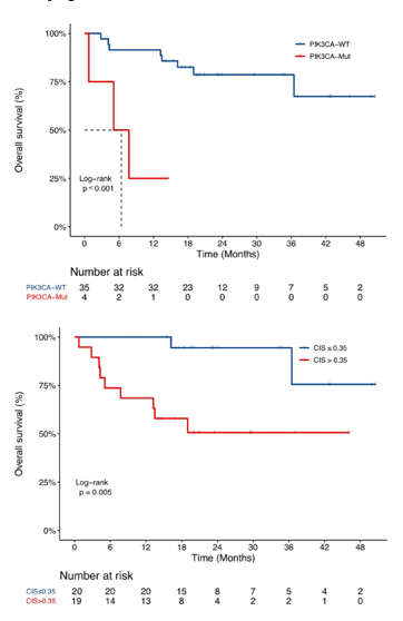
Figure 5: Genetic features affected the clinical response to treat-ment in HGSC cohort. Kaplan-Meier curves showing different effects of genetic features on patients’ OS. The effect of PIK3CA status (A) and CIS (B) in HGSC cohort. Patients were divided into two groups with or without PIK3CA mutation (A). At the same time, patients were classified into two groups according to their CIS values ordered from small to big, the cutoff is setter as 0.35 because of similar cases between above 0.35 group and the other group (B). Log-rank test was performed to inter-group comparison and p value calculation.
Discussion
In this study, comprehensive genomic profiling of four serous ovarian tumor cohorts was generated using targeted sequenc¬ing of 425 cancer-related genes. Molecular features showed high frequencies of BRAF and KRAS mutation in OSBT group, whereas TP53 was the most dominant mutation in HGSC pa¬tients which was consistent with previous studies [12-14]. Nu¬merous studies have shown that LGSC originates from OSBT and has a high prevalence of BRAF and KRAS mutations, but in our study, only KRAS was still so common in LGSC group and certain alterations of LGSC were somehow similar with that in HGSC group [15, 16]. We observed that PTK2 amplification occurred in 25.5% (12/47) cases in HGSC cohort among which 41.7% (5/12) also harbored co-amplification with MYC, a gene located close to PTK2 on chromosome 8. PTK2, located at the tip of chromosome 8q24.3 locus which has been confirmed as a susceptibility locus in serous ovarian cancer, encodes focal adhesion kinase (FAK) [12, 17-19]. Excitingly, several clinical trials (NCT01138033, NCT01943292, and NCT00787033) on FAK inhibitors were ongoing and may be benefit to HGSC pa- tients with PTK2 amplification [20].
As we known, certain germline mutations played important roles in cancer occurrence. Thence, an analysis of germline mu¬tation was performed in all four SOT cohorts and revealed that germline mutations were relatively common both in LGSC and HGSC but rare in OSA and OSBT cohorts. As the most frequent germline mutations, BRCA1 and BRCA2 which were known in mediating homologous recombination (HR), were found to be mutated at a prevalence of 21.3% and 6.4% respectively in HGSC patients. Several studies suggested that ovarian cancer patients carried BRCA1/2 germline mutations are sensitive to PARP inhibitors (PARPi) which indicated an important thera¬peutic approach for HGSC cohort [21-23]. Besides BRCA1 and BRCA2, other mutations in HR pathway such as WRN, PALB2, RAD50, BLM were also identified in our study. Inhibitors tar¬geted HR related proteins also have been exploited and are be¬ing tested in clinical trials (NCT02157792, NCT01955668) in cancer therapy.
Finally, we systematically correlated the molecular profile of HGSC patients with their clinical outcomes and found several molecular indexes that could be used as prognostic biomarkers. Numerous genetic and functional studies have clearly showed that PIK3CA gene played an important role in the PI3K-Akt pathway associated with development of neoplasia in ovarian tumors [24, 25]. In our study, we identified that patients carry¬ing PIK3CA mutation had a shorter OS. In-depth investigation of PIK3CA mutation sites showed that mutation sites of PIK-3CA occurred in the helical domain (p.E542K and p.E545K) and kinase domain (p.H1047R) in our results. A recent study on non-small-cell lung cancer patients evidenced that PIK3CA mu¬tations in helical domain (p.E542K), kinase domain (p.Y1021H and p.H1047R) and C2 domain (p.N345K) were associated with a worse progressive free survival (PFS) [11]. These findings in-dicated PIK3CA status can be considered as an efficacious bio¬marker for clinical outcomes prediction. Since the sample size in our study is small, these results are needed to be further con¬firmed in the future.
Some researches demonstrated that chromosome instability (CIN) was associated with the occurrence of tumors, the ac-quisition of multi-drug resistance and poor clinical outcome in many cancer types [26-31]. In our study, TMB and CIN analy-sis showed an increasement of prevalence in OSBT, LGSC and HGSC cohorts, which indicated that genomic instability was ex-acerbated accompanying tumor progression. In order to further elucidate whether CIN had effect on the clinical outcome, chro¬mosome instability score (CIS) was measured to evaluate the chromosome stability status of three SOT cohorts and linked to OS in HGSC cohort. Obviously, higher CIS indicated a shorter OS. Accordingly, the CIS value of chromosome instability thus may also be a useful clinical biomarker in HGSC cohort.
In conclusion, our study systematically revealed the comprehen-sive genomic profiling among four SOT cohorts. Additionally, we first correlated chromosome instability with clinical out-comes of patients and found that CIS could be a useful clinical biomarker in HGSC prognosis.
Acknowledgement
This Project Supported by The Science and Technology Foun-dation of Guizhou (No. QKHLH2015-7455, No.[2020]1Y429,) Guizhou Provincial Health Planning Commission Science and Technology Fund (No. gzwjkj2020-1-175), and The Science Foundation of Zunyi Medical University (No. 2018KY36) , and The Project from Technology and Science Bureau of Zunyi City, Guizhou Province (No. [2018]85) . This study was also sup-ported by Nanjing Geneseeq Technology Inc. China. We thank the patients for providing their samples in this study.The authors declare that there is no conflict of interest regarding the publica¬tion of this article.References
- Hyuna Sung, Jacques Ferlay, Rebecca L Siegel, Mathieu Laversanne, Jemal A, et al. (2011) Global cancer statistics. CA: a cancer journal for clinicians 61: 69-90.
- Bodurka DC, Michael T Deavers, Chunqiao Tian, Charlotte C Sun, Anais Malpica, et al. (2012) Reclassification of serous ovarian carcinoma by a 2-tier system: a gynecologic oncology group study. Cancer 118: 3087-3094.
- Malpica A, Michael T Deavers, Karen Lu, Diane C Bodurka, Edward N Atkinson, et al. (2004) Grading ovarian serous carcinoma using a two-tier system. The American journal of surgical pathology 28: 496-504.
- Seidman JD, Iren Horkayne Szakaly, Jonathan A Cosin, Hyung S Ryu, Moutaz Haiba, et al. (2006) Testing of two binary grading systems for FIGO stage III serous carcinoma of the ovary and peritoneum. Gynecologic oncology 103: 703-708.
- Seidman JD, Iren Horkayne Szakaly, Moutaz Haiba, Charles R Boice, Robert J Kurman, et al. (2004) The histologic type and stage distribution of ovarian carcinomas of surface epithelial origin. International journal of gynecological pathology 23: 41-44.
- Arend RC, Angelina I Londoño, Allison M Montgomery, Haller J Smith, Zachary C Dobbin, et al. (2018) Molecu-lar Response to Neoadjuvant Chemotherapy in High-Grade Serous Ovarian Carcinoma. Molecular Cancer Research 16: 813-824.
- Bolger AM, M Lohse, B Usadel (2014) Trimmomatic: a flexible trimmer for Illumina sequence data. Bioinformatics 30: 2114-2120.
- Li H, R Durbin (2009) Fast and accurate short read alignment with Burrows–Wheeler transform. Bioinformatics 25: 1754-1760.
- DePristo MA, Eric Banks, Ryan Poplin, Kiran V Garimella, Jared R Maguire, et al. (2011) A framework for variation discovery and genotyping using next-generation DNA sequencing data. Nature genetics 43: 491.
- Cibulskis K, Michael S Lawrence, Scott L Carter, Andrey Sivachenko, David Jaffe, et al. (2013) Sensitive detection of somatic point mutations in impure and heterogeneous cancer samples. Nature biotechnology 31: 213.
- Jin Y, Hua Bao, Xiuning Le, Xiaojun Fan, Ming Tang, et al. (2019) Distinct co-acquired alterations and genomic evolution during TKI treatment in non-small-cell lung cancer patients with or without acquired T790M mutation. Oncogene 2019: 1-14.
- Network CGAR (2011) Integrated genomic analyses of ovarian carcinoma. Nature 474: 609.
- Labidi Galy SI, Eniko Papp, Dorothy Hallberg, Noushin Niknafs, Vilmos Adleff, et al. (2017) High grade serous ovarian carcinomas originate in the fallopian tube. Nature communications 8: 1093.
- Zhong F, Tao Zhu, Xuedong Pan, Yanling Zhang, Haikun Yang, et al. (2019) Comprehensive genomic profiling of high-grade serous ovarian carcinoma from Chinese patients identifies co-occurring mutations in the Ras/Raf pathway with TP53. Cancermedicine 8: 3928-3935.
- Singer G, Robert Oldt, Yoram Cohen, Brant G Wang, David Sidransky, et al. (2003) Mutations in BRAF and KRAS characterize the development of low-grade ovarian serous carcinoma. Journal of the National Cancer Institute 95: 484-486.
- Ducie J, Fanny Dao, Michael Considine, Narciso Olvera, Patricia A. Shaw, et al. (2017) Molecular analysis of high-grade serous ovarian carcinoma with and without associated serous tubal intra-epithelial carcinoma. Nature communications 8: 990.
- Goode EL, Georgia Chenevix Trench, Honglin Song, Susan J Ramus, Maria Notaridou, et al. (2010) A genome-wide association study identifies susceptibility loci for ovarian cancer at 2q31 and 8q24. Nature genetics 42: 874.
- Schaller MD (2010) Cellular functions of FAK kinases: insight into molecular mechanisms and novel functions. J Cell Sci 123: 1007-1013.
- Zhao J, JL Guan (2009) Signal transduction by focal adhesion kinase in cancer. Cancer and Metastasis Reviews 28: 35-49.
- Yoon H, Joshua P Dehart, James M Murphy, Ssang Taek Steve Lim (2015) Understanding the roles of FAK in cancer: inhibitors, genetic models, and new insights. Journal of Histochemistry & Cytochemistry 63: 114-128.
- Yang D, Sofia Khan, Yan Sun, Kenneth Hess, Ilya Shmu-levich, et al. (2011) Association of BRCA1 and BRCA2 mutations with survival, chemotherapy sensitivity, and gene mutator phenotype in patients with ovarian cancer. Jama 306: 1557-1565.
- Bolton KL, Georgia Chenevix Trench, Cindy Goh, Siegal Sadetzki, Susan J Ramus, et al. (2012) Association betweenBRCA1 and BRCA2 mutations and survival in women with invasive epithelial ovarian cancer. Jama 307: 382-389.
- Bryant HE, Niklas Schultz, Huw D Thomas, Kayan M Parker, Dan Flower, et al. (2005) Specific killing of BRCA2-deficient tumours with inhibitors of poly (ADP-ribose) polymerase. Nature 434: 913.
- Campbell IG, Sarah E Russell, David YH Choong, Karen G Montgomery, Marianne L Ciavarella, et al. (2004) Mutation of the PIK3CA gene in ovarian and breast cancer. Cancer research 64: 7678-7681.
- Philp AJ, Ian G Campbell, Christine Leet, Elizabeth Vincan, Steven P Rockman, et al. (2001) The phosphatidylinositol 3′-kinase p85α gene is an oncogene in human ovarian and colon tumors. Cancer research 61: 7426-7429.
- Katari S, Mahmoud Aarabi, Angela Kintigh, Susan Mann, Svetlana A Yatsenko, et al. (2018) Chromosomal instability in women with primary ovarian insufficiency. Human Reproduction 33: 531-538.
- Raab M, Mourad Sanhaji, Shengtao Zhou, Franz Rödel, Ahmed El Balat, et al. (2019) Blocking Mitotic Exit ofOvarian Cancer Cells by Pharmaceutical Inhibition of the Anaphase-Promoting Complex Reduces Chromosomal Instability. Neoplasia 21: 363-375.
- Penner Goeke S, Zelda Lichtensztejn, Megan Neufeld, Jennifer L Ali, Alon D Altman, et al. (2017) The temporal dynamics of chromosome instability in ovarian cancer cell lines and primary patient samples. PLoS genetics 13: e1006707.
- Lengauer C, KW Kinzler, B Vogelstein (1998) Genetic instabilities in human cancers. Nature 396: 643.
- Baumbusch LO, Åslaug Helland, Yun Wang, Knut Liestøl, Marci E Schaner, et al. (2013) High levels of genomic aberrations in serous ovarian cancers are associated with better survival. PloS one 8: e54356.
- Etemadmoghadam D, Anna deFazio, Rameen Beroukh-im, Craig Mermel, Joshy George, et al. (2009) Integrated genome-wide DNA copy number and expression analysis identifies distinct mechanisms of primary chemoresistance in ovarian carcinomas. Clinical cancer research 15: 1417-1427.