

Advances in Bioengineering and Biomedical Science Research(ABBSR)
ISSN: 2640-4133 | DOI: 10.33140/ABBSR

Impact Factor: 1.7

Research Article - (2022) Volume 5, Issue 1
Biosafety of Polymethyl Methacrylate as Glaucoma Drainage Plate
2Faculty of Math and science Universitas, Indonesia
3William H. Morganc, Indonesia
Received Date: Feb 09, 2022 / Accepted Date: Feb 19, 2022 / Published Date: Feb 23, 2022
Copyright: ©Copyright: ©2022 Virna Dwi Oktariana Asrory, et al. This is an open-access article distributed under the terms of the Creative Commons Attribution License, which permits unrestricted use, distribution, and reproduction in any medium, provided the original author and source are credited.
Citation: Virna Dwi Oktariana Asrory, Bambang Soegijono, Ratna Sitompul, Widya Artini, William H Morgan (2022) Biosafety of Polymethyl Methacrylate as Glaucoma Drainage Plate. Adv Bioeng Biomed Sci Res 5(1): 04-09.
Abstract
This study assesses surface roughness, biomaterial alteration and chemical compound on the polymethylmethacrylate plate of glaucoma drainage device (GDD) pre and 30 days post implantation on rabbit’s eye. The implant’s surface roughness was examined in 2D and 3D by Atomic Force Microscopy (AFM). The biomaterial alteration was assessed by Fourier Transform Infrared Spectroscopy (FTIR). Any tissue adhesion was also assessed since it might affect the result of the examination. Chemical compound was measured by Gas Chromatography Mass Spectrometry (GCMS). In the preimplantation assessment of surface contour, there were some sloping and rising area, but the range of amplitudo was around 28 nm. The feature of the implant in 2D was quite smooth. There were slight roughness changes of the PMMA plate after implantation and the range of amplitudo became around 22.9 nm. The elevation that was seen might be caused by the shape of the implant (curve). The FTIR assessment showed that the wavelength transmission pre and post implantation was relatively similar, which was 98,33% with frequency 3444,41 cm-1 compared to 98,09% with frequency 3445,46 cm-1. It means that there was no polymer degradation. There were some additional compounds found on the implant after surgery, but not a toxic compound.
Introduction
Glaucoma is the second leading cause of blindness in Indone-sia, after cataract [1]. The aim of glaucoma treatment is to re-duce intraoccular pressure (IOP), which can be obtained through medications, laser or surgery, depends on the type and severity of glaucoma. In general, when medications has failed, filtration surgery is the next step taken [2-4]. The most common filtration surgery is trabeculectomy. Unfortunately, it cannot be done in high-degree fibrosis such as glaucoma in young people, glau¬coma that caused by uveitis, trauma, neovascularisation, and other glaucoma that is grouped as refractory glaucoma. In those circumstances the alternative treatment is glaucoma implant sur¬gery [3, 4].
Polymethyl methacrylate (PMMA) is known to be inert in the eye. It can be moulded. It has been used as ocular prosthesis and intraoccular lens. There is no reported adverse reactions of PMMA [5-7].
This study was designed to evaluate the alteration on the mate-rial after utilization.
Material and methods
The implantation was done in the faculty of veterinary medi-cine - Bogor Agricultural university on April-May 2015. We got the ethical approval from the ethical committee of the univer-sity no 032/KEH/SKE/IV/2015 in accordance with the ARVO Statement for the Use of Animals in Ophthalmic and Vision Re¬search. The research on PMMA plate was done in Universitas Indonesia and police laboratory.
The topography of the implant surface was assessed with Atom¬ic Force Microscopy (AFM) with contact scanning technique. The PMMA glaucoma implant was identified, along with the roughness of the surface itself.
The assessment of biomaterial alteration after implantation was done by Fourier Transform Infrared Spectroscopy (FTIR). It will assess the wavelength of the biomaterial. Any tissue adhesion was also assessed since it may affect the result of the examination.
There was possibility of compound changes of the biomaterial after the implantation. Thus the chemical compound alterations were meassured by Gas Chromatography Mass Spectrometry (GCMS).
All of the examinations were done pre surgery and 30 days after the implantation in the rabbit’s eye. The data will be compared based on pre and post assessment.
Results
The results of the surface contour meassurement of the implant material are shown in these following charts.
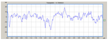
Figure 1: Assessment chart of the implant’s surface roughness before surgery.
On the assessment of the surface contour, there was some slop-ing and rising area, but the amplitudo range was around 20 nm. This data shows that the implant’s surface is quite smooth.
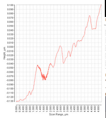
Figure 2: Assessment chart of the implant’s surface roughness 30 days after surgery.
After the surgery, the amplitudo range was ±10-40 nm. The cell attachment on the implant’s surface could interfere with the last result. That attachment was part of body response towards foreign substance. The pattern of the chart looks different pre and post implantation, allegedly related with the placement of the material, because the implant has a certain curve. But this doesn’t concern for a safety on the material, because the changes on the chart doesn’t represent a significant difference on the material surface of the implant between before and after implantation.
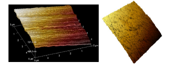
Figure 3: 3D picture of the PMMA implant before implantation
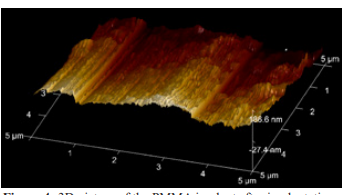
Figure 4: 3D picture of the PMMA implant after implantation
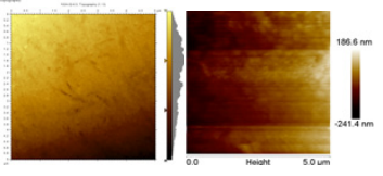
Figure 5: 2D picture of the PMMA implant’s surface before (A) and after (B) surgery
Table 1. Topography of PMMA pre and post 30th day implantation in the rabbit eye
|
|
Pre Operation 1 |
Pre Operation 2 |
Mean Pre Operation |
Post Operation |
|
Image Rq (nm) |
34.1 |
27.8 |
30.95 |
43.6 |
|
Image Ra (nm) |
28 |
23.3 |
25.65 |
22.9 |
After the implantation, the implant is not as clear as before implantation. The attachment of tissues on the implant’s surface can affect the result of the examination.
The result of AFM examination shown in table 1 that there was only slight changes. Image in Ra showed 28 nm pre surgery to 22.9 nm 30 day post implantation. The surface contour may alter the result.

Figure 6: The result of the infrared emission of the PMMA implant before implantation
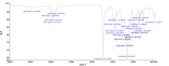
Figure 7: The result of the infrared emission of the PMMA im plant after implantation
Table 2: The results of FTIR Examination
|
No. |
FTIR |
|||
|
Pre-Operasi |
|
H+30 |
|
|
|
1 |
3444.41 cm-1 |
98.33% T |
3445.46 cm-1 |
98,09% T |
|
2 |
2995.25 cm-1 |
94.99% T |
2995.24 cm-1 |
94.76% T |
|
3 |
- |
|
2853.55 cm-1 |
96,27% T |
|
4 |
- |
|
2927.10 cm-1 |
92.51% T |
|
5 |
2951.09 cm-1 |
92.66% T |
2951.02 cm-1 |
91.51% T |
|
6 |
1723.36 cm-1 |
69.62% T |
1723.40 cm-1 |
67,11% T |
|
7 |
1634.95 cm-1 |
97.33% T |
- |
|
|
8 |
1484.66 cm-1 |
90.95% T |
1484.72 cm-1 |
89.96% T |
|
9 |
1447.04 cm-1 |
87.16% T |
1447.03 cm-1 |
85.54% T |
|
10 |
1435.24 cm-1 |
86.04% T |
1435.27 cm-1 |
84.31% T |
|
11 |
1386.39 cm-1 |
93.03% T |
1386.27 cm-1 |
91.94% T |
|
12 |
1270.12 cm-1 |
86.09% T |
1270.16 cm-1 |
84.48% T |
|
13 |
1240.34 cm-1 |
82.25% T |
1240.31 cm-1 |
80.25% T |
|
14 |
1190.41 cm-1 |
78.39% T |
1190.33 cm-1 |
76.03% T |
|
15 |
1144.07 cm-1 |
67.99% T |
1144.06 cm-1 |
64.29% T |
|
16 |
1063.17 cm-1 |
89.59% T |
1063.10 cm-1 |
87.51% T |
|
17 |
986.48 cm-1 |
87.56% T |
986.54 cm-1 |
85.78% T |
|
18 |
912.28 cm-1 |
93.36% T |
912.32 cm-1 |
92.34% T |
|
19 |
966.54 cm-1 |
88.02% T |
966.49 cm-1 |
86,39% T |
|
20 |
841.38 cm-1 |
91.02% T |
841.28 cm-1 |
89.88% T |
|
21 |
827.01 cm-1 |
93.85% T |
827.05 cm-1 |
98.01% T |
|
22 |
810.47 cm-1 |
94.40% T |
810.50 cm-1 |
93.62% T |
|
23 |
749.62 cm-1 |
87.15% T |
749.63 cm-1 |
85.63% T |
|
24 |
482.71 cm-1 |
92.76% T |
481.69 cm-1 |
91.26% T |
On the table, it is shown that the wavelength transmission be-tween before or after surgery was relatively similar. For example on number 1 (at the same point), the transmission of infrared before surgery was 98,33% with frequency 3444,41 cm-1, while after surgery the transmission of infrared was 98,09% with fre=quency 3445,46 cm-1.
Frequencies 1150 cm-1 until 1250 cm-1 were caused by the vi¬bration strain C-O-C. Frequencies 986 cm-1, 1063 cm-1, and 841 cm-1 are the characteristic of vibration absorption of PMMA. Frequencies 1386 cm-1 and 749 cm-1 are the characteristic of vibration absorption of a-methyl group. Frequencies 2995 cm-1 and 2951 cm-1 are caused by vibration strain C-H (contained in PMMA). This shows that there is no polymer degradation on the implant that was already used.
GCMS measurement on the material before and after surgery shows that there was some additional compounds on the implant after surgery. Those compounds were seen on table 5 line 1, 4 and 6. Compounds on line 2 and 5 were not altered in quality and quantity. Compound on line 3 and 7 were changed in quantity only.
Discussion
PMMA is a synthetic biomaterial and has been widely known as bioinert material, which means nontoxic and biologically inactive. Its uses as implant material or surgery aids on human has been done before, on ophtalmology, orthopaedic, and neurosurgery [8-11].
Biomaterial has physical, chemical and biological properties. Each of these properties can be analysed through a series of examination. In this research, the examination was done for the physical and chemical properties. Chemical properties in-cluding composition, bond and atomic structure, while physical properties examination including microstructure, phase, density and material porosity [8, 12]. The surface of the implant was assessed by AFM, polymer degradation was assessed by FTIR, and chemical compound before and after implantation was assessed by GCMS.
Surface topography assessment of the implant wasdone through variety of examination including optic microscope, electron microscope which is scanning electron microscope (SEM) and transmission electron microscope (TEM) also scanning probe microscope (SPM) which is scanning tunneling microscope (STM) and atomic force microscope (AFM). Optic microscope and electron microscope are only able to make assessment in 2D. Electron microscope can be affected by electron beam energy. The sample could be destroyed [12].
Scanning probe microscope is able to assess in 3D with atomic resolution. STM examination usually used on material made of metal and semi conductor or even material with electronic surface structure, while AFM is used for polymer material. The advantage of AFM is it has ability to assess mechanical contact force, Van der waals force, electromagnetic force, capillary force, chemical bond,electrostatic bond, etc [12, 13].
Table 3: Comparison of implant material’s topography and the PMMA material
|
Type of implant |
Image Ra(μm) |
|
PMMA post implantation |
0.0229 |
|
Ahmed FP7 (silicon) |
1.5 ± 0.1 |
|
Ahmed S-2 (polypropilene) |
1.3 ± 0.1 |
|
Baerveldt implant (silicon) |
0.1 ± 0.01 |
|
Molteno implant (polypropilene) |
0.07 ± 0.01 |
AFM is used to assess the topography of the implant. The structure was smooth on 2D examination, but on 3D examination, the implant looks not entirely flat. The surface of the implant seem fluctuating on micrometer measurement. Before surgery, the am¬plitudo differences is around 28 nm. The implant has curvature, by using AFM, the elevation of the surface can’t be seen
Table 4: GCMS Result on PPMA Implant Material Pre-Implantation
|
Substances on PMMA Implant |
|||
|
RT |
Area (%) |
Substances |
Quality |
|
4.045 |
10.55 |
Alumunium, tripropyl- SS Tripropylalumunium SS UN 2718 SS Trypropylalumunium |
78 |
|
|
|
Oxotri (isopropoxo) vanadium |
64 |
|
|
|
3-Butenoic acid (CAS) SS Vinylacenic acid SS.beta. –Butenoic acid |
59 |
|
4.148 |
1.38 |
Propyl acrylate SS 2-Propenoic acid, propyl ester (CAS) |
50 |
|
|
|
2-Propenoic acid, methyl ester SS Acrylic acid methyl ester |
50 |
|
|
|
2-Propenoic acid, methyl ester (CAS) SS Methyl acrylate SS Methyl propenoate |
50 |
|
4.216 |
87.27 |
2-Propenoic acid, 2-methyl-, methyl ester (CAS) SS Methyl methacrylate SS Mme |
91 |
|
|
|
2-Propenoic acid, 2-methyl-, methyl ester (CAS) SS Methyl methacrylate SS Mme |
91 |
|
|
|
2-Butenoic acid, methyl ester, (Z) – SS Crotonic acid, methyl ester, (z) - |
91 |
|
4.952 |
0.79 |
2(5H)-Furanone, 3-methyl- (CAS) SS 2-Methyl-2-butenolide |
93 |
|
|
|
2(5H)-Furanone, 3-methyl- SS.alpha.-Methyl-.gamma.-crotonolactone |
93 |
|
|
|
3-methyl - 5H - furan - 2 - on |
78 |
Table 5. GCMS Result on PMMA Implant Post Implantation
|
Substances on PMMA Implant |
|||
|
RT |
Area (%) |
Substances |
Quality |
|
2.968 |
2.66 |
-Methylamino-propylamine |
78 |
|
|
|
Amphetamine SS Aderal SS Dexedrin |
43 |
|
|
|
dl-Phenylepherine |
43 |
|
3.968 |
12.54 |
Alumunium, tripropyl- SS Tripropylalumunium SS UN 2718 SS Tripropylalumunium |
78 |
|
|
|
Oxotri (isopropoxo) vanadium |
64 |
|
|
|
3-Butenoic acid (CAS) SS Vinylacenic acid SS.beta. –Butenoic acid |
59 |
|
4.08 |
1.77 |
N-Propyl acrylate SS 2-Propenoic acid, propyl ester SS 1-propyl acrylate |
47 |
|
|
|
Propyl acrylate SS 2-Propenoic acid, propyl ester (CAS) |
47 |
|
|
|
2-Propenoic acid, methyl ester (CAS) SS Methyl acrylate SS Methyl propenoate |
43 |
|
4.131 |
23.22 |
2-Propenoic acid, 2-methyl-, methyl ester (CAS) SS Methyl metacrylate SS Mme |
91 |
|
|
|
2-Butenoic acid, methyl ester, (Z) – SS Crotonic acid, methyl ester, (Z) – |
91 |
|
|
|
Methyl 2-butenoate SS 2-Butenoic acid, methyl ester |
90 |
|
4.157 |
56.4 |
2-Propenoic acid, 2-methyl-, methyl ester (CAS) SS Methyl metacrylate SS Mme |
91 |
|
|
|
2-Propenoic acid, 2-methyl-, methyl ester (CAS) SS Methyl metacrylate SS Mme |
91 |
|
|
|
2-Butenoic acid, methyl ester, (Z) – SS Crotonic acid, methyl ester, (Z) – |
91 |
|
4.396 |
2.42 |
Methyl metacrylate SS 2-Propenoic acid, 2-methyl-, methyl ester |
64 |
|
|
|
2-Propenoic acid, 2-methyl-, methyl ester (CAS) SS Methyl metacrylate SS Mme |
64 |
|
|
|
2-Propenoic acid, 2-methyl-, 2-propenyl ester SS Methacrylic acid, allyl ester |
64 |
|
4.883 |
1 |
2(5H)-Furanone, 3-methyl- (CAS) SS 2-Methyl-2-butenolide |
91 |
|
|
|
2(5H)-Furanone, 3-methyl- SS.alpha.-Methyl-.gamma.-crotonolactone |
90 |
|
|
|
3-methyl - 5H - furan - 2 - on |
91 |
AFM surface contour measurement on the implant that has been implanted on rabbit’s eye for 30 days shown slight changes. It thought to be related with the tissue reaction on the implant and the manipulation during surgery. AFM has ability to assess any changes on the surface in nanometer. Rabbit’s tissue attached to the implant or the procedure of the surgery may affect the result of the examination. This changes need to be considered due to polymer degradation, even though it has been widely known that PMMA is a nonbiodegradable material [14].
Fourier Transform Infra Red (FTIR) Spectroscopy was used to prove any changes on the implant, which is by polymer degrada-tion or chemical compound alteration. FTIR uses wave by mod¬ulating interferometric. The signal will be captured and recorded on interferogram. The result will be calculated mathematically and the interaction between infrared radiation and natural vibra¬tion of the atom in the material will be assessed [15].
In this study, the frequency before and after surgery is similar. Chart 3 and 4 and also Table 2 shows the similarity between infrared transmission before and after surgery. Vibration on PMMA pictured with frequency 986 cm-1, 1063 cm-1 and 841 cm-1. These frequencies was from sequenced of atom on PMMA compound which contains group of C-H, C=O, CH3 and -OCH3. The result of this study is consistent with an experiment which was done by Ramesh16 et al, below:
Table 6: Vibrations and Frequencies on PMMA
|
Description of Vibrations |
Wavenumbers (cm-1) |
|
C-H stretching |
2927-2986 |
|
C=O stretching |
1700-1744 |
|
CH3 stretching |
1439 |
|
-OCH3 stretching |
1195 |
It concluded by FTIR results that the slight changes on the AFM examination was not related with the PMMA. It probably be¬cause of manipulation during the surgery.
Chemical compound on PMMA material before and after implantation was assessed with GCMS. Molecular weight was also assessed. Examination using GCMS start with changing PMMA solid structure into gas which will be ionised and fragmented into ion fragment. These ion fragment will be assessed on mass spectrometer.15 Based on the results, we could conclude although there was alterations but non toxic compounds were found.
The polymethylmethacrylate has toxic monomer in the form of liquid and gas. In a study in the rat, after exposure to methyl methacrylate concentrations of 0, 90, 437 or 2262 mg/m3 (0, 21, 104 or 538 ml/m3) by inhalation, 10 % to 20 % of the substance was deposited in the lower respiratory tract and metab-olized there (EU 2002). Irritation of the upper respiratory tract and eyes and possible CNS effects were reported in humans after exposure to methyl methacrylate concentrations of up to 250 ml/ m3 [17]. This study did not found this monomer with this high
Conclusions
There were slight roughness changes of the PMMA plate after the implantation. The wavelength transmission pre and post implantation was relatively similar. Some additional chemical compounds were found after the implantation, but not a toxic compound.
References
- Giangiacomo, A. & Coleman, A. L .(2009). Glaucoma (eds. Grehn, F. & Stamper, R.) Springer Berlin Heidelberg, 13-21.
- Sharaawy, T., Sherwood, M. & Hitchings, R .(2009). Glaucoma 1. Philadelphia: Saunder Elsevier.
- Stamper, R. L., Lieberman, M. F. & Drake, M. V. in Beck-er-Shaffer’s Diagnosis and Therapy of the Glaucomas 436-8 (Elsevier Health Sciences, 2009).
- Stamper, R. L., Lieberman, M. F. & Drake, M. V. in Beck-er-Shaffer’s Diagnosis and Therapy of the Glaucomas 466-90 (Elsevier Health Sciences, 2009).
- Eisenberg, D. L., Koo, E. Y., Hafner, G., & Schuman, J. S. (1999). In vitro flow properties of glaucoma implant devices. Ophthalmic Surgery, Lasers and Imaging Retina, 30(8), 662-667.
- Koura, Y., Fukushima, A., Nishino, K., Ishida, W., Nakaku-ki, T., Sento, M., ... & Ueno, H. (2006). Inflammatory reaction following cataract surgery and implantation of acrylic intraocular lens in rabbits with endotoxin-induced uveitis. Eye, 20(5), 606-610.
- KIM, M. S., & RHEE, S. W. (1990). Effects on the surrounding tissues and morphological changes of cellular components after implantation of PMMA and heparin sur-face modified PMMA intraocular lens in rabbit eyes. Journal of the Korean Ophthalmological Society, 161-174.
- Bandyopadhyay, A., & Bose, S. (Eds.). (2013). Characterization of biomaterials. Newnes.
- Ko, A. L., Nerva, J. D., Chang, J. J., & Chesnut, R. M. (2014). Traumatic fracture of a polymethyl methacrylate patient-specific cranioplasty implant. World neurosurgery, 82(3-4), 536-e11.
- Al-Otaibi, A. G., & Al-Qahtani, E. S. (2012). Polymethyl methacrylate intraocular lens opacification 20 years after cataract surgery: A case report in a tertiary eye hospital in Saudi Arabia. Saudi Journal of Ophthalmology, 26(1), 105-107.
- Sampath Kumar, T. S .(2013). Characterization of biomaterials. Elsevier, 11-47.
- Wang, H. & Chu, P. K .(2013). Characterization of biomaterials. Elsevier,105-174.
- Jessy, R. S., & Ibrahim, M. H. (2014). Biodegradability and Biocompatibility of Polymers with Emphasis on Bone Scaffolding: A Brief Review. Int. J. Sci. Res. Publ, 4, 7-9.
- Schmitt, J., & Flemming, H. C. (1998). FTIR-spectroscopy in microbial and material analysis. International Biodeteri-oration & Biodegradation, 41(1), 1-11.
- Choritz, L., Koynov, K., Renieri, G., Barton, K., Pfeiffer, N., & Thieme, H. (2010). Surface topographies of glaucoma drainage devices and their influence on human tenon fibroblast adhesion. Investigative Ophthalmology & Visual Science, 51(8), 4047-4053.
- Ramesh, S., Leen, K. H., Kumutha, K., & Arof, A. K. (2007). FTIR studies of PVC/PMMA blend based polymer electrolytes. Spectrochimica Acta Part A: Molecular and Biomolecular Spectroscopy, 66(4-5), 1237-1242.
- MAK Commission. (2014). The MAK-collection part I, MAK value documentations 2014. The MAK collection for Occupational Health and Safety. Wiley-VCH Verlag GmbH & Co KGaA, 320.