

Advances in Bioengineering and Biomedical Science Research(ABBSR)
ISSN: 2640-4133 | DOI: 10.33140/ABBSR

Impact Factor: 1.7

Research Article - (2022) Volume 5, Issue 3
Bioresonance According to Paul Schmidt (BaPS) and its Beneficial Effects on the Integrity of the Intestinal Barrier in vitro
2University of West Bohemia, Faculty of Electrical Engineering, Universitni 8, 30100 Pilsen, Czech Republic. Supervisor: Prof. Dr. Thomas Blecha, Germany
Received Date: Jul 18, 2022 / Accepted Date: Jul 22, 2022 / Published Date: Aug 03, 2022
Copyright: ©Copyright: ©2022 Peter C. Dartsch. This is an open-access article distributed under the terms of the Creative Commons Attribution License, which permits unrestricted use, distribution, and reproduction in any medium, provided the original author and source are credited.
Citation: Peter C. Dartsch, Dietmar Heimes (2022) Bioresonance According to Paul Schmidt (BaPS) and its Beneficial Effects on the Integrity of the Intestinal Barrier in vitro. Adv Bioeng Biomed Sci Res 5(3): 180-185.
Abstract
The intestinal epithelium, which is only one cell layer thick, has two essential tasks. The first is to create a physical barrier between the contents of the intestinal lumen and the rest of our body. The second is to ensure an efficient absorption of essential nutrients from the gut lumen and to produce mucus, anti-microbial peptides and cytokines with both protective and immune-regulatory properties. Thus, a reduced barrier function may have consequences, not only for intestinal, but also for systemic health. In this study we investigated whether the use of a Mini-Rayonex device, which has been developed to emit a bioresonance frequency spectrum according to Paul Schmidt (BaPS), might be able to promote and maintain the integrity of the intestinal barrier. Intestinal epithelial cells (IPEC-J2) were cultivated on microporous transwell plates to build up an intestinal barrier after exposure to a Mini-Rayonex device with the dipole antenna system (= verum). Corresponding control cultures were treated in the same manner but were exposed to a non-working Mini-Rayonex device without the dipole antenna system (= placebo). The transepithelial electrical resistance was measured as an indicator of intestinal barrier integrity. Thereafter, the achieved intestinal barrier from both Mini-Rayonex devices was exposed to 500 and 1,000 µM H2 O2 for 30 hours and the transepithelial electrical resistance was measured again. Moreover, the effect of both Mini-Rayonex devices (verum and placebo) on the regeneration process of intestinal epithelial cells at conditions of oxidative stress was investigated via examination of the closure of a cell-free area. Exposure of the cells to the actively emitting Mini-Rayonex device strengthened the intestinal barrier more than 30 % in comparison to the non-emitting Mini-Rayonex device. Moreover, the strengthened intestinal barrier was significantly more resistant by 20 % towards 500 µM H2 O2 and about 30 % towards 1,000 µM H2 O2 after exposure to the actively emitting Mini-Rayonex device than the “normal” intestinal barrier after exposure to the non-emitting device. In accordance with this observation was the result that the regeneration process after traumatization or injury of intestinal epithelial cells was increased by the actively emitting Mini-Rayonex device by more than 12 % in direct comparison with the non-emitting device. We conclude that the use of the actively emitting Mini-Rayonex device in vivo might also result in an improved intestinal epithelial barrier integrity, function and regeneration, which might improve and maintain well-being and systemic health.
Keywords
Bioresonance According to Paul Schmidt (BaPS); Intestinal Barrier, IPEC-J2 cells; Transepithelial Electrical Resis¬tance (TEER); Oxidative Stress; Cell Regeneration; Cell Culture.
Introduction
The intestine is the primary organ for food digestion, absorption and metabolism, which also acts as an essential physical and immunological barrier. Its physiological functions include nutri¬ent absorption, pathogen sensing and intestinal homeostasis [1]. Thus, a reduced barrier function might have unwanted consequences, not only for intestinal, but also for systemic health [2].
There is some evidence that increased intestinal permeability underlies the pathogenesis and/or maintenance of some autoim¬mune disorders such as Crohn’s disease and coeliac disease as well as type 1 diabetes [3-5]. Food components and gut-derived bacteria can affect intestinal barrier integrity [6-9]. Oxidative stress as an imbalance between the production and elimination process of reactive oxygen species plays another essential factor for many intestinal, cardiovascular and neurodegenerative dis-eases [10-16].
Regeneration is another essential point which assures the integ-rity of the intestinal epithelial barrier. Regeneration of complex structures such as the intestinal barrier after traumatization or injury requires dramatic changes in cellular behaviour. Regen-erating tissues initiate a program that includes diverse processes such as wound healing, cell death and dedifferentiation. More-over, newly regenerated tissues must integrate into preexisting cellular structures [17-19].
The term „bioresonance“ includes two words; the first is the word „bio“, which refers to the naturalness of a procedure and the second term „resonance“ describes the ability of two systems to oscillate at the same frequency, i.e. to be in resonance [20]. As early as 1975, engineer Paul Schmidt researched the effect of frequencies on humans and animals. He used a frequency gen-erator and applied the generated frequencies to the organism via the skin and was able to see that this accelerated the healing be¬havior of various organs. Finally, he developed a dipole antenna system, today’s dipole antenna system of bioresonance accord¬ing to Paul Schmidt (BaPS), which achieved the same regulatory effects in the organism. There are many studies about the dipole antenna system which is integrated in each bioresonance device according to Paul Schmidt. The most important one is a clin¬ical prospective, double-blind, randomized, placebo-controlled study, which demonstrated the reproducible effect of the dipole antenna system regarding pain reduction and which was pub¬lished at the German register for clinical studies [21].
Prompted by this background we investigated whether the use of a Mini-Rayonex device, which has been developed to emit a bioresonance frequency spectrum according to Paul Schmidt (BaPS), might be able to promote and maintain the integrity of the intestinal barrier.
Material and methods
Bioresonance Device According to Paul Schmidt (BaPS) For application of bioresonance according to Paul Schmidt (BaPS) on the cells, a so-called Mini-Rayonex device was used. The device was kindly provided by Rayonex Biomedical GmbH, D-57368 Lennestadt, Germany, for the duration of the experi-ments. The Mini-Rayonex device has been manufactured by this company for more than 30 years as a product to harmonize en¬dogenous and exogenous stress situations in the organism. Guid¬ed by the principles of bioresonance according to Paul Schmidt (BaPS), the fundamental frequency value 12.50 was integrated into the Mini-Rayonex using a dipole antenna system (Fig. 1). In line with bioresonance according to Paul Schmidt (BaPS), 12.50 is one of the most important fundamental frequency values that has a very positive effect on the regulation ability of the organism when it is exposed to any kind of stress.

Figure 1: Dipole antenna system from a Mini-Rayonex device developed by engineer Paul Schmidt and used in the present study for application of bioresonance according to Paul Schmidt (BaPS) on intestinal epithelial cells. The construction is based on a fixed rod plane and an equally elaborated, but rotatable rod plane above it. The individual rods of the two different rod planes are each designed as a dipole antenna. The figure illustrates the inner design of the Mini-Rayonex as a portable bioresonance device according to Paul Schmidt.
For this study, two different kinds of Mini-Rayonex devices were used: (1) Verum, i.e. an actively emitting Mini-Rayonex device with a dipole antenna system and (2) placebo, i.e. a non-emitting Mini-Rayonex device without a dipole antenna system as corresponding control.
Cultivation of Intestinal Epithelial Cells
The investigations were conducted with IPEC-J2 cells (ACC-701; Leibniz Institut, DSMZ, Braunschweig, Germany). The cells were routinely grown as mass cultures in Dulbecco’s Modification of Eagle‘s Medium and Ham’s F12 medium (1+1) supplemented with 10 % growth mixture and 0.5 % gentamycin and cultivated in an incubator at 37 °C in an atmosphere of 5 % CO2 and 95 % air at nearly 100 % humidity. The cells were regularly subcultured twice a week.
For the experiments, cells were taken from 80-90 % conflu-ent mass cultures and exposed to the actively emitting and the non-emitting Mini-Rayonex devices in two separated mini-incu¬bators (distance from each other was about 10 meters in different rooms to avoid any interactions of the Mini-Rayonex devices) at 37 °C without gassing. In order to keep the pH constant at 7.4 at these incubation conditions, another culture medium (medium # 2) was used for the exposure periods in the mini-incubators. This medium was a mixture of Leibowitz L-15 and RPMI 1640 (2+1) supplemented with 10 % fetal calf serum, 20 mM HEPES buffer and 0.5 % gentamycin.
Transepithelial Electrical Resistance (TEER)
TEER is broadly used as an experimental readout for measuring the integrity of epithelial monolayers cultured under static con¬ditions in vitro. IPEC-J2 cells were grown on the surface of a 0.4 µm porous membrane (transwell plate, Corning, Sigma-Aldrich, Deisenhofen, Germany) which yields two separated compartments within the cell culture dish. As a matter of fact, the cells covering the surface of the membrane (= directed towards the intestinal lumen) represent a physical barrier against the low¬er compartment (= blood). TEER was measured by placing one electrode in the culture medium in the upper compartment and another electrode in the lower compartment. Electrical resis-tance was measured by a portable voltmeter (Millicell ERS-2 Voltmeter, Millipore/Merck, Darmstadt, Germany) as described in detail elsewhere [22-24].
Epithelial cells were grown for 7 days with two culture medium exchanges during this cultivation period to an electrical resis-tance of about 1,000 to 1,500 Ω/cm2 representing an impaired intestinal barrier. Then, culture medium was changed to medium # 2 and cell layers were exposed for another 3 days to the active-ly emitting and non-emitting Mini-Rayonex devices in separated mini-incubators. Thereafter, TEER was measured again and data were compared with each other. Three independent experiments were conducted.
Intestinal Barrier and Oxidative Stress
After the final measurement of TEER of the intestinal barrier, the epithelial cell layers were exposed to 500 and 1,000 µM H2O2 for 30 hours in medium # 2 to the actively emitting and non-emitting Mini-Rayonex devices in separated mini-incu-bators. Then, TEER was measured again as already described above and the results for both Mini-Rayonex devices were com-pared. Moreover, cell cultures representing the intestinal barrier were fixed with 100 % methanol, stained with Giemsa’s azur eosin methylene blue solution (Merck, Darmstadt, Germany) and air-dried. Three independent experiments were conducted.
Cell Regeneration
The granulation phase, characterized by the occurrence of mi-gration and proliferation of epithelial cells for closing a defect, was simulated. For this purpose, intestinal epithelial cells were seeded at a density of 100,000 cells/ml into the four individual compartments of a silicone 4 well-culture insert (ibidi, Gräfelf-ing, Germany) [17,18]. The single compartments of the inserts were separated by a 500 μm thick silicone bar with an outer sil-icone frame of 700 μm. Due to the special adhesion area, an insert adheres firmly to the bottom of a culture dish and forms a distinct cell-free area (artificial wound), which the cells can colonize by migration and proliferation.
Upon reaching confluency within 48 hours after cell seeding, the silicon frames were removed with tweezers to achieve a sharp edge of the cell-free area between the compartments. The intes¬tinal epithelial cells in the dishes were allowed to migrate and proliferate for 6 hours in medium # 2 and exposed to the actively emitting and non-emitting Mini-Rayonex devices in separated mini-incubators. Finally, cell cultures were fixed with 100 % methanol, stained with Giemsa’s azur eosin methylene blue solution (Merck, Darmstadt, Germany) and air-dried. The clo-sure of the cell-free area was examined by micrographs (4 mi-crographs for each cell sample) and calculated by a specialized software with artificial intelligence from KML Vision, Graz, Austria (IKOSA AI software). Three parallel experimental se-ries were conducted.
Data Presentation and Statistical Analysis
Data presented here are given as mean values ± standard deviations. For statistical analysis the non-parametric two-tailed Wilcoxon-Mann-Whitney rank sum test was used. Statistical significance was assumed at p ≤ 0.01.
Results
TEER of the intestinal barrier was measured to be 3,470 ± 269 Ω/cm2 for the actively emitting Mini-Rayonex device and 2,605 ± 120 Ω/cm2 for the non-emitting Mini-Rayonex device. This difference is statistically significant (p ≤ 0.01) demonstrating that the actively emitting Mini-Rayonex device strengthened the intestinal barrier by more than 30 % in comparison to the non-emitting device. As a reference, TEER of the porous mem¬branes without any cellular barrier was measured to be 150 to 180 Ω/cm2.
As shown in Fig. 2, the strengthened intestinal barrier after exposure to the actively emitting Mini-Rayonex device was more resistant against oxidative stress when compared to the non-emitting Mini-Rayonex device. While TEER was reduced to 83,8 ± 4.9 % (for 500 µM H2O2) and 79.3 ± 8.2 % (for 1,000 µM H2O2) when exposed to the actively emitting Mini-Rayon¬ex device, it was reduced to 64.3 ± 5.4 % (for 500 µM H2O2) and 40.5 ± 6.7 % (for 1,000 µM H2O2) after exposure to the non-emitting Mini-Rayonex device. The difference was statis¬tically significant (p ≤ 0.01). This result was also documented microscopically on epithelial cell cultures representing the intes¬tinal barrier after fixation and staining (Fig. 3).
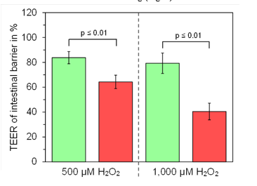
Figure 2: Presentation of the effect of oxidative stress on intestinal barrier integrity by the addition of either 500 µM H2O2 or 1,000 µM H2O2 for 30 hours. The green columns represent the strengthened intestinal barrier by use of the actively emit¬ting Mini-Rayonex device in comparison to a non-emitting Mini-Rayonex device (red columns). The strengthened intestinal barrier can resist oxidative stress significantly better (p ≤ 0.01, Wilcoxon-Mann-Whitney test) than the untreated intestinal bar¬rier. Data represent mean values ± standard deviations of three independent experiments.
Figure 3: Representative micrographs of the integrity of the intestinal epithelial barrier after exposure to 1,000 µM H2O2 for 30 hours and after fixation and staining. (A) Intestinal barrier by use of the actively emitting Mini-Rayonex device and (B) intestinal barrier by use of the non-emitting Mini-Rayonex device. Note that the cell density as visualized by the number of red stained cell nuclei is much higher in (A) than in (B), which also reflects the higher TEER for the intestinal barrier of the actively emitting Mini-Rayonex device as depicted in Fig. 2.
Regeneration of intestinal epithelial cells as visualized and de-termined by colonization of a cell-free space was significantly increased by exposure to the actively emitting Mini-Rayonex in comparison to the non-emitting Mini-Rayonex device. We mea¬sured a significantly increased closure of the cell-free area for the cells exposed to actively emitting Mini-Rayonex device In com- parison to the non-emitting Mini-Rayonex device (Fig. 4). The closure was 95.8 ± 2.1 % for the actively emitting Mini-Rayonex device and 83.3 ± 3.0 % for the non-emitting Mini-Rayonex de¬vice (p ≤ 0.01). The result is also depicted in the micrographs of epithelialcell cultures after 6 hours of regeneration time (Fig. 5).

Figure 4: Summarized presentation of the single measurement values of three independent experiments on cell regeneration with exposure to the actively emitting Mini-Rayonex device (green circles) in comparison to the non-emitting Mini-Rayonex device (red circles). The mean values of the single values are indicated in green or red dashed lines. It can be seen that the closure of the cell-free area is much higher after exposure to the actively emitting Mini-Rayonex device when compared with the non-emitting Mini-Rayonex device.

Figure 5: Representative micrographs of the regeneration process after fixation and staining of intestinal epithelial cells after 6 hours of regeneration. (A) Closure of the cell-free area of a representative cell culture after exposure to the actively emitting Mini-Rayonex device. (B) Closure of the cell-free area of the cells exposed to the non-emitting Mini-Rayonex device. Note the markedly increased closure of the cell-free area in the left micrograph after exposure to the actively emitting Mini-Rayonex device. Micrographs were taken with an Olympus IX 50 inverted microscope equipped with an Olympus Planachromate 10x and an Olympus E-10 digital camera at 4 megapixel resolution and bright field illumination.
Discussion
The main problem of studies on whole multi-cellular organisms such as experimental animals or volunteers is the complexity of the test systems. There are numerous unknown variables which are difficult to establish. In contrast, cultivation of eukaryotic cells can be standardized and provides the opportunity to vary different factors depending on the experimental needs. There¬fore, cell culture studies which focus on one single aspect of cell behaviour such as the integrity of the intestinal barrier in combination with epithelial cell regeneration are meaningful approaches to describe the effects of oxidative stress on the epithelial cell layer.
In the present study this approach was performed by using cul-tured intestinal epithelial cells which are able to differentiate and build an intestinal barrier of high integrity when the culture con-ditions are adequate. The IPEC-J2 cell line was chosen, because “the IPEC-J2 cell line is unique as it is derived from the small intestine and is neither transformed nor tumorigenic in nature. IPEC-J2 cells mimic the human physiology more closely than any other cell line of non-human origin” [25]. The cells were originally isolated in 1989 by Helen Berschneider at the University of North Carolina [26]. The advantage of the IPEC-J2 cell line as an in vitro model originates from its morphological and functional similarities with intestinal epithelial cells in vivo [27]. The epithelial cells of the intestinal barrier have a high turnover rate, because they are quite sensitive against alterations of their endogenous environmental conditions involving a deficiency of the epithelium and immune/inflammation mediating cells [11].
We were able to demonstrate that the building of an intestinal barrier on microporous membranes via primary proliferation and secondary differentiation of IPEC-J2 cells was significantly increased by use of the actively emitting Mini-Rayonex device in comparison to the non-emitting Mini-Rayonex device. This means that the fundamental frequency value 12.50 of the biore¬sonance according to Paul Schmidt (BaPS), which was integrat- ed into the actively emitting Mini-Rayonex device by using a dipole antenna system, was able to promote the development of an intestinal epithelial barrier with a higher integrity. At the same time, this strengthened intestinal epithelial barrier was able to resist oxidative stress much better than an intestinal barrier which was not exposed to this specific frequency value 12.50 as emitted by the actively emitting Mini-Rayonex device. From that point of view, the statements of bioresonance according to Paul Schmidt (BaPS) have been fully confirmed in the present in vitro-study.
In addition, regeneration is another essential point which assures the integrity of the intestinal epithelial barrier. When regenera¬tion is promoted, the integrity of the intestinal epithelial barrier is reconstructed much earlier after traumatization. As observed in the present study, the exposure to an actively emitting Mini-Ray¬onex device resulted in an increased intestinal epithelial regener¬ation which might also increase the turnover rate of cells in vivo.
We conclude that the use of the actively emitting Mini-Rayonex device in vivo might also result in an improved intestinal epi¬thelial barrier integrity, function and regeneration, which might improve and maintain well-being and systemic health.
References
- Segrist, E., & Cherry, S. (2020). Using diverse model systems to define intestinal epithelial defenses to enteric viral infections. Cell host & microbe, 27(3), 329-344.
- Lea, T. (2015). Epithelial cell models; general introduction. The Impact of Food Bioactives on Health, 95-102.
- Liu, Z., Li, N., & Neu, J. (2005). Tight junctions, leaky intestines, and pediatric diseases. Acta paediatrica, 94(4), 386-393.
- Silva, M. A. (2009). Intestinal dendritic cells and epithelial barrier dysfunction in Crohn’s disease. Inflammatory bowel diseases, 15(3), 436-453.
- Bosi, E., Molteni, L., Radaelli, M. G., Folini, L., Fermo, I.,Bazzigaluppi, E., ... & Paroni, R. (2006). Increased intestinal permeability precedes clinical onset of type 1 diabetes. Diabetologia, 49(12), 2824-2827.
- Visser J, Rozing J, Sapone A, Lammers K, Fasano A (2009) Tight junctions, intestinal permeability and autoimmunity; the celiac disease and type 1 diabetes paradigms. Ann N Y Acad Sci 1165: 195-205
- Lammers KM, Lu R, Brownley J et al (2008) Gliadin induces an increase in intestinal permeability and zonulin release by binding to the chemokine receptor CXCR3. Gastroenterology 135: 194-204.
- El Asmar, R., Panigrahi, P., Bamford, P., Berti, I., Not, T., Coppa, G. V., ... & Fasano, A. (2002). Host-dependent zonulin secretion causes the impairment of the small intestine barrier function after bacterial exposure. Gastroenterology, 123(5), 1607-1615.
- Tomasello, G., Mazzola, M., Leone, A., Sinagra, E., Zum-mo, G., Farina, F., ... & Carini, F. (2016). Nutrition, oxidative stress and intestinal dysbiosis: Influence of diet on gut microbiota in inflammatory bowel diseases. Biomed Pap Med Fac Univ Palacky Olomouc Czech Repub, 160(4), 461-466.
- Hussain, T., Tan, B., Yin, Y., Blachier, F., Tossou, M. C., & Rahu, N. (2016). Oxidative stress and inflammation: what polyphenols can do for us?. Oxidative medicine and cellular longevity, 2016.
- Bhattacharyya A, Chattopadhyay R, Mitra S, Crowe SE (2014). Oxidative stress: an essential factor in the pathogenesis of gastrointestinal mucosal diseases. Physiol Rev 94: 329-354.
- Wang, Y., Chen, Y., Zhang, X., Lu, Y., & Chen, H. (2020). New insights in intestinal oxidative stress damage and the health intervention effects of nutrients: A review. Journal of Functional Foods, 75, 104248.
- Finkel, T., & Holbrook, N. J. (2000). Oxidants, oxidative stress and the biology of ageing. nature, 408(6809), 239-247.
- Kim, S., & Sieburth, D. (2018). Sphingosine kinase regulates neuropeptide secretion during the oxidative stress-response through intertissue signaling. Journal of Neuroscience, 38(38), 8160-8176.
- Patel, R. S., Ghasemzadeh, N., Eapen, D. J., Sher, S., Ar-shad, S., Ko, Y. A., ... & Quyyumi, A. A. (2016). Novel bio-marker of oxidative stress is associated with risk of death in patients with coronary artery disease. Circulation, 133(4), 361-369.
- Riaz Rajoka, M. S., Thirumdas, R., Mehwish, H. M., Umair, M., Khurshid, M., Hayat, H. F., ... & Barba, F. J. (2021). Role of food antioxidants in modulating gut microbial communities: Novel understandings in intestinal oxidative stress damage and their impact on host health. Antioxidants, 10(10), 1563.
- Witte, M. B., & Barbul, A. (1997). General principles of wound healing. Surgical Clinics of North America, 77(3), 509-528.King, R. S., & Newmark, P. A. (2012). The cell biology of regeneration. Journal of Cell Biology, 196(5), 553-562.
- Sharif, M. F. Carlson, BM (2007). Principles of Regenerative Biology.Dietmar Heimes .(2014). Bioresonance according to Paul Schmidt. 4th edition, Spurbuchverlag, Baunach.
- Clinical-prospective, doubleblind-randomised, placebocon-trolled study about pain reduction with bioresonance devices according to Paul Schmidt. German register for clinical studies (2019).
- Lea, T. (2015). Epithelial cell models; general introduction. The Impact of Food Bioactives on Health, 95-102
- Geens, M. M., & Niewold, T. A. (2011). Optimizing culture conditions of a porcine epithelial cell line IPEC-J2 through a histological and physiological characterization. Cytotech-nology, 63(4), 415-423
- Bernardini, C., Algieri, C., Mantia, L. D., Zannoni, A., Sal-aroli, R., Trombetti, F., ... & Nesci, S. (2021). Relationship between serum concentration, functional parameters and cell bioenergetics in IPEC-J2 cell line. Histochemistry and Cell Biology, 156(1), 59-67
- Vergauwen, H. (2015). The IPEC-J2 cell line. The Impact of Food Bioactives on Health, 125-134.
- Berschneider, H. M. (1989). Development of normal cultured small intestinal epithelial cell lines which transport Na and Cl. Gastroenterology, 96(Suppl. Pt 2), A41.
- Schierack, P., Nordhoff, M., Pollmann, M., Weyrauch, K. D., Amasheh, S., Lodemann, U., ... & Wieler, L. H. (2006). Characterization of a porcine intestinal epithelial cell line for in vitro studies of microbial pathogenesis in swine. Histochemistry and cell biology, 125(3), 293-305.