



Review Article - (2026) Volume 9, Issue 3
Age-Dependent Nonlinear Protein Aggregation: A Mathematical Model of Gut-Brain Axis Mediated Neurodegeneration

Received Date: Jun 12, 2026 / Accepted Date: Jul 10, 2026 / Published Date: Jul 20, 2026
Copyright: ©2026 Himadri B. Bohidar, et al. This is an open-access article distributed under the terms of the Creative Commons Attribution License, which permits unrestricted use, distribution, and reproduction in any medium, provided the original author and source are credited.
Citation: Amishi, Bohidar, H. B. (2026). Age-Dependent Nonlinear Protein Aggregation: A Mathematical Model of Gut-Brain Axis Mediated Neurodegeneration. Adv Neur Sci, 9(3), 01-09.
Abstract
Structural changes caused by misfolding, aggregation and clumping of proteins can manifest in serious pathological conditions. These also have a significant bearing on the biological activity of intrinsically disordered proteins (IDPs). Developing a general understanding of the neurodegenerative diseases by moving beyond the isolated age-based paradigms is one of the challenges of molecular biophysics. Herein, we propose a simple dynamic system level model that incorporates hormonal control, genetic predisposition, immune decline and the gut-brain axis as well as their interactions, to show that aggregation accelerates through critical tipping points rather than progressing steadily over time. Most strikingly, the model identifies a universal convergence around midlife, near the age of 47 years, when gender- specific protections fade and disease risk equalizes, and reveals gut dysbiosis as a dominant driver that can enhance protein clumping between age of 45 - 65 years. These findings shift the therapeutic focus from late-stage symptom management to proactive, stage-specific intervention, highlighting early immune support, midlife gut restoration, and late-stage clearance enhancement as key opportunities. Ultimately, the work presented in this study positions the gut- brain axis as an important lever to prevent the development of neurodegeneration decades prior to the onset of clinical symptoms. Furthermore, the generalized and simple model proposed herein can be modified to address an array of problems associated with the IDPs by suitably choosing the lever parameters.
Keywords
Protein Aggregation, Nonlinear Kinetics, Gut–Brain Axis, Systems Biology, Computational Modelling, Neurodegeneration
Introduction
The basic idea of traditional structural function paradigm in molecular biology is that a protein must fold into a stable, three-dimensional native structure to perform its biological role, which has been significantly expanded by the recognition of intrinsically disordered proteins (IDPs) [1]. IDPs possess intrinsically disordered regions don’t have a single stable structure under physiological conditions, instead they are dynamic ensembles of interconverting arrangements. This causes IDPs to structural changes caused by misfolding, aggregation, and clumping. This protein aggregation is a sign of various pathological conditions, including neurodegenerative diseases like Alzheimer’s, Parkinson’s, and Huntington’s disease. Healthy cellular function maintenance depends on proteostasis which is defined as critical balance between protein synthesis, correct folding, and the efficient clearance of damaged or misfolded proteins [2]. This balance is critically examined by the biological complexity of aging for IDPs. As we age several protective systems of the body begin to deteriorate.
Thymic involution reduces immune regulation and clearance efficiency starting in early adulthood, proteasomal activity declines due to oxidative stress, and melatonin production decreases sharply after middle age. Moreover, genetic factor, like the fragility factor (F) in carrier states, can set a higher baseline risk for protein clumping, which makes individuals more volatile to these age-related declines. The self-organization of proteins and polypeptides into biologically active secondary structures, post their biosynthesis, is mediated by several factors. This is a thermodynamic process replete with several metastable states until the equilibrium state with minimum free energy is achieved, which is the native state of the protein. For the minimization of the free energy hydrophobic forces play a major role which entails localization of nonpolar residues in the core of the molecular structure, and simultaneous placement of hydrophilic residues on the surface thereby balancing the hydrophobic-hydrophilic interactions with water. Furthermore, hydrogen bonding, van der Waals forces and screened Coulombic interactions play a key role in stabilizing the secondary structure. In addition, the environmental factors like pH, ionic strength, temperature and presence of other moieties contribute to this stabilization. However, any imbalance in the aforesaid molecular interactions may lead to misfolding, aggregation or clumping of proteins that causes neurodegenerative diseases like amyloidosis, Alzheimer’s, Parkinson’s and prion diseases. Cell stress induced by the misfolded insoluble fibrillar protein molecules are often responsible for neuronal death. A general correlation between protein aggregation dynamics and neurodegeneration needs to be established which is the objective of this work
Current comprehension of neurodegeneration is predicated on fragmented, age-centric viewpoints that do not incorporate multifactorial nonlinear biological mechanisms [3,4]. This study provides a simplistic systems level modelling that highlights the dynamic relationship between aging variables, hormone control, genetic predisposition, and the gut-brain axis. With the application of a constitutive differential equation for protein clumping kinetics, this research introduces the "Gut Factor (G(t)) as a significant kinetic multiplier. This factor defines the condition of the intestinal barrier and microbial health, where gut health balance and the release of pro-inflammatory endotoxins like LPS (lipopolysaccharides) serve as major drivers of protein misfolding, and thus causing diseases. In addition to pointing out the key factors, this conceptual framework provides a solution to the issues associated with inherent abnormalities in proteins and polypeptides.
Methodology
The IDP Vulnerability Factor
The old protein models’ assumptions revolve around the stability of ‘lock and key’. However, this model focuses on the Disorder-Function perspective [5]. Considering IDPs state is largely dependent on the molecular physics of their surrounding environment, they appear as dynamic structural arrays (or protein clouds). This fundamental shift in research is crucial because IDPs cannot achieve a single stable structure under physiological circumstances. They adopt many exchangeable states, instead. Organized proteins have amino acid composition with higher hydrophobic content in polar and charged residues than IDPs which further leads to their environmental sensitivity [6]. IDPs are resistant to near boiling temperatures and acidic pH conditions because of their residue composition whereas regular proteins denature under this condition [7].
Modelling Approach
a) Kinetic Framework
The major insight of this study is the formulation of a non-linear differential equation that goes beyond simple linear aging models [8]. We model the change in the concentration of clumped/ aggregated protein (PC) over time (t) using a systems level differential framework which serves as the constitutive equation given by

where each parameter represents a biologically relevant factor (or lever) namely,
• PC : Concentration of clumped protein which accumulates when formation exceeds clearance
•kprod and kclear : Rate constants for aggregated protein production and clearance. For aggregation to dominate in the system, kprod« kclear.
• F: Fragility Factor, represents genetic carrier state where F>1 (e.g., F≈1.5), which indicates increased likelihood of misfolding and aggregation due to reduced functional protein reserve [9]. (Figure 1)

Figure 1: Study of Protein Clumping based on Fragility Factor
• A: Age/Inflammation Factor, increases with age due to thymic involution and inflammaging processes.
• H: Hormonal/Gender Factor, it is modulated by estrogen, testosterone, and other hormones.
• M(t): Melatonin/Clearance Factor, decreases with age, representing declining efficiency of antioxidant defence and waste removal efficacy.
• G(t): Gut Factor, acts as a bidirectional catalyst that amplifies aggregation by 180–200% in the age range of 45–65 years.
• ptotal = Pc + Pfree: Total protein concentration, where Pfree represents the sum of monomers and oligomers
b) Time-Dependent Parameter Specification
The model recognizes that the age-related factors are inherently time-dependent levers.
• A(t): Grows exponentially during early life (0–40 years), accelerating most significantly during young adulthood and middle age, then plateaus after age of 60 years as maximum values approach saturation. Hence the risk of IDPs being clumped due to thymic involution increases exponentially after 20 years as the thymus gland’s protective immunological and regulatory functions that peak during early development and begin to decline and ultimately vanish in early 20s [10]. (Figure 2)

Figure 2: Protein Clumping Risk dependency on age
M(t): Demonstrates strong negative correlation with age, starting at peak at birth, melatonin levels consistently decline throughout the lifespan, reaching near zero at the age of 100 years due to lifestyle and stress. All of which in total are influenced by hormones as well [11]. (Figure 3)

Figure 3: Protein Clumping Risk based on Melatonin Factor
f(A,H) (The Proteostatic Buffer): Since this function depends on both age and hormones and both being time dependent, it is itself time dependent. This function accounts for the simultaneous protection of estrogen and the thymus. In youth, this function acts as a kinetic suppressant, holding the aggregation flux at a negligible baseline and neutralizing the pro-aggregatory potential of IDPs, and with age it fluctuates, but equalizes for both gender around the age of 47 years [12,13],(Figure 4).

Figure 4: Protein Clumping Risk based on age, gender and hormone dependence.
• G(t) (The Gut-Multiplier): A high G(t) (representing dysbiosis and LPS leakage) lowers the entropic barrier for IDP aggregation while simultaneously poisoning the proteasomal clearance pathways.
c) Equilibrium Analysis: At equilibrium, when formation rate equals the clearance rate

This equilibrium equation reveals that the total clumping profile follows a sigmoid curve, showing sharp transitions around the age of 35 years that represent global state transitions between proteostatic phases (Figure 5).

Figure 5: Equilibrium dynamics of the total clumping profile across the human lifespan derived from Eq. (2).
From equation (2) it is easy to be understood that that proteostatic clumping does not move in a simple straight line, instead it moves in a S-shaped path, showing a phase-dependent condition. During the first thirty years of life, the profile remains near-zero, that means a strong proteostatic balance which inhibits aggregation formation. However, the curve shows an important universal state transition at age of 35 years, at which point the system splits. This sudden alteration shows a proteostatic instability due to stress, which results in the fast nonlinear growth of protein aggregates. Following this the growth profile approaches a saturation level, which defines equilibrium at the specific age of 35 years that demarcates the boundary between accurate protein stabilization, and its instability zones.

Figure 6: This figure shows kinetics of protein aggregation and self-assembly considering all the factors discussed in this work
Figure 6 gives a consolidated summary of relative contributions of all biological factors to the total aggregation of proteins with respect to age. The overall clumping corresponds to a S- shaped sigmoid curve, commencing at almost 100% and declining weirdly around the age of 35 years, signifying a global state change from one phase to another. The low clumping perspective is governed by thymic level reaches its highest point at birth and its significance begins to deplete at around 25 years of age before progressively diminishing further. The estrogen hormone factor acts as a shield to inhibit protein clumping, and it exhibits a sinusoidal oscillatory variation with exponential decay, displaying hormonal cycles that stabilize and diminish post-menopause. The melatonin factor exhibits a pronounced Gaussian peak at the age of 22 years followed by a rapid decline at age of 40 years, signifying factors active solely during peak physical and reproductive maturity in both males and females. Interestingly, the estrogen protective nature shows a dual-phase behaviour with a Gaussian peak at 26 years and a piecewise shift at the age of 35 years, which infers the change towards the onset of menopause. An important step function escalating to 55% at the age of 45 years clearly establishes a pathological limit. The model uses a bifurcation analysis where age 45 acts as a phase transition. By setting a 55% activity threshold, the point where linear metabolic decline (1%/year) meets the exhaustion of hormonal buffers, we demonstrate that the age of 47 years represents the mathematical 'Point of No Return' where proteostatic failure becomes statistically inevitable. The gut factor G(t) is an important health factor rising between the age of 55-60 years to indicate gut imbalance and increased biological noise inside the human body. At last, protein clumping major dependency on G(t) gives an important conclusion, showcasing how gut health effects protein aggregation by forming a hump that decreases at the age of 70 years.
d) Gut-Brain Axis as a Kinetic Multiplier
In the constitutive equation, the Gut Factor (G) represents the state of the intestinal barrier and microbial health through the Gut-Brain correlation [14]. It is a time-dependent variable

LPS stays trapped in the intestines and is excreted. It acts as the primary driver of formation (kprod) of the following:
• Systemic Alarm: Once in the blood, LPS triggers a massive immune response.
• Neuroinflammation: LPS can cross a weakened and directly activate microglia, which creates an environment where proteins (like Alpha-synuclein) misfold and clump much faster.
• The Vicious Cycle: High LPS levels further damage the gut lining, leading to more LPS leakage.
On the other hand, SCFAs are the primary beneficial metabolites produced when good gut bacteria ferment dietary fibres [15]. The essence of the present work lies in the observation of aggregation amplification observed between age of 45-65 years. In the context of IDPs, we model the gut factor as a source of macromolecular crowding. Other salient features of the model are: (i) mechanism protocol that entails when the intestinal barrier fails, the systemic influx of inflammatory markers increases the density of the intracellular environment, and (ii) for the IDPs this increased density forces the disordered monomers to collapse into seeds of nucleation. This explains why GI symptoms precede clinical neurodegeneration by approximately 15 to 20 years with the gut provides the kinetic energy for the initiation of IDP miss-steps. Accumulation outpaces the clearance capacity, resulting in a rapid shift from a soluble monomeric state to an insoluble aggregated fibrillar phase. An intact intestinal barrier and a balanced gut microbiota, characterized by robust production of short-chain fatty acids, mitigate neuroinflammation and preserve brain functioning, thereby counteracting the aggregation cascade [16,17]
v. Modelling Universal Tipping Points
The model reveals a critical phenomenon of the ‘risk equalization’ clearly defined as the following:
• During Phase 1 (Age 0-25 years), high thymic defence and melatonin levels helps to maintain a Low Clumping conditions inside the human body.
• During Phase 2 (25-45 years), gender-specific division occurs. Females are benefitted from estrogen whereas males have a sharper 85% drop in melatonin, leading to faster threshold crossings.
• Around the age of 47 years, the model predicts a universal proteostatic breakdown when protective layers start to degrade, specifically estrogen levels and gut stability decreases (Phase 3). This change is caused by the excluded volume effects of macromolecular overcrowding, which limits the space available for normal protein folding, and as a result, abnormal intermolecular interactions ensue leading to uncontrolled aggregation [18]. As aggregation kinetics speed up, the system reaches at an intersection point. At this point, the rate of aggregate formation exceeds the rate of clearance, and the system transits from a stable state into a self-harming condition of instability. This results in a positive feedback loop where protein clumps trigger microglial cells and neuroinflammation, which in turn promotes protein aggregation, that in turn replicates the conditions seen in neurodegenerative disorders [19].
IDP-Specific Characterization Methodology:
i. Experimental Characterization Framework: The kinetic modelling was validated through experimental characterization of IDPs which used standard biophysical techniques that takes advantages on the distinct conformational behaviour of IDPs compared to globular proteins. These techniques include circular dichroism spectroscopy to detect the characteristic lack of secondary structure, nuclear magnetic resonance for residue-specific conformational dynamics, and fluorescence-based methods to monitor hydrodynamic radius and compaction states.
Environmental Sensitivity Assessment
Important aspect of this model is understanding IDP response to environmental disturbances:
• pH Effects: Neutralizing acidic groups at lower pH which helps to reduces the net charge on IDPs (or regions), leading to the increased solubility and a more compact structural state. This contrasts with the globular proteins, in which it is found that acidic conditions cause protonation of negatively charged side chains, that leads to charge imbalances and aggregate formation [20].
Temperature Effects: While high-temperature conditions reveal the hydrophobic core of ordered proteins that leads to aggregation, IDPs shows resistance to increased temperatures due to their lower hydrophobic residue content. Instead, increased temperature often enhances the conformational dynamics of IDPs, potentially aiding in the sampling of various structural states, including those that might facilitate functional interactions [21].
Functional Characterization Integration
The kinetic model incorporates IDP functional categories relevant to aggregation and disease:
• Molecular Assemblers: IDPs with high disorder content interact with multiple binding partners to encourage formation of higher-order complexes. The disorder content increases with the size of the protein complexes, and upon binding to partners, disordered assemblers maintain their open structure, allowing multiple proteins to bind to a single IDR. This multivalent binding capacity is structurally facilitated by the extended conformational behaviour of IDPs, which possess hydrodynamic volumes significantly larger than globular proteins of equivalent mass, thereby maximizing the accessible surface area for intermolecular interactions [22]. This inherent flexibility and lack of fixed three-dimensional structure enable IDPs to participate in diverse binding mechanisms, including coupled folding and binding, as well as highly dynamic and multivalent interactions [23].
• Scaffolding Function: IDPs can regulate spatiotemporal assembly of signalling partners by acting as scaffolds, which includes formation of biomolecular condensates. Scaffolding regions contain the highest degree of disorder among functional categories [24]. This functional characterization aligns with the D2 (Disorders in Disorders) concept, as IDPs are associated with neurodegenerative diseases including Alzheimer's disease (deposition of α-synuclein, tau, and amyloid-β proteins) and Parkinson's disease (α-synuclein accumulation) where the accumulation of aggregates containing intrinsically disordered proteins serves as a defining pathological feature [25].
Results and Discussion
This study of the kinetics of self-aggregation proposed in this work reveals a complex, non-linear environment that goes beyond traditional models of simply aging framing neurodegeneration as a systemic collapse of proteostatic balance. It becomes clear that the tilt to a pathological state is given by opposed pleiotropy, by analysing the concentration of aggregated protein (Pc) through an algorithm that explains for the tension between production and clearance. In this regime, certain alleles that provide reproductive advantages early in life via stress-response mechanisms pathways later drive harmful buildup during post-reproductive stages. The kinetic trajectory displays a sigmoid profile with a critical turning point around age 35, where the statistical bottleneck for IDP clustering is lowered while at the same time compromising proteasomal clearance pathway. This transition is accelerated by the age-dependent decrease of the Melatonin/Clearance Factor (M), which shows a sharp decrease from birth, reaching near-zero by age 100. This decline significantly reduces the usefulness of antioxidant defences as well as the lymphatic elimination systems required for the clearance of disorganized oligomers.
An important component of this research is the finding of the Gut-Brain Axis as an essential kinetic multiplier (G(t)), especially throughout the midlife window of 45–65 years. During this phase,poor intestinal barrier integrity permit for the systemic import of inflammatory signals, which results in macromolecular saturation. This biophysical activity limits the excluded volume becomes available for natural protein folding, causing disordered monomers to collapse and start nucleation, thus increasing the aggregation [26]. This basic increase results in a global proteostatic turnaround at nearly around age of 47 years, a point of equilibrium where unique gender-based risk profiles disappear. Although the Hormonal Factor (H) is regulated by estrogen and testosterone, which acts as a good kinetic suppressant in youth, the midlife drop in these endocrine buffers, together with a 85% decline in melatonin levels by age 50, which drives the system toward a 55% clumping threshold. At this splitting point, the pace of aggregate accumulation surpasses the rate of cellular clearance, resulting in a positive feedback loop that stimulates microglial cells and causes inflammation in the brain. The subsequent uncontrolled aggregation weakens IDPs' critical fundamental roles, which are required for the creation of higher-order complexes and spatiotemporal regulation of biomolecular condensates. By analysing these physiological shifts, the model creates a 15-20 year preclinical window where gastrointestinal dysbiosis and endocrine volatility act as early indicators of proteostatic failure, allowing a significant lead time for therapeutic intervention before the onset of irreversible motor or memory. The salient features of various relevant models are presented in Table-1. Data for previous models are taken from the literature cited in this work.
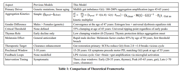
Evidence of important nonlinear changes in the molecular structure of proteins is rapidly challenging the view of human aging as a continuous linear decrease. While recent research3 has identified a generic molecular change around age of 44 years, in this work a more detailed and mechanistically integrated view of the tippling point near the age of 47 years is presented. Unlike other omics-based observation that focus on various metabolic markers, our model identifies the convergence of gender-specific protections and the initiation of widespread proteostatic decline. It also recognises how the gut imbalance acts as a prominent driver of protein clumping specifically between the ages of 45 and 65 years, that has not been adequately investigated in the general multi-omics literature. Earlier studies8 have identified generic turning points for neurodegeneration at the age of 40 years, while our research gives a predicted mathematical framework that explains risk equalization at the age of 47 years governed by the vanishing of the estrogen protective and thymic regulation factors simultaneously.
Finally, identifying these tipping points moves the treatment focus away from late-stage symptom treatment and towards proactive, stage-specific therapies. Recognition of age 47 years as a universal convergence point enables targeted midlife gut regeneration and early immunological assistance, which has the potential to prevent neurodegeneration decades before symptoms appear. This systems-level approach bridges the gap between solitary metabolic measurements and the complex relationship of factors that influence human health.
A schematic depiction of various lever parameters influencing protein aggregation kinetics, and their biophysical consequences are given in Scheme 1.

Scheme 1: Progressive transition of proteins into pathological aggregates driven by the nonlinear interplay of biological stress that act as levers.
Conclusion
With the help of transfer of the attention from late-stage protein accumulation to midlife intestinal imbalance as the primary cause of disease pathway, the outcomes of this study essentially reshape the development of neurodegeneration. In this simplistic, and yet predictive model, it is shown that the path to neurodegeneration is not a linear progression of aging, but it lies between the ages of 45 and 65 years, there is a systemic kinetic surge of protein aggregation. The age of 35 years marks an important transition point where proteostatic stability collapses, initiating the transition into a permanent state of protein instability and aggregation. Next to this shift, the estimation of the Gut-Brain Kinetic multiplier is critical to this perspective shift, with a surge from 1.0 to 2.8 serving as the primary catalyst for breaking the 55% clumping threshold which identifies age-adjusted imbalance as the major pathogenic factor, despite early-life genetic or gender-specific characteristics. While males have an earlier melatonin driven vulnerability and females have a post-menopausal, these diverse trajectories converge due to universal midlife microbiome alterations, this model clarifies the merging of pathological risk at age of 47 years.
This study provides an important biophysical roadmap for preventative medicine by establishing a 15-20 year preclinical window in which gastrointestinal symptoms and G(t) peaks precede motor disorder. Approaches that target the lipopolysaccharide (LPS)-driven vicious circle through gut restoration and Short-Chain Fatty Acid (SCFA) intake, such as butyrate supplementation, provide a theoretical mechanism for resetting the G(t) multiplier to 1.0 and breaking the feedback loop before irreversible neurological damage occurs. Finally, these findings suggest that proactive midlife gut stabilization, rather than downstream aggregate clearance, is the most effective strategy for preventing neurodegenerative onset, allowing for a shift from reactive symptomatic management to true disease-modifying interventions. Based on this model's three unique biological windows, future research and clinical trials should move away from a one-size-fits-all approach to neuroprotection. Instead, we propose a tiered method that focuses on the dominating kinetic driver at each life stage. Table-2 summarizes the key results of our work.
|
Phase |
Focus years |
Biological Driver |
Strategy |
|
Primordial |
20–35 |
Thymus & Sex Hormones |
Immunological/Endocrine buffering |
|
Preventative |
45–65 |
Gut Imbalance (G(t)) |
LPS reduction & SCFA restoration |
|
Stabilizing |
70+ |
Impaired Clearance |
Clearance enhancement & G(t) recovery |
Table 2: Key Ages When Major Biological Changes occur in Human Body.
We can shift the clinical targets from managing the symptoms to maintaining the integrity of a healthy gut-brain axis, by adopting this chronobiological framework. Developing a general understanding of the neurodegenerative diseases by moving beyond the isolated age-based paradigms is one of the remaining challenges of molecular biophysics. The current work was a step in that direction.
Statement of Usage of Artificial Intelligence
No AI tools were used in this work.
Author Contribution
The research problem was conceptualized jointly by both authors, Amishi performed the numerical analysis, results were discussed jointly, and the manuscript was written together. Both the authors consent to submit this work for publication.
Conflict of Interest
Authors declare no conflict of interest.
Data Availability
Since no raw data was created in this work, none is available.
Funding Information
This work did not receive funding from any source.
Acronyms:
1. PC:- Protein Clumping
2. a.u :- Arbitrary Units
3. SCFA:- Short Chain Fatty Acids
4. LPS:- Lipopolysaccharide
5. IDP:- Intrinsically Disordered Protein
6. IDR:- Intrinsically Disordered Regions
References
- M. Varadi, W. Vranken, M. Guharoy, P. Tompa, Computational approaches for inferring the functions of intrinsically disordered proteins, Front. Mol. Biosci. 2 (2015) 1–8.
- C.L. Klaips, G.G. Jayaraj, F.U. Hartl, Pathways of cellular proteostasis in aging and disease, J. Cell Biol. 217 (2018) 51–63.
- V. Voicu, C. Toader, M. È?erban, R.-A. Covache-Busuioc, A.V. Ciurea, Systemic neurodegeneration and brain aging: Multi-omics disintegration, proteostatic collapse, and network failure across the CNS, Biomedicines 13 (2025) 2025.
- J. Kosyakovsky, The neural economics of brain aging, Sci. Rep. 11 (2021) 12167.
- R. Trivedi, H.A. Nagarajaram, Intrinsically disordered proteins: An overview, Int. J. Mol. Sci. 23 (2022) 14050.
- V.N. Uversky, Targeting intrinsically disordered proteins in neurodegenerative and protein dysfunction diseases: Another illustration of the D2 concept, Expert Rev. Proteomics 7 (2010) 543–564.
- A.L. Darling, V.N. Uversky, Intrinsic disorder and posttranslational modifications: The darker side of the biological dark matter, Front. Genet. 9 (2018) 158.
- J. Wei, Y. Zhou, H. Liu, X. Chen, Y. Zhang, J. Wang, Kinetic models reveal the interplay of protein production and aggregation, Chem. Sci. 15 (2024) 8430–8442.
- P. Zhao, C. Wang, S. Sun, X. Wang, W.E. Balch, Tracing genetic diversity captures the molecular basis of misfolding disease, Nat. Commun. 15 (2024) 3333.
- N. Yayon, E. Kenigsberg, A. Tanay, I. Amit, A spatial human thymus cell atlas mapped to a continuous tissue axis, Nature 635 (2024) 708–718.
- M. Shukla, R. Duangrat, C. Nopparat, A. Sotthibundhu, P. Govitrapong, Melatonin augments the expression of core transcription factors in aged and Alzheimer's patient skin fibroblasts, Biology 13 (2024) 698.
- T.S. Wiley, J.T. Haraldsen, Cyclic E2 and P4 on Alzheimer's disease pathways, Adv. Alzheimers Dis. 6 (2017) 32–44.
- H.P. Priyanka, R.S. Nair, Neuroimmunomodulation by estrogen in health and disease, AIMS Neurosci. 7 (2020) 401–417.
- N. Molinero, A. Antón-Fernández, F. Hernández, J. Ávila,B. Bartolomé, M.V. Moreno-Arribas, Gut microbiota, an additional hallmark of human aging and neurodegeneration, Neuroscience 518 (2023) 141–161.
- P. Padhi, C. Worth, G. Zenitsky, H. Jin, K. Sambamurti, V. Anantharam, A. Kanthasamy, A.G. Kanthasamy, Mechanistic insights into gut microbiome dysbiosis-mediated neuroimmune dysregulation and protein misfolding and clearance in the pathogenesis of chronic neurodegenerative disorders, Front. Neurosci. 16 (2022) 836605.
- N.U. Mahbub, M.M. Islam, S.-T. Hong, H.-J. Chung, Dysbiosis of the gut microbiota and its effect on α-synuclein and prion protein misfolding: consequences for neurodegeneration, Front. Cell. Infect. Microbiol. 14 (2024) 1348279.
- S.K. Chakrabarti, D. Chattopadhyay, The gut–brain–immune triad in neurodegeneration: An integrated perspective, J. Transl. Gastroenterol. (in press).
- T.B. Thompson, P. Chaggar, E. Kuhl, A. Goriely, Protein-protein interactions in neurodegenerative diseases: A conspiracy theory, PLoS Comput. Biol. 16 (2020) e1008267.
- K.P. Kepp, A quantitative model of human neurodegenerative diseases involving protein aggregation, Neurobiol. Aging 80 (2019) 46–55.
- F.-X. Theillet, A. Binolfi, B. Bekei, A. Martorana, H.M. Rose, M. Stuiver, S. Verzini, D. Lorenz, M. van Rossum, D. Goldfarb, P. Selenko, Physicochemical properties of cells and their effects on intrinsically disordered proteins (IDPs), Chem. Rev. 114 (2014) 6661–6714.
- R. Appadurai, J. Nagesh, A. Srivastava, High resolution ensemble description of metamorphic and intrinsically disordered proteins using an efficient hybrid parallel tempering scheme, Nat. Commun. 12 (2021) 958.
- M. Arbesú, G. Iruela, H. Fuentes, J.M.C. Teixeira, M. Pons, Intramolecular fuzzy interactions involving intrinsically disordered domains, Front. Mol. Biosci. 5 (2018) 39.
- T. Orand, M.R. Jensen, Binding mechanisms of intrinsically disordered proteins: Insights from experimental studies and structural predictions, Curr. Opin. Struct. Biol. 90 (2025) 102958.
- V.N. Uversky, A decade and a half of protein intrinsic disorder: Biology still waits for physics, Protein Sci. 22 (2013) 693–724.
- M.M. Babu, The contribution of intrinsically disordered regions to protein function, cellular complexity, and human disease, Biochem. Soc. Trans. 44 (2016) 1185–1200.
- M.S. Hipp, S.-H. Park, F.U. Hartl, Proteostasis impairment in protein-misfolding and -aggregation diseases, Trends Cell Biol. 24 (2014) 506–514.