

Advances in Bioengineering and Biomedical Science Research(ABBSR)
ISSN: 2640-4133 | DOI: 10.33140/ABBSR

Impact Factor: 1.7

Review Article - (2018) Volume 1, Issue 3
A Review of the Involvement of Disturbed Treg/CTC Ratios in the Sustained Proliferation of Psoriasis with a Brief Gender Distribution Analysis
Received Date: Oct 23, 2018 / Accepted Date: Nov 04, 2018 / Published Date: Dec 28, 2018
Copyright: ©Copyright: ©2018 Aurangzeb Pirzada. This is an open-access article distributed under the terms of the Creative Commons Attribution License, which permits unrestricted use, distribution, and reproduction in any medium, provided the original author and source are credited.
Abstract
Aim: To provide evidence from current literature that psoriasis is an autoimmune disorder involving a highly proliferated effector response in relation to a suppressed, inactivated and outnumbered regulatory component.
Disease Background Pathophysiology: Discusses the octet phenotypes, histological analyses, epidemiological data, additional components followed by briefly reflecting on its psychoanalytical aspects.
Evidence Based Review: Provides a historical perspective of Treg research and spans Treg details across cellular, molecular and genetic details mentioning its subtypes, TCR signalling comparison and marker diversity moving onto bringing FOXP3 under spotlight to discuss its structure, pathways followed by establishing a link between Treg and psoriasis.
Materials and Methods: Discusses laboratory techniques namely, flow cytometry, Western Blot, quantitative-RT-PCR and culturing techniques (invitro assays) for T cells and cytokines.
Results: Focus on disease relationship with IFN/TNFs/CCRs/FOXP3 and Shh to some extent providing figures and tables for ease of quantification.
Discussion: Critically evaluates the listed research followed by an easy to follow explanation per figure/table listed in Results section.
Conclusion: Entailed with discussion is a brief overview of current treatments in light of which new theories will be summed up in this section in light of all of the contents of this article.
Disease Background and Pathophysiology
Psoriasis is a complex chronic inflammatory dermatological disease characterized by erythematous scaling plaques that spans across almost all age groups and can be divided into childhood and adult age periods. For the early part during its research it has been dealt as a disease of epidermal keratinocytic hyperproliferation associated with secondary cutaneous inflammation.
Current research is aims at molecular dissection of psoriasis and integrating genetics and biology to enhance the efficacy of its treatment by putting new range of therapies on test which has induced a shift towards higher resolution analysis. It has begun to be identified as an autoimmune inflammatory disorder.
Psoriasis Phenotypes
According to Langley, et al. 2005 psoriasis expresses itself in an octet of phenotypes namely
(a) Plaque Psoriasis
(b) Guttate Psoriasis
(c) Erythroderma
(d) Generalised Pustular Psoriasis
(e) Inverse Psoriasis
(f) Psoriatic Nail Disease
(g) Psoriatic Arthritis
(h) Scalp Psoriasis
Due to symptomatic homology author has chosen only the 2 most epidemiologically common types mentioned below:
Plaque Psoriasis (Psoriasis Vulgaris)
It is known to be the most frequent occurrence of psoriasis where the patient develops erythroderma with silvery scales ranging from circular to oval shaped red plaques that sometimes itch. Location is generally on high trauma areas such as elbows, knees, or trunk/ scalp. This type of psoriasis is persistent and the patient does not experience remission.
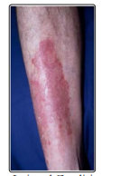
Figure P1: Cited by Lui and Zandi in a web article edited by Stoppler, C.M for www.emedicinehealth.com shows a case of plaque psoriasis on leg courtesy of Hon Pak MD US Army
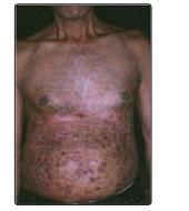
Figure P2: Cited by Lui and Zandi in a web article edited by Stoppler, C.M for www.emedicinehealth.com shows a case of abdominal plaque psoriasis courtesy of Hon Pak MD US Army
Guttate Psoriasis
Appearing as be small red bumps on skin which are covered by a fine silvery scale which is much thinner than that observed in plaque variety. It covers a large percentage of body and is often self-limiting in a few weeks but quite a lot of cases are persistent and require a wide range of available treatments such as topical therapies to subcutaneous/ IV injections or narrow band UV b phototherapy. Epidemiologically it is rated as a second most frequent occurrence and is more prevalent amongst age groups below 30 years of age. It has a very clear aetiological linkage with streptococcal throat infection that is followed by an outburst of guttate. It may be persistent and express repeated episodes amongst individuals who are permanent strep carriers [1]. It also occurs on palms with similar symptoms and intensity. It may also occur on finger tips and it is termed as acrodermatitis.
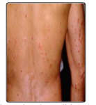
Figure P3A: Cited by Lui and Zandi in a web article edited by Stoppler, C.M for www.emedicinehealth.com shows a case of guttate psoriasis on the back courtesy of Hon Pak MD US Army
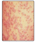
Figure P3B: Cited by Lui and Zandi in a web article edited by Stoppler, C.M for www.emedicinehealth.com shows a closeup of Figure P3A case of guttate psoriasis on the back courtesy of Hon Pak MD US Army
Histological Analysis
Laboratory diagnosis of psoriasis may require aspiration of inflamed skin exudate or lesion samples itself. Disease clinical features include inflammation, increased vascularity, scaling epidermis, epidermal hyperproliferation. Due to abnormal maturation rates there is visible hyperkeratosis coupled with parakeratosis accompanied by epidermal acathosis and rete ridge elongation due to hyperproliferative state. Skin undergoing psoriatic pathophysiology expresses hyperplasia of keratinocytes and their failure to differentiate which can be identified by using different combinations of skin stains [2].
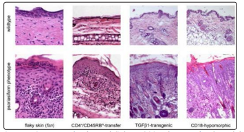
Figure P4: By Boehncke and Schon, 2007 shows animal model based investigation of histopathologic reaction patterns. All 4 types once stimulated by different reactants exhibit features of human psoriasis accompanied by hyperproliferative inflammatory cutaneous lesions
One of the symptoms includes poorly developed granular layer and its gradual loss with a low recovery rate. Skin lesions consist of progressive proliferative dermatitis dominated by dermo-epidermal infiltrates of an increased number of mast cells and neoangiogenesis. However lesional analysis varies broadly due to the disease subtype diversity.
Epidemiology
It mainly affects Caucasians and people of European ancestry with an incidence of approximately 2.5% in UK according to UKGPRD with it being less common amongst other ethnic groups [3].
According to National Psoriasis Foundation – USA 6 to 7 million people are affected in USA with maximal frequency expressed in European origin populous and minimal attributed to Samoan origin. In UK it is present amongst all age groups but 30-69 year age group is most captured by it [4].
Prevalence
Measures existing clinical cases which at any given time, net amount is around 2 to 2.25% of US population and slightly higher for UK populous.
Rate of incidence
It is at about 250,000 new cases registered in US annually. Despite rigorous statistical monitoring and population based studies majority of mild cases are seldom or never reported.
Additional Components
Geography and ethnicity are directly related to disease incidence and evidence suggests psoriasis frequency is elevated in cooler regions in countries such as Australia and Norway. Data expresses slight deviations showing a higher incidence amongst females.
Lifestyle choices such as smoking exert a positive correlation with psoriasis occurrence whereas data on drug abuse has not provided any definitive evidence however data on alcohol consumption produces conflicting evidence. Psoriatic patients show a 30% higher risk for cancer whereas obesity and CV diseases are exacerbated by chronic psoriasis. HLA class 1 involvement is due to B13, B17, B37, B57 and Cw6 whereas from class 2, only DR4 and DR7 are linked with psoriatic expressivity. However, gene mutation loci are identified to be as follows 17q25 = SLC9ARI 3q = SLC12A8 19p = Jun Proteins.
Psychosocial Consequences
Socially perceived to be contagious, it can lead to the individual being socially isolated with none or very limited support network increasing mental distress, chronic anxiety, depression and early onset patients of younger age are more vulnerable than adults.
Improvement of QoL is key in treatment effectiveness. It has a negative impact on patient’s health and workplace productivity [5]. Qualitative questionnaires and statistical evaluation will improve physicians’ insight in patients’ lifestyle and improve their approach to treatment.
Evidence Based Research Review
Whether it is economic model or physiological, importance of an immune system and regulatory component of that immune system is undeniable.
Historical Perspective
Psoriasis is a relatively new addition to the list of autoimmune diseases. That it is indeed deserving of such a position is well-supported by evidence suggesting the involvement of effector-mediated damage and nascent regulatory components of the immune system. Research shows that there is a strong correlation between the state of the regulatory component of the immune system and the occurrence of psoriasis. For this reason, it is necessary to briefly describe some of the research on q Following Miyara & Sakaguchi 2008, Research on
Tregs Can Be Divided into 5 Major Periods: 1969 – 1982
Claman, et al. 1966 initiated research on different lymphocyte subsets. Late 1960s saw Nishizuka and Sakakura in parallel with Gershon and Kondo in 1970-71 attempting a successful experimental approach at studying splenocytes involving animal models and thymectomy based investigations; further confirmed by Penhale, et al. 1973 which added irradiation to the existing protocols.
1982 – 1995
Murine modelling based thymic deletion/induction research flourished until 1986-87 when introduced clonal deletion and put forward the Th1/Th2 subset classification [6,7]. This put breaks on Treg research however research on autoimmunity continued.
1995 – 2000
Sakaguchi, et al. 1995 not only discovered CD25 marker but established its importance by conducting murine model comparative investigations providing evidence that addition of CD4+CD25+Tregs in autoimmune mouse models, prevented autoimmunity. They clarified the relationship of thymectomy and generation of CD4+CD25+Tregs. Shevach, et al. and Sakaguchi, et al. progressed to elucidate CD4+CD25+Treg relationship with ILs via invitro investigation.
2000 – 2003
Neuropilin, CD103, GPR83, CD62-L, CTLA-4, GITR all of whom were ubiquitous on the rest of T cell populations were discovered, however CD25 although expressed on all T populous, was found in a higher frequency on CD4+CD25+Tregs.
2003 – Present
IPEX models led to the findings of a TF FoxP3 and it was due to the gene expressivity deficiency of this TF that was involved in disease progression. Powel, et al. 1982, Godfrey, et al. 1991 and Schubert, et al. 2001 provided evidence of FoxP3 expression necessary for CD4+CD25+Treg induction and forced expression of which induced Treg functionality in other subsets aswell, invitro and invivo.
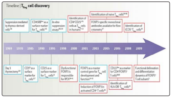
Figure E1: Shows a brief timeline created to sequencialise the rate and order of progress in field of regulatory T cells by Sakaguchi, et al. 2010
Functionality/Properties
About 5% of the total T cell count, CD4+CD25+Tregs are α/βTCR type. RAG – 2 genes is reported to be controlling the α/βTCR expression. They are driven by the expression of TF FoxP3. Although mostly CD4+CD25+ phenotype, not all Treg subsets are but any given T cells under correct environmental influence may act as suppressor e.g. the suppressive activity of CD8+ which unlike Treg is irreversible.
They originate from thymus following sequential positive and negative selection. Former occurring in cortical epithelium with MHC Ag whereas latter takes place policed by DCs in thymic medullary epithelium. This phase is estimated to be between day 3 to day 7 following thymic development as elicited by several mouse model investigations
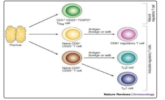
Figure E2: Shows a thymic hierarchichal developmental sequence for CD4+CD25+naturalregulatory T cells that end up in the periphery to module the effector responses by Mills, 2004
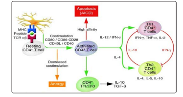
Figure E3: By Jiang and Chess, et al. 2004, presents a schematic of how the antigen activated CD4T cells undergo stimulation and proliferation for the maintenance of peripheral autoimmunity. In addition, superimposed on these intrinsic mechanisms are control mechanisms mediated by distinct subsets of NKT, CD4+, and CD8+ regulatory (suppressor) T cells
Table E4: Published by Jiang and Chess, et al. 2004 provides a brief overview of all subsets of T cells which are involved in regulatory mechanisms around the body
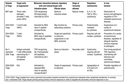
They are then trained by intrathymic generation owing to stimulation by strong agonist ligands mainly thymic CD80 or haematopoietic APCs. Apostolou, et al. 2002 provides data about the longevity of thymically educated Tregs which remain in peripheral circulation for a longer period than their induced counterparts. Although anergic and suppressive in nature, they express numerous CAMs such as LFA and ICAM-1 in addition to NGFR and OXO40 and 4-1BB superfamily. Kretschmer, et al. 2005 mentions the rigorous TCR dependent selection that is required before maturation and peripheral exposure
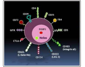
Figure E5: Published by Thornton, et al. 2010 presents an artist’s image of all the receptors and markers that Treg cell lines have to offer to the immune system for immunomodulatory purposes
Using murine autoimmunity models and invitro/invivo analyses, 3 mechanisms have been suggested by Asseman, et al. 1999, Annacker, et al. 2001 and Nakamura, et al. 2001.
Direct cell to cell mode is one where all those CAMs come in handy whereby they induce suppressive signals via their surface receptor molecules. They may also physically hinder or exclude CD4+CD25-cells or lastly APC modulation specifically targeting the CD80 and CD86 on them may be the key to net suppression of Teff.
FOXP3
Emerging from the ashes of IPEX model studies, FOXP3 became the single most important determinant of Treg functional viability and centre of academic research for mapping Treg phenomenology. Belonging to the forkhead family of TFs where its main function involves repression along with infrequent co-repressor or promoter activity, it has C and N termini and 11 exons are involved in its encoding. N terminus provides quintessential sites for repressive activity whereas the much more complicated C terminus harbours forkhead binding DNA domains which endow it to repress IL2 mRNA transcription by binding to IL2 promoter.
FOXP3 expressivity detection did not assume permanency in routine Treg identification/cell culture testing protocols but is now proving to be immensely convenient due to marker ubiquitination. However FOXP3 detection comes at an irreversible cost of cell permeation for testing which renders live cell action impossible for now. It has now been identified as the only component, lack of expressivity of which yields ineffective Tregs which in turn lead to the inevitability of autoimmunity [8].
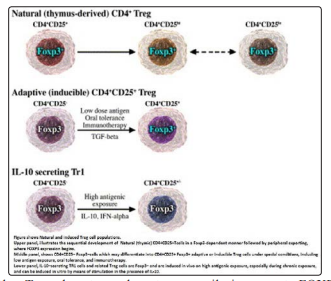
Other Treg phenotypes do not necessarily incorporate FOXP3 expression from the beginning; however forced retroviral transduction may provide the means to do so. It is part of a much larger much intricate network of TFs, co-repressors and co-activators synergizing to bring about immune regulation or in other cases suffering from malfunction leading to autoimmune disease.
A co-repressor in reality, it has recently been characterised as acetylated and phosphorylated protein complex which endows it with the kinase interaction capacity. Main involvement in silencing genes that are switched on following Teff activation gives it a characteristic methylation pattern which although time consuming to map and identify is unique to it.
Nonetheless it displays a remarkable co-repressing affinity for TCR repertoire generation and recombination which is why Tregs express a slow responsiveness to agonist ligand or Ag stimulation but this is compensated by EGFs invivo.
Most recent research focuses on molecular mechanisms influencing Treg functionality where FoxP3 represses IL2, IL4 and IFN-γ by interacting with NFAT, AML1, Runx1 protein.
Current research has differentiated Tregs into different species of T cells differing in proliferative capacity, cytokine productivity and expression of complement proteins providing strong evidence that molecular signalling pathways differ as well depending on downstream or upstream signalling events.
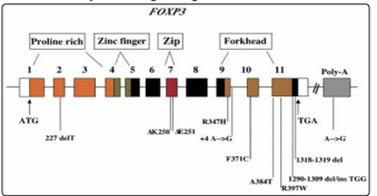
Figure: Shows FOXP3 gene organization and corresponding mutations in IPEX. This gene consists of composed of 11 coding exons and 2 upstream noncoding exons not shown here. The positions of the translational start and stop codons (ATG and TGA) are indicated. The predicted functional domains include a proline-rich N-terminal region, a zinc finger, leucine zipper (zip), and forkhead domains
exons and 2 upstream noncoding exons not shown here. The positions of the translational start and stop codons (ATG and TGA) are indicated. The predicted functional domains include a proline-rich N-terminal region, a zinc finger, leucine zipper (zip), and forkhead domains
Cell Line Molecular Signalling Comparison Teff TCR Signalling
An experimental investigation by Chan, et al. 1995 and detailed meta-analysis by Aguado, et al. 2006 describes it as a bimodal signalling responder phenomenon where TCR and APC (co-stim) signals are vital to IL2 proliferative generation. Single signalling incidences lead to anergy. However in normal circumstances, TCR/ CD4 interaction yields peptide MHC complex formation causing src kinase family mediated ITAM phosphorylation leading to recruitment and phosphorylation of ZAP70 which intern phosphorylates LAT that is the final key towards activating a myriad of different signalling pathways under different cascades such as P13K, calcium dependent NFAT and MAPKs.
Treg TCR Signalling
Although largely unchartered owing to limitations in cell surface marker reliability, cell numbers, isolation/extraction techniques and invitro proliferative capacity; there is evidence suggesting that Treg are designed to inhibit Teff TCR signalling cascades i.e. their signalling is to inhibit Teff TCR signalling.
MAPK
Initial phases of research produced evidence of defective pathways being responsible Treg phenotype which was further backed up by evidence provided by Tsang, et al. and Hickman, et al. 2006 but Crellin, et al. 2007 and Koonpaew, et al. 2006 conducted investigations on human cord blood which negated the earlier findings [9]. This pathway in Tregs appears decelerated many a fold in contrast to defective expression or lack of cascade functional components. Furthermore, reverse testing clinical trials revealed invivo Treg mechanisms differed slightly than those undergoing invitro testing but calcium signalling became apparent nonetheless as reported by Su, et al. 2004 and Gavin, et al. 2002.
NFAT
An independent anergy inducing co-promoter element, expressed in T cell lines, its unhindered activity accelerates the rate of anergic Teff cell lines [10]. AP1 is NFAT antagonist, complexing with whom leads to timely deactivation. FOXP3 undergoes heterodimerisation with NFAT and AP1 to repress IL2 transcription and increase CTLA4/ CD25 expression. Further research from Rudensky laboratories (NY) is confirming the evidence.
PK13 Borlado, et al. 2000 and Parsons, et al. 2001 suggest PK13 to be critical for Treg expression. Crellin, et al. in 2007 confirmed this situational conclusion by displaying Treg incapability of activating AKT. Patton, et al. 2006 expanded upon it to demonstrate that such Tregs lack suppressive function. Like all other pathways, it has its negative regulators and promoter elements.
NFkB
Sequential AKT activation is the key to NFkB expression which is repressed by FOXP3. NFkB has a complex relationship with FOXP3 and is it is confusing to deduce a direct or inverse interaction of the two. Researchers have slightly different opinions on it. Generally FOXP3 behaves as an NFkB agonist.
These molecular signalling mechanisms lead to the production of IL10 and TGFβ which are mediators of anergic induction in effector T cells but when defective these pathways are ineffective in bringing about positive regulatory phenotype in Tregs. IL2,4,6 are now confirmed as anti-suppressive and have been suspicious for decades [11].
<img src="https://www.opastpublishers.com/scholarly-images/453-6ad83fc3bbe8f-a-review-of-the-involvement-of-disturbed-tregctc-ratios-in-t.png" width="300" height="200">
Figure E8: Published by Haigi, et al. 2011 is a depiction of complexity in which these signalling cascades are interwined and that is where the impact upon macro cellular dynamics is generated. Signalling pathways, TFs and structure of the immune Foxp3 gene. The promoter and CNSs (conserved non-coding sequence) in the introns are shown in purple; CNS1 is the intronic enhancer 1 that contains the TGF-β-responsive element and binding sites for TF CREB and STAT5; CNS3 region contains binding site for TF c-Rel. Diverse signalling intermediates and TFs are represented in diverse colours
Psoriasis; a T Cell Disease
Past decades have seen a rise in research on psoriasis as an autoimmune disorder especially through the use of animal models.
<img src="https://www.opastpublishers.com/scholarly-images/453-6ad83fe37ed66-a-review-of-the-involvement-of-disturbed-tregctc-ratios-in-t.png" width="300" height="200">
Bar chart illustrates the rising number of publication output for murine modelled psoriasis have shown consistently increasing numbers over the past 4 decades.
Erythematous scaling plaques have undergone immense histological investigation revealing the existence of inflammatory infiltrates containing high number of T cells. This has been confirmed by Bata – Czorgo, et al. 1995 and Baadsgaard, et al. 1990 further investigating using animal models and assessment of anti-TNF therapies.
In addition to that blood samples from psoriasis patients are tested revealing an unusually high number of Tregs which is proportional to disease activity index. Bovenschen, et al. 2006 provided evidence Bar chart illustrates the rising number of publication output for murine modelled psoriasis have shown consistently increasing numbers over the past 4 decades.
Erythematous scaling plaques have undergone immense histological investigation revealing the existence of inflammatory infiltrates containing high number of T cells. This has been confirmed by Bata – Czorgo, et al. 1995 and Baadsgaard, et al. 1990 further investigating using animal models and assessment of anti-TNF therapies.
In addition to that blood samples from psoriasis patients are tested revealing an unusually high number of Tregs which is proportional to disease activity index. Bovenschen, et al. 2006 provided evidence
of a similar Treg scenario in skin lesions. Criteria for identifying the Tregs are their expressivity of FOXP3. Both of these locations have a higher than normal Treg cell count as confirmed by Yan, et al. 2010.
As high as the Treg cell count maybe, the balance tilts in favour of Teff which outnumber the Treg, by a difference large enough to render their presence ineffective and reduce their 3 methods of influence negligible, continuing to mount inflammatory damage.
Not only Tregs are outnumbered and their functional capacity dampened down in both blood and psoriatic skin lesions, the Teff cells have a stronger expression of their
These claims have been verified by therapeutic trials using monoclonal antibodies and monitoring of different cytokines such as IL4, IL6 and IL10. However these trials brought forward the fact that Treg in those loci expressed a higher recombinant frequency of TCR repertoire than in other locations.
Sugiyama, et al. 2005 elaborated upon the current scenario where Treg functional impairment in blood and lesional skin is compounded by enhancement of Teff phenotypes elevating the disease activity index, both of which express IL6R.
According to Goodman, et al. 2009 and Pasare & Medzhitov, 2003 IL6 not only enhances Teff function but it also provides resistance against Treg suppressive control and it is found in all the psoriatic lesions indicating that this is one of the primary factors in disease progression.
Methods And Materials
Fontenot, et al. 2006; Chan, et al. 2006; Chauhan, et al. 2009; Sun, et al. 2012; Eriksen, et al. 2005; Kagami, et al. 2010 and Sugiyama, et al. 2005 have been consulted to sieve out the most important and frequently employed techniques in conducting research upon regulatory T cells and establishing, checking, reverse engineering their correlation with autoimmunity in every disease example.
Culture Assays
(a) T cell Assays
For proliferation, they are usually CFSE labelled followed by CD3 stimulation. They undergo tryphan blue exclusion viability testing. Glass fibre scintillation counting is also used. Suppression assays generally involve Teff and Treg isolation via LN draining and are co-cultured with Tcell depleted syngeneic splenocytes and CD3mab for 72hrs. 2 groups i.e. 1= [Teff + Treg] and 2 = [Teff + CD3]. Rate of growth in both wells is proliferative capacity of Teff and suppressive capacity of Tregs.
(b) Cytokine Assays
ELISA and ELISPOT kits with 96 to 1536 titre well capacity are suitable for this purpose usually targeting TGF-β1 and IL-10. Other variants require Ab mediated bioactivity monitoring new magnetic bead array technology is showing signs of promise.
Molecular Biology
a. Western Blot Using
chemiluminescent enzyme conjugates, treated proteins from isolated Treg cell lines are run on SDS-PAGE and incubated overnight under the influence of anti-FoxP3 antibodies. It facilitates sample differentiation and imaging techniques can be used to interpret band intensity and what it signifies per patient i.e. acceptors and rejecter.
b. QRT PCR
It is an efficient quantification system for nucleic acid sequences, their rate of synthesis and detection of mutation in endogenous or transfected genes of T cell line. It is useful for receptor mapping and monitoring intracellular components or the changes therein. It is completely automated and run by trained laboratory personnel. Full procedure is available from most UK hospital laboratories on request.
Flow Cytometry
It is used for multicolour, multiparametric analysis of LN drained single cell suspension coupled with FACS and sometimes preceded by MACS to perform antibodies staining, cell sorting data analysis, cell topography and cell DNA content analysis.
Results
Presenting comparative quantitative analysis in favour of the importance of regulatory elements and the lack thereof is the focal point of this section. Autoimmunity spans across three magnitudes namely molecular where genes and encoded protein determine the cellular mechanics, cellular which is driven by environmental stimuli and finally epidemiological scale where the former two factors are presented in terms of population based analysis. Evidence from 5 articles will cover these levels briefly giving reader an idea of multi factorial influence on autoimmunity.
Animal Model Studies by Langley etal, 2005
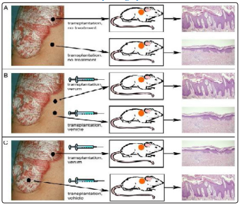
Figure R1: Shows a comparative analysis of psoriatic and healthy human skin of a psoriasis patient by grafting different skin segments onto a psoriasis SCID mouse model
A. Grafting from healthy skin segment and psoriatic lesion performed upon SCID mouse model show no changes in phenotype of each graft following transplant hence retaining their qualities
B. Healthy skin segment from psoriasis patient is useful for studying the trigger factors of psoriasis, much of what remains a mystery or confusing at best
C. Psoriatic lesional skin segment provides a useful experimental model to test therapeutics and other treatment options in test and their benefits/side effects
Role of Th1, Th17 and Th22 in the Pathogenesis of Psoriasis Elaborated by Kagami, et al. 2010
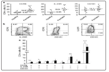
Figure R2: shows data analysis illustrating the correlation of psoriasis DSI with the rate of expressivity of circulating CCR6+, IL-23R+, and Cd161+ markers
(a) The percentage of CD4+ cells expressing CCR6, IL-23R, and CD161 among unstimulated cells from 17 healthy individuals and 21 untreated psoriatic (horizontal bars, mean; each circle, single donor). In all 3 cases, receptor expressivity is higher for CD4+ T cells in psoriatic matrix in comparison to non-psoriatic CD4+ T cells
(b) Representative dot plot analyses shows CCR6, IL-23R, and CD161 expression in circulating unstimulated CD4+ cells for same patient
(c) The percentages of each subset divided by the total number of circulating CCR6, IL-23R, and CD161 -positive cells in unstimulated CD4+ cells of healthy individuals (white bars) and psoriatics (black bars). Data expressed as mean±SD; *P<0.05, **P<0.01, and ***P<0.001
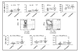
Figure R3: Displays an increment in the circulatory frequency of Th17, Th1, and Th17/Th1 cell pairs in psoriasis
(a) In single-color flow analyses, circulating IL-17A+, IL-22+, IFN-g+, (TNF)-alpha+ cells are increased in psoriasis patients
(b) Representative two-color dot plot analyses of IL-17A and IFN-Y expression in stimulated CD4+ cells from a healthy volunteer and a psoriasis patient
(c) The percentages of circulating Th17 (IL-17A+IFN-Y−), Th1 (IL-17A-), and Th17/Th1 (IL-17A+IFN-Y+) cells among stimulated CD4+ cells from healthy individuals and psoriatics. For a and c, each circle, single donor, horizontal bars, mean; and *P<0.05, **P<0.01, and ***P<0.001
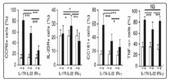
Figure R4: Presents results when IL-17A− cells, IL-22−cells, IFN-Y− and from psoriatics were gated and the percentage of cells coexpressing CCR6, IL-23R, CD161, or TNF-α was determined (white bars). Data expressed as mean±SD; NS, not significant; *P<0.05, **P<0.01, and ***P<0.001
Role of Sonic Hedgehog Signalling in Treg Conditioning by Stewart, et al.
Although not psoriasis specific but this exciting research avenue has introduced better understanding of immunogenetics of autoimmunity. These experimental investigations can be extrapolated upon psoriasis to predict treatment options and sequential targeting
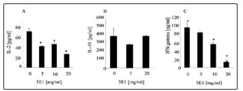
Figure R5: Presents data from an investigation where the Ab mediated neutralization of Shh via anti-Shh Ab (5E1) led to eventual down-regulation of cytokine production
Increasing concentrations of Anti-Shh Ab (5E1) was added (5-20 µg/ml) at the initiation of cultures of purified CD4+ T cells which were activated by immobilized anti-CD3 (1 µg/ml) and soluble anti-CD28 (5 µg/ml) Abs
Supernatants were collected at 72 h, and the level of IL-10 (A), IL-10 (B), and INF-y (C) were measured by ELISA and compared with cultures of anti-CD3/28-activated T cells without added 5E1. ***, p<0.001; **p< 0.01
This is a classic example of comparative proliferative assay as mentioned in Methods and Materials section of this paper
FoxP3 Article Results Put Forward by Sun, et al. 2012 as Part of His Research
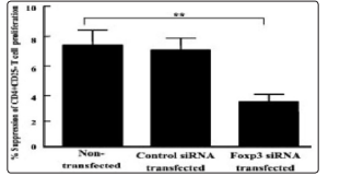
Figure R6: From the abstract of Sun, et al. 2012 shows that reverse transduction of FOXP3 deficient/mutated regulatory T cell lines fromgetting suppressed whereby they continue to proliferate unhindered
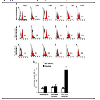
Figure R7: Presents data that provides evidence of the effect of Foxp3 knockdown upon nTreg phenotype
(A) Stained nTregs undergo gated detection and FACS analysis for 6 markers all of which are ubiquitous not only on regulatory T cells rather almost all other subsets. Gates were set on CD4+CD25 + cells and Foxp3 and cell surface marker expression marker was shown as the percentage of CD4+CD25 + cells co-expressing individual nTreg marker examined. Data presented is representative FACS histogram of three independent experiments with nTregs from three individual donors
(B) Effect of Foxp3 knockdown on nTreg nonresponsive to allogeneic simulation was performed by co-culturing non-transfected and Foxp3- or control-siRNA transfected nTregs, respectively with (stimulated) or without (non-stimulated) irradiated allogeneic PBMC for 72 h for proliferation assay using the reagent WST-1. The data are presented as mean ± SD of three independent experiments with nTregs from three individual donors. *p<0.05, comparison between stimulated and non-stimulated nTregs transfected with Foxp3 siRNA
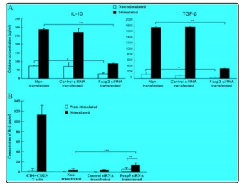
Figure R8: Presents data to quantitate the effects of Foxp3 knockdown of cytokine profile in nTregs. The cytokine profile in Foxp3 siRNA transfected nTregs was examined by ELISA measurement of amount of cytokines IL-10, TGF-β and IL-2 in nTreg cultures
(A) Supernatants were collected from the stimulated co-cultures and from cultures of non-stimulated nTregs for measurement of IL-10 and TGF-β. Data are presented as mean ± SD of four independent experiments with nTregs from four individual donors. *p<0.05, Foxp3 siRNA transfected nTregs cultured alone vs. non-transfected nTregs cultured alone; **p <0.01, stimulated Foxp3 siRNA transfected nTregs vs. stimulated non-transfected nTregs
(B) Supernatants collected from cultures of nTregs stimulated with (stimulated) or without (non-stimulated) PMA and ionomycin were used for assessment of IL-2 production. Supernatants from CD4+CD25 – T cells cultures with or without PMA and ionomycin were used as positive controls for IL-2 measurement. Data are presented as mean ± SD of three independent experiments using nTregs from three individual donors. *p<0.01, comparison between stimulated and non-stimulated Foxp3 siRNA transfected nTregs; ***p < 0.001, comparison between stimulated Foxp3 siRNA transfected and stimulated non-transfected nTregs
Table R9: Shows standard primer sequences used in laboratory for running QRTPCT during clinical investigation upon Tregs
|
Gene Seuuence of PCR primers |
|
|
Foxp3 Sense |
5′- CAC CTG GCT GGG AAA ATG G-3′ |
|
Antisense: |
5′-GGA GCC CTT GTC GGA TGA T-3′ |
|
CTLA-4 Sense: |
5′-CCGMCTMCTGCTGCAAGGA- 3′ |
|
Antisense: |
5′-CCCAGATTTATGTAATTGATCCAGM-3′ |
|
GITR Sense: |
5′-G 1 1 1 1GGCTTCCAGTGTATCGA-3′ |
|
Antisense: |
5′-MCACAGTGAGAMCCCGMCT-3′ |
|
TGF- Sense: |
5′-TGGAMCCCACMCGAMTC-3′ |
|
Antisense: |
5′-GGGTTCAGGTACCGCTTCTC-3′ |
|
IL-10 Sense: |
5′-TGAGMCAGCTGCACCCACT-3′ |
|
Antisense: |
5′-GGCMCCCAGGTMCCCTTA-3′LAG3 |
5. Role of IFN in Aggravating Psoriasis Put Forward by Eriksen, et al. 2005
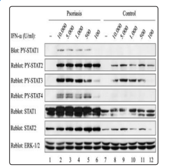
Figure R10: Is a Western Blot of psoriasis patients PBLs showing increased interferon (IFN) alpha -induced signalling. PBL frompsoriasis patients and healthy control donors were stimulated with IFN- as indicated for 10 min and lysed. Total cell lysates were subjected to western blotting using the indicated Abs
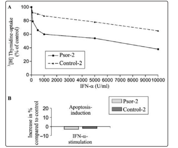
Figure R11: Presents a graphical analysis of increased growth inhibition in psoriatic T cells
(A) Psor-2 and Control-2 cells were cultured with interferon (IFN)-as indicated in culture medium with IL-2 (1000 U per mL) for 96 h. Cell cultures were subsequently subjected to [3H]thymidine uptake detected in cells incubated without IFN-
(B) Psor-2 and Control-2 cells were cultured with IFN- (10,000 U/mL) for 24 h. Cells were then subjected to quantification of apoptotic cells using the fluorescent DNA-binding dye, 7-amino-actinomycin D. Mean fluorescence intensity (MFI) values from IFN- -stimulated cells are depicted as change in percent of MFI values detected in cells incubated without IFNalpha
Discussion
This paper presents the opinion that it is due to defective or outnumbered Treg cell lines that autoimmunity continues unchallenged supported by the effector branch of immune system. Evidence is available from numerous animal model studies suggesting the above mentioned theory which started as a hypothesis.
However the results in this paper are selected to point out that the problem is much more intricate than just number or functional impedance.
Complexities exist at molecular signalling levels which affect the cellular behaviour and psoriasis being an autoimmune disorder cannot be isolated for being a dermatological disease rather, information from other diseases such as RA, IBD, MS and SLE can be applied and extrapolated onto it for investing in improving the current drug batch available to patients. Another aim for is to use these results and aim to shift the return of psoriasis which happens for almost every patient, towards complete remission and make this remission irreversible.
Studies from other disease models have indicated that the Treg number, functional, proliferation defects arise from intrinsic or extrinsic cellular factors. This imbalance shifts the Treg subset of the patients into a state of inactivation whereby their suppressive potential is removed due to disturbance in the equilibrium of above mentioned factors. Secondly the myriad of proinflammatory cytokines in conjunction with reciprocating APCs enable a reduction in the suppressive capacity of these otherwise incapacitated Treg cell lines whilst simultaneously providing resistance to the effector immune media.
Therefore even though the Treg: Teff ratio may be physiologically favourable for suppressive control but this inactivation and anergic Treg cell line provides prolonged inflammatory damage to the skin causing which responds by hyperproliferating and vascularising.
Logically it is the molecular signalling damage that should affect the Treg functionality much more than simply an imbalance of cell lines because genetic defects are irreversible and require intervention to correct the mutation. Rate of loss of tolerance is higher when triggered by molecular damages rather than physiological factors which may be temporary or self-limiting.
However one confusion in this scenario is the phenomenon of peripheral plasticity which in case of psoriasis may favour an increment in aggressive Teff attack.
References for this paper indicate an ever growing strong genetic linkage involving TFs etc. Much of these genetic elements are known as well as their role in conjunction with Treg functionality. In most cases the fault is identifiable but the means to achieve an upper hand in correcting that defect and the use of appropriate technology is one of the multiple limiting factors that molecular biologists, immunologists and biochemists face.
Focus upon improving the assaying techniques remains open due to the inability of technicians to facilitate invivo mimicking which is key in understanding the dynamics of interaction. Moreover a parallel upgrading of imaging techniques will be considered essential by most perfectionists and rightfully so.
R1
The fact that transfer of lesion does not attenuate onto the murine body or subside in its anti-inflammatory intensity is indicative of the presence of cellular components that are inducing this proinflammatory state.
0R2
T helper cell lines are very useful in providing a comparative analysis of the markers they express under normal and proinflammatory states and which markers are increased under each condition. Hence this method acquires its credibility due to situational associative interpretation which provides a chance to carry out assays to test the hypothesis.
R3
It is a similar mode but with increased variables. This keeps helper T cell lines as constant but tests IL and TNF secretion rate in, around and during psoriasis in patients.
R4
Combinational analysis is used in this case whereby the IFN and IL secretory cells are also checked for their expressivity of chemokine receptors both of these factors on their own and in combination have undergone proliferation in psoriatic lesions and it can be hypothesised that these factors are responsible for prolonged inflammatory damage.
R5
Shh was blocked by progressive anti-Shh Ab in IL2/IL10/IFN-γ positive environments and its progressively decreasing concentrations were used to identify the correlation between the three ILs and IFNγ. IL10 shows least consistent correlation however IL2 indicated a net decline with minor fluctuations but IFNγ exhibited a proportional relationship with Shh decrease.
R6
It is very straight forward as it depicts the importance of FOXP3 as an IC marker as a resisting force against environmental pressures leading to suppression whereas
R7
involves flow cytometry for checking the expression of 6 different types of surface markers part B involves their responsiveness to allogeneic transplantation once injected with nontransfected Tregs.
R8
Compares the expressivity of different ILs by stimulated and unstimulated Treg subsets to check for the effects of SiRNA transfection and what quantitate their IL2, IL10 and TGFb expression.
Table R9 shows a standard sequence length for different markers used for analysis in QRTPCR.
R10
It shows that there is a proportional correlation between IFNalpha signalling and psoriasis disease severity index which is further confirmed by the figure R11 where IRMA, shows that when Tregs are cultured in IFNalpha, their uptake counts have a negative gradient coefficient due IFN’s inhibitory effect. Part B shows an increase in apoptotic induction due IFNalpha and it is because IFNalpha has been known to aggravate Th1 reaction profile but it requires the coordination of STAT and src to induce autoimmune state. Increase in apoptosis is similar to inducing autoimmune damage.
Current Treatments
Strober, et al. 2009 (employing Delphi analysis to generate combinational therapies) and Mendonca and Griffiths 2005 (classifying treatments into groups based upon targets or source) summarise current treatments mentioned below. However latest list of drugs and treatments undergoing multilevel clinical trials is available from the National Foundation of Psoriasis USA.
T Cell Targeting
Therapies Biologic therapy offers drugs such as Efalizumab and Alefecept which are highly effective in patients.
Cytokine Modulators
Since psoriatic lesional analysis shows Th1 cytokine presence, Th2 cytokines IL4 and IL10 would be best to counter their influence. • Infliximab • Etanercept
PDT (light therapy), Lasers and Radiation
No longer a novice it is administered in clinics routinely using different sets of wavelengths and photosensitizers although research continues on finding optimum combinations.
• Narrow UVB
• Broadband UVB
• Narrow UVA
• Excimer laser Use of radiotherapy is still controversial due to conflicting results using Tregs as targets.
Anti TNF Therapy
TNF are proinflammatory elements which accelerate disease severity. This treatment knocks out TNF from psoriatic lesions.
• TNF inhibitor
• Efalizumab
• Alfacept
MTX Family
This is used in chemotherapy and autoimmune diseases due to its antifolate and antimetabolite properties. It can applied in combination with TNF inhibitor, cyclosporine or on its own.
Corticosteroids (Glucocorticoids)
It is the most basic treatment for psoriasis. It does have immunosuppressive properties accompanied by its side effects. Their main classes are:
• Topical •
Intralesional
• Intra-articular
Natural “Remedies”
(a) Coal tar
(b) Relaxation therapy
(c) Dead sea treatment
Metabolite Specific Drugs
a. COX – 2 inhibitors
b. NSAIDs
c. Hydroxychloroquine
d. Hydroxyurea
e. Mercaptopurine + thioguanine
f. Leflunomide
g. Sulfasalazine
h. Cyclosporine and azathioprine
i. Mycophenolate mofetil
Summary
Broadly range of treatments is divided into monoclonal antibodies (assume multiple roles such as anti-TNF or T cell mediator, etc.), routine immunosuppressive drugs, corticosteroids, natural remedies, light therapy and biologic agents.Conclusion
This endeavour of studying, controlling and manipulating the regulatory components for avoiding autoimmunity has been an exciting journey and probably the most important landmark in the history of modern immunology.
Results section focussed on investigations centred on ILs, IFN, TNFs and FOXP3 but monitoring other newly identified factors may lead to better prospects of gene therapy improving patient QoL by bringing down DSI.
Although it is still unclear how Treg suppression functions at molecular scale, there are still some questions such as what actually triggers the initiation of psoriasis. Is it trauma, a high frequency/ consistent trauma at one skin location that renders effector response into permanent activation. B cell therapy might be able to answers some of these questions and although a useful tool in monitoring and curbing the effector damage, it is only as good as the level of B – T cell interaction. Dr Michael Dustin from NIH Immunology Laboratories has suggested the involvement and discussed the importance of PKCtheta pathways that is Multi-planar association of following factors poses.
(I) Treg deficiency or ratio imbalance renders the Tregs ineffective for the most part of their interaction.
(II) Multifactorial resistance to Treg function has been proven following trials and counter trials with their respective receptors to certify their involvement.
(III) Functional incapacity is synonymous to the tip of the iceberg as it reveals a myriad of pathway misalignment, cell marker involvement and site specific interactions.
Dr Michael Dustin (NIH Immunology Laboratories) puts emphasis upon Treg cellular communication (calcium signalling), mechanobiology, in understanding these threatening defects intervening through mechanotransduction. Newly emerging PKCtheta and its role in controlling Treg function can be used to create super-Tregs by employing systemic therapy to hit effector targets with immune-compromising the patient as well inducing resistance to phenotype conversion in Treg subsets. This set of techniques has a healthy prospect in contrast to just an ineffective time dependent Treg accumulation in psoriatic sites [12-125].
References
-
-
- Tefler NR, Chalmers RJG, Whale K, Colman G (1992) The role of streptococcal infection in the initiation of guttate psoriasis. Arch Dermatol 128: 39-42.
- Canavese M, Altruda F, Silengo L, Castiglioni V, Scanziani E, et al. (2011) Clinical, pathological & immunological features of psoriatic-like lesions affecting keratin 14 vascular endothelial growth factor transgenic mice. Journal of Histology & Histopathology 26: 285-296.
- Fontenot JD, Gavin MA, Rudensky AY (2003) Foxp3 programs the development & function of CD4+CD25+ regulatory T cells. Nature Immunology 4: 330-336.Fry L, Baker SB, Powles VA (2007) Psoriasis-A Possible Candidate for vaccination. Autoimmunity Reviews 6: 286-289.
-
-
-
- Langley BGR, Krueger GG, Griffiths MEC (2005) Psoriasis: epidemiology, clinical features, & quality of life. Annals of Rheumatic Disease 64: 18-23.
- Bhosle JM, Kulkarni A, Feldman RS, Balkrishnan R (2006) Quality of Life in patients with psoriasis. Health & Quality of Life Outcomes 4: 1-7.
- Kappler JW, Roehm N, Marrack P (1987) T cell tolerance by clonal elimination in the thymus. Cell 49: 273-280.
- Mosmann TR, Coffman RL (1989) TH1 and TH2 cells: different patterns of lymphokine secretion lead to different functional properties. Annual Reviews in Immunology 7: 145-173.
- Zhou Z, Xiaomin S, Berezov A, Li B, Greene IM (2009) Structural Aspects of the FOXP3 regulatory complex as an immunopharmacological target. InternationalImmunopharmacology 9: 518-520.
- Appleman LJ, Zachanis D, Grader-Beck T, vanPuigenbroek AA, Boussoitis VA (2001) Helper T cell anergy: from biochemistry to cancer Pathophysiology & therapeutics. Journal of Molecular Medicine 78: 673-683
- Lopes EJ, Zeigler FS (2000) Foxp3: More than just a marker of regulatory T cells; A review of the role of FoxP3 in processes including immune homeostasis & development. [online] Benaroya Research Institute. (http://www.abcam.com/index. html?pageconfig=resource&rid=11528&pid=10629) [3rdMarch 2012].Macian F, Garcia-Cozar F, Im SH, Horton HF, Byrne MC, et al. (2002) Transcriptional mechanisms underlying lymphocyte tolerance Cell 109: 719-731.Mak HKR, Hundhausen C, Nestle OF (2009) Progress in Understanding the Immunopathogenesis of Psoriasis. Actas Dermo-Sifiliográficas 100: 2-13.
- Waldman TA (2006) The biology of interleukin-2 & interleukin-15: implications for cancer therapy & vaccine design. Nature Reviews Immunology 6: 595-601
- Aguado E, Martinez-Florensa M, Aparicio P (2006) Activation of T lymphocytes and the role of adapter LAT. Transplant Immunology 17: 23-26
- Apostolou I, Sarukhan A, Klein L, von Boehmer H (2002) Origin of regulatory T cells with known specificity for antigen. Nature Immunology 2: 301-306
- Asadullah K, Sterry W, Stephanek K, Jasulaitis D, Leupold M, et al. (1998) IL-10 is a key cytokine in psoriasis. Proof of principle by IL-10 therapy: a new therapeutic approach. Journal of Clinical Investigation 101: 783-794
- Asadullah K, Volk H, Sterry W (2002) Novel Immunotherapies for psoriasis. Trends in Immunology 23: 47-53.
- Ash RZ, Ilaria Tinazzi I, Gallego CC, Kwok C, Wilson C, et al. (2011) Psoriasis patients with naildisease have a greater magnitude of underlying systemic subclinical enthesopathy than those with normal nails. Annals of Rheumatic Disease 71: 4553-4556.
- Bacchetta R, Gambineri E, Roncarolo GM (2007) Role of regulatory T cells & FOXP3 in human diseases. Journal of Allergy & Clinical Immunology 120: 227-235.
- Baker H (1976) Corticosteroids & pustular psoriasis. Br J Dermatol 12: 83-88.
- Balato A, Unutmaz D, Gaspari AA (2009) Natural Killer T Cells: An Unconventional T-Cell Subset with Diverse Effector & Regulatory Functions. Journal of Investigative Dermatology 129: 1628-1642.
- Bata-Csorgo Z, Hammerberg C, Voorhees JJ, Cooper KD (1995) Intralesional T-lymphocyte activation as a mediator of psoriatic epidermal hyperplasia. J Invest Dermatol 105: 89-94S.
- Bata-Csorgo Z, Cooper KD, Ting KM, Voorhees JJ, Hammerberg C (1998) Fibronectin & alpha5 integrin regulate keratinocyte cell cycling. A mechanism for increased fibronectin potentiation of T cell lymphokine-driven keratinocyte hyperproliferation in psoriasis. Journal of Clinical Investigation 101: 1509-1518.
- Benderitter M, Linard C (2011) Abdominal γ-radiation induces an accumulation of function-impaired regulatory T cells in the small intestine. International Journal of Radiation Oncology Biology Physics 80: 869-876.
- Berth-Jones J (2005) Psoriasis. Medicine 33: 50-55.
- Bettelli E, Dastrange M, Oukka M (2005) FoxP3 interacts with nuclear factor of activated T cells &NF kappa B to represscytokine gene expression & effector function of Th cells. Proceedings ofthe National Academy of Sciences of the United States of America 102: 138-143.
- Boehncke WH, Schon MP (2007) Animal Models of Psoriasis. Clinics in Dermatology 25: 596-605
- Boyd AS, Menter A (1989) Erythrodermic psoriasis. J Am Acad Dermatol 21: 1985-1991
- Braverman IM, Sibley BA (1982) Role of the microcirculation in the treatment & pathogenesis of psoriasis. J Invest Dermatol 78: 12-17.
- Buckner HJ (2010) Mechanisms of impaired regulation by CD4+CD25+FOXP3+ regulatory T cells in human autoimmune diseases. Nature Reviews Immunology 10: 849-859.
- Burger RA, Torres AR, Warren RP, Caldwell VD, Hughes BG (1997) Echinacea-induced cytokine production by human macrophages. International Journal of Immunopharmacology 19: 371-379.
- Campbell JD, Zeigler FS (2007) FoxP3 modifies the phenotypic & functional properties of regulatory T cells. Nature Immunology Reviews 7: 305-310.
- Chan AC, Dalton M, Johnson R, Kong GH, Wang T, et al. (1995) Activation of ZAP-70 kinase activity by phosphorylation of tyrosine 493 is required for lymphocyte antigen receptor function. The EMBO Journal 14: 2499-2508.
- Chan FSV, Chau S, Tian L, ChenY, kwong YK, et al. (2006) Sonic hedgehog promotes CD4+ T lymphocyte proliferation & modules the expression of a subset of CD28-targetted genes. International Immunology 18: 1627-1636.
- Chandran V, Raychaudhuri PS (2010) Geoepidemiology and environmental factors of psoriasis and psoriatic arthritis. The Environment, Geoepidemiology and Autoimmune Disease 34: J314-J321.
- Chatila AT (2005) Role of Treg in human diseases. Journal of Allergy & Clinical Immunology 116: 949-959.
- Chauhan KS, Saban RD, Hyung KL, Dana R (2009) Levels of Foxp3 in regulatory T cells reflect their functional status in transplantation. Journal of Immunology 182: 148-153.
- Choilean NN, Redmond PH (2006) Regulatory T cells & Autoimmunity. Journal of Surgical Research 130: 124-135.
- Chong FB, Wong KH (2007) Immunobiologics in the treatment of psoriasis. Clinical Immunology 123: 129-138.
- Christen U, vonHerrath GM (2004) Initation of autoimmunity. Current Opinion in Immunology 16: 759-767.
- Christopher E (2001) Psoriasis-Epidemiology & clinical symptom. Clinical & Experimental Dermatology 26: 314-320.
- Claman HN, Chaperon EA, Triplett EF (1966) Thymus marrow-cell combinations Synergism in antibody production. Proceedings of the Society for Experimental Biology & Medicine 122: 1167-1171.
- Coutinho A (2002) Immunology at the crossroads. EMBO Reports 3: 1008-1011.
- Crellin NK, Garcia RV, Porter SB, Ge Y (2007) Altered activation of AKT is required for the suppressive function of human CD4+CD25+regulatoryTcells. Blood 109: 2014-2022.
- Crompton T, Outram SV, Buckland J, Owen MJ (1997) A transgenic T cell receptor restores thymocyte differentiation in IL7Rα chain-deficient mice. Eur J Immunol 27: 100-104.
- Crompton T, Outram SV, Hager-Theodorides AL (2007) Sonic hedgehog signaling in T-cell development & activation. Nat Rev Immunol 7: 726-735.
- Editorial (2005) Essence of Harmony. Nature Immunology6: 325.
- Ellis CN, Fradin MS, Messana JM, Brown MD, Siegel MT, et al. (1991) Cyclosporine for plaque-type psoriasis. Results of a multidose, double-blind trial. N Engl J Med 324: 277-284.
- Fehervari Z, Sakaguchi S (2004) CD4+ Tregs and immune control. Journal of Clinical Investigation 114: 1209-1217.
- Foca-Suciu N, Manavalan SJ, Conrtesini R (2003) Generation & function of antigen-specific suppressor & regulatory T cells. Transplant Immunology 11: 235-244.
- Fontenot DJ, Rasmussen PJ, Williams ML, Dooley LJ, Farr GA, et al. (2006) Regulatory T Cell Lineage Specification by the Forkhead Transcription Factor FoxP3. Immunity 22: 329-341.
- Gavin MA, Clarke SR, Negrou E, Gallegos A, Rudensky AY (2002) Homeostasis and anergy of CD4+CD25+suppressor T cells in vivo. Nature Immunology 3: 33-41.
- Gershon RK, Kondo K (1970) Cell interactions in the induction of tolerance: the role of thymic lymphocytes. Immunology 18: 723-737.
- Gershon RK, Kondo K (1971) Infectious immunological tolerance. Immunology 21: 903-914.
- Gershon RK, Cohen P, Hencin R, Leibhaber SA (1972) Suppressor T cells. Journal of Immunology 21: 903-914.
- Godfrey VL, Wilkinson JE, Russell LB (1991) X-linked Lymphoreticular disease in the scurfy (sf) mutant mouse. American Journal of Pathology 138: 1379-1387.
- Goldsmith AL (2012) Ensuring Dermatology’s Future. Journal of Investigative Dermatology 132: 747-750.
- Green DR, Webb DR (1993) Saying the “S” word in public. Immunology Today 14: 523-525.
- Grozdev I, Kast D, Cao L, Carlson D, Pujari1 P, et al. (2012) Physical and Mental Impact of Psoriasis Severity as Measured by the Compact Short Form-12 Health Survey (SF-12) Quality of Life Tool. Journal of Investigative Dermatology 132: 1111-1116.
- Gudjonsson EJ, Johnston A, Ellis NC (2012) Novel systemic drugs under investigation for the treatment of psoriasis. J Am Acad Dermatol 67: 139-147.
- Hager-Theodorides A, Dessens JT, Outram SV, Crompton T (2005) The transcription factor Gli3 is involved in the differentiation of fetal, but not adult, CD4-CD8- Double Negative thymocytes, Blood 106: 1296-1304.
- Hager-Theodorides AL, Rowbotham NJ, Outram SV, Dessens JT, Crompton T (2007) Beta-selection: abundance of TCRbeta-/ gammadelta- CD44- CD25- (DN4) cells in the foetal thymus. Eur J Immunol 37: 487-500.
- Hickman SP, Yang J, Thomas RM, Wells AD, Turka LA (2006) Defective activation of protein kinase C & Ras-ERK pathways limits IL-2 production & proliferation by CD4+CD25+regulatoryTcells. Journal of Immunology 177: 2186-2194.
- Hoxtermann S, Nuchel C, Altmeyer P (1998) Fumaric acid esters suppress peripheral CD4- & CD8-positive lymphocytes in psoriasis. Dermatology 196: 223-230.
- Jiang S (2008) Regulatory T cells & Clinical Application. NY: Springer p3-154.
- Jiang H, Chess L (2004) An integrated view of suppressor T cell subsets in immunoregulation. Journal of Clinical Investigation 114: 1198-1208.
- Johanne E, Gudjonson MD, James T, Elder MD (2007) Psoriasis Epidemiology. Clinics in Dermatology 25: 535-546.
- Jonuleit H, Schmitt E, Stassen M, Tuettenberg A, Knop J, et al(2001) Identification & functional characterization of human CD4(+)CD25(+) T cells with regulatory properties isolated from peripheral blood. J Exp Med 193: 1285-1294.
- Jorge Neto J, Fracasso JF, Camargo Neves M Do CL, Dos Santos LE, Banuth VL (1996) Treatment of varicose ulcer and skin lesions with Calendula officinalis L. or Stryphnodendron barbadetiman (Vellozo) Martius. Revista de Ciencias Farmaceuticas 17: 181-186.
- Kaplan RP, Russell DH, Lowe NJ (1983) Etretinate therapy for psoriasis: clinical responses, remission times, epidermal DNA & polyamine responses. J Am Acad Dermatol 8: 95-102.
- Kretschmer K, Apostolou I, Hawiger D, Khazaie K, Nussenzweig CM, et al. (2005) Inducing & expanding regulatory T cell populations by foreign antigen. Nature Immunology 6: 1219-1227.
- Leung DYM, Travers JB, Giomo R, Norris DA, Skinner R, et al. (1995) Evidence for streptococcal superantigen-driven process in acute guttate psoriasis. J Clin Invest 96: 2106-2112.
- Levings KM, Allan S, D’Hennezel E, Piccirillo AC (2006) Functional Dynamics of Naturally Occurring Regulatory T Cells in Health and Autoimmunity. Advances in Immunology 92: 119-155.Li B, Saouaf JS, Samanta A, Shen Y, Hancock WW, et al. (2007) Biochemistry & therapeutic implications of mechanisms involved in FOXP3 activity in immune suppression. Current Opinion in Immunology 19: 583-588.
- Li L, Godfrey WR, Porter SB, Ge Y, June CH, et al. (2007) CD4+CD25+regulatoryTcell lines from human cord blood have functional & molecular properties of T cell anergy. Blood 106: 3068-3073.
- Matzinger P (2002) The danger model: A renewed sense of self. Science 296: 301-305.
- Mendonca C, Griffiths MEC (2005) Psoriasis: Future Drugs. Medicine 33: 56-57.
- Meola T Jr, Soter NA, Lim HW (1991) Are topical corticosteroids useful adjunctive therapy for the treatment of psoriasis with ultraviolet radiation? Arch Dermatol 127: 1708-1713.
- Miyara M, Sakaguchi S (2007) Natural regulatory T cells: mechanisms of suppression. Trends in Molecular Medicine 13: 108-116.
- Miyara M, Gorochov G, Ehrenstein M, Musset L, Sakaguchi S, et al. (2011) Human FoxP3+ regulatory T cells in systemic autoimmunes diseases. Autoimmunity Reviews 10: 744-755.
- Miyara M, Sakaguchi S (2008) Regulatory T cells & the Control of Auto-immunity; from day3 thymectomy to FoxP3+ Regulatory T cells. In: Shiuping Jiang Regulatory T cells & Clinical Application. London: Springer p3-16.
- Molin L, Cutler TP, Helander L, Nyfors B, Downes N (1997) Comparative efficacy of calcipotriol (MC 903) cream & betamethasone 17-valerate cream in the treatment of chronic plaque psoriasis. A randomized, doubleblind, parallel group multicenter study. Calcipotriol Study Group. Br J Dermatol 136: 89-93.
- Mrowietz U, Christophers E, Altmeyer P (1998) Treatment of psoriasis with fumaric acid esters: results of a prospective multicentre study. German Multicentre Study. British Journal of Dermatology 138: 456-460.
- Nagel A, Hertl M, Eming R (2009) B – Cell Directed Therapy for Inflammatory Skin Diseases. Journal of Investigative Dermatology 129: 289-301.
- Neimann LA, Porter BS, Gelfand MJ (2006) The epidemiologyof psoriasis. Expert Opinion in Dermatology 1: 63-75.
- NIshizuka Y, Sakakura T (1969) Thymus & reproduction: sex-linked dysgenesia of the gonad after neonatal thymectomy in mice. Science 166: 753-755.
- Ochs HD, Zeigler SF, Torgerson TR (2005) FoxP3 acts as a rheostat of immune response. Immunological Reviews 203: 156-164.
- Outram SV, Owen MJ (1994) The helix-loop-helix containing transcription factor USF activates the promoter of the CD2 gene. J Biol Chem 269: 26525-26530.
- Outram SV, Hager-Theodorides AL, Shah DK, Sacedon R, Shrimpton RE, et al. (2002) Bone morphogenetic protein 2/4 signaling regulates early thymocyte differentiation. J Immunol 169: 5496-5504.
- Outram SV, Varas A, Pepicelli CV, Crompton T (2000) Hedgehog signalling regulates differentiation from double negative to double positive thymocyte. Immunity 13: 187-197.
- Outram SV, Crompton T, Buckland J, Owen MJ (1998) Distinct roles for the interleukin 7 receptor (chain in foetal & adult thymocyte development revealed by analysis of Interleukin 7 receptor alpha deficient mice. Eur J Immunol 28: 1859-1866.
- Outram SV, Amess J, Horton MA (1988) Erythromyeloid lineage fidelity is conserved in erythroleukaemia. Leuk Res 12: 651-657.
- Outram SV, Crompton T, Merida I, Varas A, Martinez-Á C (2001) Diacylglycerol kinase α activity is required for survival of CD4+CD8+ thymocytes during development. Immunology 105: 391-398.
- Outram SV, Grimwade D, Crompton T (2001) Repression of CD2 Gene Expression Is Mediated by an AP-2 Related Factor. Biochemical & biophysical research communications 281: 409-415.
- Ouyang W, Beckett O, Ma Q, Paik J, DePinho AR, et al. (2010) Foxo proteins cooperatively control the differenciation of FoxP3+ regulatory T cells. Nature Immunology 11: 618-627.
- Penhale WJ, Farmer A, McKenna RP, Irvine WJ (1973) Spontaneous thyroiditis in thymectomised and irradiated Wistar rats. Clinical and Experimental Immunology 15: 225-236.Powell BR, Buist NR, Stenzel
- P (1982) An X-linked syndrome of diarrhoea, polyendocrinopathy and fatal infection in infancy. Journal of Paediatrics 100: 731-737.
- Roujeau JC, Bioulac-Sage P, Bourseau C, Guillaume JC, Bernard P, et al. (1991) Acure generalized exanthematous pustulosis. Analysis of 63 cases. Arch Dermatol 127: 1333-1338.
- Rowbotham NJ, Hager-Theodorides AL, Cebecauer M, Shah DK, Drakopoulou E, et al. (2007) Activation of the Hedgehog signaling pathway in T-lineage cells inhibits TCR repertoire selection in the thymus & peripheral T-cell activation. Blood 109: 3757-3766.
- Rudensky YA, Gavin MA, Zheng Y (2006) FOXP3 & NFAT; Partners in Tolerance. Cell 126: 253-256.Rudensky AY, Sakaguchi S (2003) The Role of Regulatory T Cells in Controlling Immunologic Self-To
- lerance. International Review of Cytology 225: 1-32.
- Sacedon R, Nunez V, Diez B, Hernandez-lopez C, Gutierrezde Frias C, et al. (2005) Sonic hedgehog is produced by follicular dendritic cells & protects germinal center B cells from apoptosis. J Immunol174: 1456-1461.
- Sacedon R, Vicente A, Hernandez-Lopez C, Gutierrez C, Outram SV, et al. (2003) Hedgehog proteins, expression & function in the thymus. Immunologia 22: 117-126.
- Sakaguchi S, Fukuma K, Kuribayashi K, Masuda T (1985) Organ-specific autoimmune diseases induced in mice by elimination of a T cell subset. I. Evidence for the active participation of T cells in natural self tolerance: deficit of a T cell subset as a possible cause of autoimmune disease. Journal of Experimental Medicine 161: 72-87
- Sakaguchi S, Sakaguchi N, Asano M, Itoh M, Toda M (1995) Immunologic self-tolerance maintained by activated T cells expressing IL-2 receptor-chains. Journal of Immunology 155: 1151-1164.
- Sakaguchi S, Takahashi T, Nishizuka Y (1982) Study on cellular events in post-thymectomy autoimmune oophoritis in mice. I. Requirement of Lyt-1 effector cells for oocytes damage after adoptive transfer. Journal of Experimental Medicine 156: 1565-1576.
- Schlaak JF, M Buslau, W Jochum, E Hermann, M Girndt, et al. (1994) T cells involved in psoriasis vulgaris belong to the Th1 subset. Journal Investigative Dermatology 102: 145-149.
- Schon PM, Boehncke WH (2005) Psoriasis. New England Journal of Medicine 352: 1899-1912.
- Schubert LA, Jeffrey E, Zhang Y, Ramsdell F, Zeigler SF (2001) Scurfin (FOXP3) acts as a repressor of transcription and regulates T cell activation. Journal of Biological Chemistry 276: 37672-37679.
- Shah DK, Hager-Theorides A, Outram SV, Ross S, Varas A, et al. (2004) Reduced thymocyte development in Sonic Hedgehog Knockout embryos. J Immunol 172: 2296-2306.
- Sharma R, Fu MS, Ju ST (2011) IL-2: A two faced master regulator of autoimmunity. Journal of Autoimmunity 36: 91-97.
- Stern RS, Laird N, Melski J, Parrish JA, Fitzpatrick TB, et al. (1984) Cutaneous squamous-cell carcinoma in patients treated with PUVA. N Engl J Med 310: 1156-1161.
- Stern RS, Nichols KT, Vakeva LH (1997) Malignant melanoma in patients treated for psoraisis with methoxsalen (psoralen) & ultraviolet A radiation (PUVA). N Engl J Med 336: 1041-1045.
- Strober B, Berger E, Cather J, Cohen D, Crowley JJ, et al. (2009) A series of critically challenging case scenarios in moderate to severe psoriasis; A Delphi consensus approach. J Am Acad Dermatol 61: S1-S46.Stern RS (1988) The benefits, costs & risks of topical tar preparations in the treatment of psoriasis: considerations of cost effectiveness. Ann Acad Med Singapore 17: 473-476.
- Stewart AG, Lowrey AJ, Wakelin JS, Fitch MP, Lindey S, et al. (2002) Sonic Hedgehog Signaling Modulates Activation of and Cytokine Production by Human Peripheral CD4+ T Cells. The Journal of Immunology 169: 5451-5457.
- Strange P, KD Cooper, ER Hansen, G Fisher, JK Larsen, et al. (1993) T-Lymphocyte clones initiated from lesional psoriatic skin release growth factors that induce keratinocyte proliferation. J. Invest. Dermatol 101: 695-700.
- Gavin MA, Torgerson TR, Houston E, DeRoos P, Ho WY, et al. (2006) Single cell analysis of normal & FoxP3-mutant human T cells: FoxP3 expression without regulatory T cell development. Proc Natl Acad Sci USA 103: 6659-6664.
- Sugiyama H, Guylai R, Toichi E, Garaczi E, Shimada S (2005) Dysfunctional Blood and Target Tissue CD4+CD25 high Regulatory T Cells in Psoriasis: Mechanism Underlying Unrestrained Pathogenic Effector T Cell Proliferation. Journal of Immunology 174: 164-173.
- Sun L, Wu J, Yi S (2012) Foxp3 is critical for human natural CD4+CD25+regulatory T cells to suppress alloimmune responses. Transplant Immunology 26: 71-80.
- Tai X, Cowan M, Feigenbaum L, Singer A (2005) CD28 costimulation of developing thymocytes induces FoxP3 expression & regulatory T cell differenciation independently of interleukin-2. Nature Immunology 6: 152-162.
- Tang Q, Henriksen KJ, Bi M, Finger EB, Szot G, et al. (2004) In vitro-expanded antigen-specific regulatory T cells suppress autoimmune diabetes. J Exp Med 199: 1455-1465.
- Thomas R (2012) Autoimmunity: innovation & pathogenesis in therapy. Immunology & Cell Biology 90: 255.
- Tsang JY, Camara NO, Eren E, Schneider H, Rudd C, et al. (2006) Altered proximal T cell receptor (TCR) signalling in human CD4+CD25+ regulatoryTcells. Journal Leukocyte Biology 80: 145-151.
- Umetsu TD, Dekruyff HR (2010) The regulatory role of natural killer T cells in the airways. The International Journal of Biochemistry & Cell Biology 42: 520-534.
- Vollmer S, A Menssen, P Trommler, D Schendel, JC Prinz (1994) T lymphocytes derived from skin lesions of patients with psoriasis vulgaris express a novel cytokine pattern that is distinct from that of T helper type 1 & T helper type 2 cells. Eur J Immunol 24: 2377-2382.
- vonBubnoff A (2007) Regulatory T cells suppress immune responses & researchers are now working to determine precisely how, but their role in HIV pathogenesis is still unclear. IAVI Report 11(4).
- Zeigler SF (2006) FoxP3 of mice & men. Annual Reviews of Immunology 24: 209-226.
- Zöller M, McElwee KJ, Vitacolonna M, Hoffmann R (2004) Apoptosis resistance in peripheral blood lymphocytes of alopecia areata patients. J Autoimmun 23: 241-256.
- Zöller M, McElwee KJ, Vitacolonna M, Hoffmann R (2004) The progressive state, in contrast to the stable or regressive state of alopecia areata, is reflected in peripheral blood mononuclear cells. Exp Dermatol 13: 435-444.
-