

Advances in Bioengineering and Biomedical Science Research(ABBSR)
ISSN: 2640-4133 | DOI: 10.33140/ABBSR

Impact Factor: 1.7

Research Article - (2024) Volume 0, Issue 0
Using Viscoplastic Energy Model of GH-Method: Math-Physical Medicine to Validate Specific Glucose Dynamics in Type 2 Diabetes Pathophysiological Phenomena (No. 1045, VMT #443)
Received Date: Mar 15, 2024 / Accepted Date: Apr 04, 2024 / Published Date: Apr 05, 2024
Copyright: ©©2024 Gerald C. Hsu. This is an open-access article distributed under the terms of the Creative Commons Attribution License, which permits unrestricted use, distribution, and reproduction in any medium, provided the original author and source are credited.
Citation: Hsu, G. C. (2024). Using Viscoplastic Energy Model of GH-Method: Math-Physical Medicine to Validate Specific Glucose Dynamics in Type 2 Diabetes Pathophysiological Phenomena (No. 1045, VMT #443). Adv Bioeng Biomed Sci Res, 7(2), 1-9.
Abstract
Increased body weight, particularly through the accumulation of adipose tissue like visceral fat, is strongly linked to insulin resistance. As time progresses, this insulin resistance can cause fasting plasma glucose (FPG) levels to rise due to a diminishing insulin efficiency.
The interplay between fasting plasma glucose levels and the insulin-producing capabilities of pancreatic beta-cells is crucial for blood sugar regulation and understanding the mechanisms behind diabetes, especially type 2 diabetes. Continuous high fasting plasma glucose levels may induce glucose toxicity, impairing beta-cell functionality and potentially leading to beta-cell burnout over time. Fasting Plasma Glucose reflects the equilibrium between glucose production, mainly from the liver, and its consumption by bodily tissues. The capacity to normalize fasting glucose levels post-meal is indicative of effective glucose management.
Additionally, inadequate sleep can exacerbate insulin resistance, challenging glucose control. Conversely, regular exposure to moderate variations in temperature may boost metabolic adaptability, aiding in glucose regulation across different scenarios, including postprandial periods.
The researcher compiled personal data spanning from May 1, 2015, to March 17, 2024, encompassing 10,007 meals. This data includes an average Fasting Plasma Glucose of 106 mg/dL, Postprandial Plasma Glucose (PPG) of 113 mg/ dL, 13.6 grams of carbohydrates and sugar intake, 4,180 steps walked after meals, an average of 6.4 hours of sleep, and an averaged ambient temperature of 73.8 degrees Fahrenheit. He utilized the Viscoplastic Medicine Theory (VMT) to analyze the energy levels of his PPG in relation to these five influential factors mentioned.
Key Message
Reducing body weight enhances the insulin-producing function of pancreatic beta cells, which is evident in fasting glucose levels. Moreover, managing carbohydrate and sugar consumption, along with engaging in post-meal walking activity, also helps in lowering post-meal glucose levels. Additionally, sleep duration and quality, along with maintaining a comfortable ambient temperature, are secondary factors that influence glucose formation.
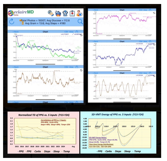
Viscoelastic Medicine theory (VMT #443) Using viscoplastic energy model of GH-Method: Math-Physical Medicine to validate specific glucose dynamics in Type 2 diabetes pathophysiological phenomena (No. 1045)
Introduction
Increased body weight, particularly through the accumulation of adipose tissue like visceral fat, is strongly linked to insulin resistance. As time progresses, this insulin resistance can cause fasting plasma glucose (FPG) levels to rise due to a diminishing insulin efficiency.
The interplay between fasting plasma glucose levels and the insulin-producing capabilities of pancreatic beta-cells is crucial for blood sugar regulation and understanding the mechanisms behind diabetes, especially type 2 diabetes. Continuous high fasting plasma glucose levels may induce glucose toxicity, impairing beta-cell functionality and potentially leading to beta-cell burnout over time.
Fasting Plasma Glucose reflects the equilibrium between glucose production, mainly from the liver, and its consumption by bodily tissues. The capacity to normalize fasting glucose levels post-meal is indicative of effective glucose management.
Additionally, inadequate sleep can exacerbate insulin resistance, challenging glucose control. Conversely, regular exposure to moderate variations in temperature may boost metabolic adaptability, aiding in glucose regulation across different scenarios, including postprandial periods.
The researcher compiled personal data spanning from May 1, 2015, to March 17, 2024, encompassing 10,007 meals. This data includes an average Fasting Plasma Glucose of 106 mg/ dL, Postprandial Plasma Glucose (PPG) of 113 mg/dL, 13.6 grams of carbohydrates and sugar intake, 4,180 steps walked after meals, an average of 6.4 hours of sleep, and an averaged ambient temperature of 73.8 degrees Fahrenheit.
He utilized the Viscoplastic Medicine Theory (VMT) to analyze the energy levels of his PPG in relation to these five influential factors mentioned.
Biomedical and Engineering or Technical Information
The following sections contain excerpts and concise information on meticulously reviewed by the author of this paper. The author has adopted this approach as an alternative to including a conventional reference list at the end of this document, with the intention of optimizing his valuable research time. It is essential to clarify that these sections do not constitute part of the author's original contribution but have been included to aid the author in his future reviews and offer valuable insights to other readers with an interest in these subjects.
Pathophysiological Explanations of Relationship Between Body Weight and Fasting Plasma Glucose
The relationship between body weight and fasting plasma glucose is multifaceted and involves complex physiological mechanisms. Here is an overview of some key pathophysiological explanations for this relationship:
1. **Insulin Resistance**: A higher body weight, especially with an increase in adipose tissue (fat), is closely associated with insulin resistance. Insulin is a hormone produced by the pancreas that allows cells to take in glucose from the bloodstream. In insulin resistance, the body's cells do not respond effectively to insulin, requiring the pancreas to produce more insulin to achieve the desired effect of lowering blood glucose levels. Over time, this can lead to higher fasting plasma glucose levels as the effectiveness of insulin continues to decrease.
2. **Inflammation**: Adipose tissue, particularly visceral fat (the fat surrounding the internal organs), is not inert; it produces various substances, including inflammatory markers that can contribute to systemic inflammation. Chronic inflammation has been linked to the development of insulin resistance, as it interferes with the signaling pathways of insulin, thus impairing glucose uptake by cells and leading to higher fasting plasma glucose levels.
3. **Adipokines**: Adipose tissue secretes a variety of hormones and cytokines known as adipokines, such as leptin and adiponectin, which play roles in regulating insulin sensitivity and metabolism. Obesity often results in an altered adipokine profile, such as high leptin (leading to leptin resistance) and low adiponectin levels, both of which are associated with insulin resistance and elevated fasting plasma glucose.
4. **Beta-Cell Dysfunction**: The pancreas' beta cells are responsible for insulin production. Chronic insulin resistance demands these cells work harder to produce more insulin, which can lead to beta-cell exhaustion over time. This reduced capacity to produce insulin can lead to an inability to adequately control fasting plasma glucose levels, resulting in higher levels of glucose in the blood.
5. **Lipotoxicity**: Excess fatty acids and their metabolites can accumulate in non-adipose tissues, such as the liver, pancreas, and muscle, leading to lipotoxicity. This accumulation can impair the function of these tissues, including the insulin-producing beta cells of the pancreas and the liver's ability to regulate glucose production, contributing to elevated fasting plasma glucose levels.
6. **Gut Microbiota**: Emerging research suggests that the composition of the gut microbiota can influence body weight and insulin sensitivity. Changes in the gut microbiota composition can affect the production of short-chain fatty acids (SCFAs), bile acid metabolism, and the integrity of the gut barrier, all of which can influence systemic inflammation and insulin resistance.
7. **Genetic and Epigenetic Factors**: Genetic predisposition can influence both body weight and the regulation of fasting plasma glucose. Additionally, epigenetic changes, which can be influenced by diet, physical activity, and other environmental factors, can affect gene expression related to glucose metabolism and fat storage, contributing to the relationship between body weight and fasting plasma glucose. These mechanisms are interconnected, and often, multiple factors contribute to the observed relationship between body weight and fasting plasma glucose. This complexity underlines the importance of a multifaceted approach to managing and preventing conditions like obesity and type 2 diabetes.
Pathophysiological Explanations of Relationship Between Fasting Plasma Glucose and Pancreatic Beta Cell’s Insulin Functions
The relationship between fasting plasma glucose levels and pancreatic beta-cell insulin functions is central to the regulation of blood sugar and the pathophysiology of diabetes, particularly type 2 diabetes. Here's how these components interact and influence each other from a pathophysiological perspective:
1. **Beta-Cell Function and Insulin Secretion**: Pancreatic beta cells are responsible for the synthesis, storage, and release of insulin in response to blood glucose levels. In a healthy individual, increased fasting plasma glucose levels trigger these cells to secrete insulin, which facilitates the uptake of glucose by tissues (such as muscle and fat tissue), thereby lowering blood glucose levels to a normal range.
2. **Glucose Toxicity**: Chronically elevated fasting plasma glucose levels can lead to a phenomenon known as glucose toxicity, which adversely affects beta-cell function. High glucose levels can cause oxidative stress and endoplasmic reticulum (ER) stress within beta cells, impairing their ability to produce and secrete insulin. This can create a vicious cycle where impaired insulin secretion leads to further increases in blood glucose levels.
3. **Insulin Sensitivity**: The relationship between fasting plasma glucose and beta-cell function is also influenced by insulin sensitivity. When the body's tissues are more sensitive to insulin, a smaller amount of insulin is required to facilitate glucose uptake, reducing the demand on beta cells. Conversely, insulin resistance (a reduced sensitivity to insulin) increases the demand on beta cells to produce more insulin, leading to beta-cell stress and potentially beta-cell failure over time.
4. **Beta-Cell Adaptation and Failure**: In the early stages of insulin resistance, beta cells compensate by increasing insulin production and secretion to maintain normal glucose levels. However, prolonged insulin resistance can lead to beta-cell exhaustion. As beta cells fail to keep up with the increased demand for insulin, fasting plasma glucose levels rise, leading to prediabetes and eventually type 2 diabetes.
5. **Amyloid Deposition**: In type 2 diabetes, islet amyloid polypeptide (IAPP), also known as amylin, can accumulate in the pancreatic islets as amyloid deposits. These deposits can contribute to the dysfunction and death of beta cells, further impairing insulin production and exacerbating hyperglycemia (high blood sugar levels).
6. **Genetic and Epigenetic Factors**: Genetic predisposition can influence beta-cell function and susceptibility to dysfunction under stress (like hyperglycemia or lipotoxicity). Epigenetic changes, which may be influenced by the environment, lifestyle, and metabolic state, can also affect the expression of genes involved in beta-cell function and insulin production.
7. **Inflammatory Cytokines**: Chronic low-grade inflammation, often present in obesity and type 2 diabetes, can affect beta-cell function. Inflammatory cytokines can directly impair insulin secretion and induce beta-cell apoptosis (programmed cell death). Understanding the intricate relationship between fasting plasma glucose and pancreatic beta-cell insulin functions is crucial for developing strategies to prevent and treat diabetes. This includes interventions aimed at improving insulin sensitivity, preserving beta-cell function, and managing blood glucose levels.
Pathophysiological Explanations of Relationship Between Postprandial Plasma Glucose and Fasting Plasma Glucose
The relationship between postprandial (after a meal) plasma glucose and fasting plasma glucose is central to understanding glucose homeostasis and the pathophysiology of dysglycemia, including conditions like prediabetes and diabetes. Here is a detailed explanation of how these two measurements relate to each other and the underlying pathophysiological mechanisms:
1. **Basic Concepts**: Fasting plasma glucose (FPG) represents the baseline level of glucose in the blood after a period of fasting, usually overnight. FPG reflects the balance between glucose production, primarily by the liver, and its use by the body's tissues. Postprandial plasma glucose refers to the level of glucose in the blood after eating, representing the body's response to the intake of carbohydrates. The ability to return to baseline fasting glucose levels after a meal indicates effective glucose regulation.
2. **Insulin Response and Sensitivity**: After a meal, the pancreas releases insulin to help cells take in glucose, reducing postprandial glucose levels. Insulin sensitivity affects how effectively this process occurs. In individuals with high insulin sensitivity, glucose is efficiently taken up by the cells, leading to a quick return to fasting glucose levels. In contrast, in insulin resistance, the effectiveness of insulin is diminished, leading to prolonged elevated postprandial glucose levels and, over time, elevated fasting glucose levels as well.
3. **Glucose Absorption and Clearance**: The speed at which glucose is absorbed from the digestive tract and the rate at which it is cleared from the blood (primarily by the liver and skeletal muscles) also influence the relationship between postprandial and fasting plasma glucose. Delayed glucose clearance can contribute to higher postprandial glucose levels, which, if chronic, can lead to an adaptive increase in basal glucose production by the liver, elevating fasting plasma glucose levels.
4. **The Role of the Liver**: The liver plays a critical role in maintaining fasting glucose levels through gluconeogenesis (the production of glucose from non-carbohydrate sources) and glycogenolysis (the breakdown of glycogen into glucose). Insulin resistance can lead to inadequate suppression of these processes after meals, contributing to an increase in fasting glucose levels over time as the liver continuously produces more glucose than necessary in response to perceived insulin insufficiency.
5. **Beta-Cell Function**: Over time, chronic exposure to elevated postprandial glucose levels can impair pancreatic beta-cell function. Beta cells may become "exhausted" from the constant demand to produce high levels of insulin, leading to a decline in insulin production and secretion. This decline contributes to the elevation of both fasting and postprandial glucose levels, as the pancreas is less able to respond to glucose intake effectively.
6. **Incretin Effect**: Incretins are hormones released from the gut in response to food intake, which enhance insulin secretion. In conditions like type 2 diabetes, the incretin effect can be diminished, reducing the postprandial insulin response and thereby affecting the relationship between postprandial and fasting plasma glucose. A reduced incretin effect can lead to higher postprandial glucose levels and, over time, contribute to an increase in fasting glucose levels as well.
7. **Dietary Patterns and Glycemic Load**: The nature of the diet, particularly the glycemic load of meals, directly influences postprandial glucose levels. High-glycemic foods cause rapid spikes in glucose levels, requiring greater insulin release to return to baseline. Repeated spikes can strain beta-cell function and contribute to insulin resistance, linking high postprandial glucose levels with eventual increases in fasting glucose levels.
In summary, the relationship between postprandial and fasting plasma glucose levels is influenced by factors including insulin sensitivity and secretion, glucose absorption and clearance rates, liver function, and dietary habits. Dysregulation in any of these areas can lead to a disruption in the balance between postprandial and fasting glucose levels, contributing to the pathophysiology of glucose intolerance and diabetes.
Pathophysiological Explanations of Relationship Between Postprandial Plasma Glucose and Carbohydrate/Sugar Intake, Post-Meal Exercise, Sleep Conditions, And Ambient Temperatures
The relationship between postprandial plasma glucose levels and factors like carbohydrate/sugar intake, post-meal exercise, sleep conditions, and ambient temperatures involves a complex interplay of physiological responses. Here is how each of these factors influences postprandial glucose levels:
1. Carbohydrate/Sugar Intake - **Immediate Impact**: The type and amount of carbohydrates consumed directly influence postprandial glucose levels. Simple sugars and high-glycemic index (GI) foods lead to rapid spikes in blood glucose, while complex carbohydrates and low-GI foods result in a slower, more gradual increase in glucose levels. - **Mechanisms**: Upon carbohydrate digestion, glucose is absorbed into the bloodstream, raising blood glucose levels. This triggers insulin secretion from pancreatic beta cells, facilitating glucose uptake by tissues. The efficiency of this process depends on the body's insulin sensitivity and the glycemic load of the meal.
2. Post-Meal Exercise - **Glucose Uptake**: Exercise increases muscle glucose uptake independently of insulin, helping to lower postprandial glucose levels. This is particularly effective shortly after eating when glucose levels are higher. - **Enhanced Insulin Sensitivity**: Regular exercise improves insulin sensitivity, making the body more efficient at using insulin to lower blood glucose levels not only immediately but also in the long term.
3. Sleep Conditions - **Sleep Quality and Duration**: Poor sleep quality or short sleep duration can lead to higher cortisol levels and increased insulin resistance. This hormonal imbalance can affect glucose metabolism, leading to higher fasting and postprandial glucose levels. - **Circadian Rhythm**: Disruptions in the circadian rhythm, such as those experienced by shift workers, can impair glucose tolerance and insulin sensitivity, affecting how the body manages postprandial glucose levels.
4. Ambient Temperatures - **Cold Exposure**: Exposure to cold temperatures has been shown to increase insulin sensitivity and glucose uptake by brown adipose tissue (BAT). This thermogenic response can potentially lower postprandial glucose levels. - **Heat Exposure**: Heat stress can increase peripheral blood flow, potentially improving insulin sensitivity and glucose disposal. However, extreme heat can also lead to dehydration, which may concentrate blood glucose and temporarily raise glucose levels.
Integrating These Factors
The net effect of these factors on postprandial glucose levels is the result of their interactions within the body's metabolic and regulatory systems. For instance:
A high-carbohydrate meal might lead to a significant spike in postprandial glucose levels, but this spike could be mitigated by post-meal exercise due to increased glucose uptake by muscles.
Poor sleep might worsen insulin resistance, making it harder for the body to manage glucose spikes effectively after carbohydrate intake. - Regular exposure to moderate cold or heat might improve metabolic flexibility, enhancing the body's ability to regulate glucose levels under various conditions, including after meals.
Understanding these relationships is crucial for developing personalized dietary and lifestyle recommendations to manage or prevent hyperglycemia and diabetes. It underscores the importance of a holistic approach to health, considering not just diet but also physical activity, sleep hygiene, and environmental factors.
MPM Background
To learn more about his developed GH-Method: math-physical medicine (MPM) methodology, readers can read the following three papers selected from his published 760+ papers.
The first paper, No. 386 (Reference 1) describes his MPM methodology in a general conceptual format. The second paper, No. 387 (Reference 2) outlines the history of his personalized diabetes research, various application tools, and the differences between biochemical medicine (BCM) approach versus the MPM approach. The third paper, No. 397 (Reference 3) depicts a general flow diagram containing ~10 key MPM research methods and different tools.
The Authors Diabetes History
The author was a severe T2D patient since 1995. He weighed 220 lb. (100 kg) at that time. By 2010, he still weighed 198 lb. with an average daily glucose of 250 mg/dL (HbA1C at 10%). During that year, his triglycerides reached 1161 (high risk for CVD and stroke) and his albumin-creatinine ratio (ACR) at 116 (high risk for chronic kidney disease). He also suffered from five cardiac episodes within a decade. In 2010, three independent physicians warned him regarding the need for kidney dialysis treatment and the future high risk of dying from his severe diabetic complications.
In 2010, he decided to self-study endocrinology with an emphasis on diabetes and food nutrition. He spent the entire year of 2014 to develop a metabolism index (MI) mathematical model. During 2015 and 2016, he developed four mathematical prediction models related to diabetes conditions: weight, PPG, fasting plasma glucose (FPG), and HbA1C (A1C). Through using his developed mathematical metabolism index (MI) model and the other four glucose prediction tools, by the end of 2016, his weight was reduced from 220 lbs. (100 kg) to 176 lbs. (89 kg), waistline from 44 inches (112 cm) to 33 inches (84 cm), average finger-piercing glucose from 250 mg/dL to 120 mg/dL, and A1C from 10% to ~6.5%. One of his major accomplishments is that he no longer takes any diabetes-related medications since 12/8/2015.
In 2017, he achieved excellent results on all fronts, especially his glucose control. However, during the pre-COVID period, including both 2018 and 2019, he traveled to ~50 international cities to attend 65+ medical conferences and made ~120 oral presentations. This hectic schedule inflicted damage to his diabetes control caused by stress, dining out frequently, post-meal exercise disruption, and jet lag, along with the overall negative metabolic impact from the irregular life patterns; therefore, his glucose control was somewhat affected during the two-year traveling period of 2018-2019.
He started his COVID-19 self-quarantined life on 1/19/2020. By 10/16/2022, his weight was further reduced to ~164 lbs. (BMI 24.22) and his A1C was at 6.0% without any medication intervention or insulin injection. In fact, with the special COVID-19 quarantine lifestyle since early 2020, not only has he written and published ~500 new research articles in various medical and engineering journals, but he has also achieved his best health conditions for the past 27 years. These achievements have resulted from his non-traveling, low-stress, and regular daily life routines. Of course, his in-depth knowledge of chronic diseases, sufficient practical lifestyle management experiences, and his own developed high-tech tools have also contributed to his excellent health improvements.
On 5/5/2018, he applied a continuous glucose monitoring (CGM) sensor device on his upper arm and checks his glucose measurements every 5 minutes for a total of 288 times each day. Furthermore, he extracted the 5-minute intervals from every 15-minute interval for a total of 96 glucose data each day stored in his computer software.
Through the author’s medical research work over 40,000 hours and read over 4,000 published medical papers online in the past 13 years, he discovered and became convinced that good life habits of not smoking, moderate or no alcohol intake, avoiding illicit drugs; along with eating the right food with well-balanced nutrition, persistent exercise, having a sufficient and good quality of sleep, reducing all kinds of unnecessary stress, maintaining a regular daily life routine contribute to the risk reduction of having many diseases, including CVD, stroke, kidney problems, micro blood vessels issues, peripheral nervous system problems, and even cancers and dementia. In addition, a long-term healthy lifestyle can even “repair” some damaged internal organs, with different required time-length depending on the particular organ’s cell lifespan. For example, he has “self-repaired” about 35% of his damaged pancreatic beta cells during the past 10 years.
Energy Theory
The human body and organs have around 37 trillion live cells which are composed of different organic cells that require energy infusion from glucose carried by red blood cells; and energy consumption from labor-work or exercise. When the residual energy (resulting from the plastic glucose scenario) is stored inside our bodies, it will cause different degrees of damage or influence to many of our internal organs.
According to physics, energies associated with the glucose waves are proportional to the square of the glucose amplitude. The residual energies from elevated glucoses are circulating inside the body via blood vessels which then impact all of the internal organs to cause different degrees of damage or influence, e.g. diabetic complications. Elevated glucose (hyperglycemia) causes damage to the structural integrity of blood vessels. When it combines with both hypertension (rupture of arteries) and hyperlipidemia (blockage of arteries), CVD or Stroke happens. Similarly, many other deadly diseases could result from these excessive energies which would finally shorten our lifespan. For an example, the combination of hyperglycemia and hypertension would cause micro-blood vessel’s leakage in kidney systems which is one of the major cause of CKD.
The author then applied Fast Fourier Transform (FFT) operations to convert the input wave from a time domain into a frequency domain. The y-axis amplitude values in the frequency domain indicate the proportional energy levels associated with each different frequency component of input occurrence. Both output symptom value (i.e. strain amplitude in the time domain) and output symptom fluctuation rate (i.e. the strain rate and strain frequency) are influencing the energy level (i.e. the Y-amplitude in the frequency domain).
Currently, many people live a sedentary lifestyle and lack sufficient exercise to burn off the energy influx which causes them to become overweight or obese. Being overweight and having obesity leads to a variety of chronic diseases, particularly diabetes. In addition, many types of processed food add unnecessary ingredients and harmful chemicals that are toxic to the bodies, which lead to the development of many other deadly diseases, such as cancers. For example, ~85% of worldwide diabetes patients are overweight, and ~75% of patients with cardiac illnesses or surgeries have diabetes conditions.
In engineering analysis, when the load is applied to the structure, it bends or twists, i.e. deform; however, when the load is removed, it will either be restored to its original shape (i.e, elastic case) or remain in a deformed shape (i.e. plastic case). In a biomedical system, the glucose level will increase after eating carbohydrates or sugar from food; therefore, the carbohydrates and sugar function as the energy supply. After having labor work or exercise, the glucose level will decrease. As a result, the exercise burns off the energy, which is similar to load removal in the engineering case. In the biomedical case, both processes of energy influx and energy dissipation take some time which is not as simple and quick as the structural load removal in the engineering case. Therefore, the age difference and 3 input behaviors are “dynamic” in nature, i.e. time-dependent. This time-dependent nature leads to a “viscoelastic or viscoplastic” situation. For the author’s case, it is “viscoplastic” since most of his biomarkers are continuously improved during the past 13-year time window.
Time-Dependent Output Strain and Stress of (viscous input*output rate)
Hooke’s law of linear elasticity is expressed as:
Strain (ε: epsilon )
= Stress (σ: sigma) / Young’s modulus (E)
For biomedical glucose application, his developed linear elastic glucose theory (LEGT) is expressed as:
PPG (strain) = carbs/sugar (stress) * GH.p-Modulus (a positive number) + post-meal walking k-steps * GH.w-Modulus ( a negative number)
Where GH.p-Modulus is reciprocal of Young’s modulus E.
However, in viscoelasticity or viscoplasticity theory, the stress is expressed as:
Stress
= viscosity factor (η: eta) * strain rate (dε/dt)
Where strain is expressed as Greek epsilon or ε.
In this article, in order to construct an “ellipse-like” diagram in a stress-strain space domain (e.g. “hysteresis loop”) covering both the positive side and negative side of space, he has modified the definition of strain as follows:
Strain =
(body weight at certain specific time instant)
He also calculates his strain rate using the following formula:
Strain rate =
(body weight at next time instant) - (body weight at present time instant)
The risk probability % of developing into CVD, CKD, Cancer is calculated based on his developed metabolism index model (MI) in 2014. His MI value is calculated using inputs of 4 chronic conditions, i.e. weight, glucose, blood pressure, and lipids; and 6 lifestyle details, i.e. diet, drinking water, exercise, sleep, stress, and daily routines. These 10 metabolism categories further contain ~500 elements with millions of input data collected and processed since 2010. For individual deadly disease risk probability %, his mathematical model contains certain specific weighting factors for simulating certain risk percentages associated with different deadly diseases, such as metabolic disorder-induced CVD, stroke, kidney failure, cancers, dementia; artery damage in heart and brain, micro-vessel damage in kidney, and immunity-related infectious diseases, such as COVID death.
Some of explored deadly diseases and longevity characteristics using the viscoplastic medicine theory (VMT) include stress relaxation, creep, hysteresis loop, and material stiffness, damping effect based on time-dependent stress and strain which are different from his previous research findings using linear elastic glucose theory (LEGT) and nonlinear plastic glucose theory (NPGT).
Results
Figure 1 shows Data table, TD and SD results.
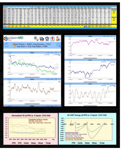
Figure 1: Data table, TD and SD results
Conclusions
In summary, his VMT-based five PPG energies are:
FPG = 27% (the highest)
Carbs/Sugar grams = 25%
Walking Steps = 16.3%
Sleep Jours = 16.0% (secondary)
Temperature = 15.7% (secondary)
The ratio of carbohydrates to steps is 1.53, aligning closely In summary, his VMT-based five PPG energies are: FPG = 27% (the highest) Carbs/Sugar grams = 25% Walking Steps = 16.3% Sleep Jours = 16.0% (secondary) Temperature = 15.7% (secondary) with the diabetes pathophysiological pathways ratio of 1.6. This is derived from dividing the eight dietary pathways by the five exercise pathways.
Furthermore, time-zone energy distributions are:
Y2015-Y2019 = 71%
Y2920-Y2024 = 29%
His initial 5-year condition was 2.5 times worse than his more recent 5-year condition, a comparison drawn by examining the ratio of 71% to 29%.
From 2015 to 2024, the individual maintained an average body weight of 171 lbs (BMI 25), leading to a FPG level of 106 mg/ dL, which is slightly above the normal range (below 99 mg/dL), and a PPG level of 113 mg/dL. This glucose differential of 7 mg/dL is attributed to the intake of 13.5 grams of carbohydrates and sugar, along with 3,878 walking steps taken after meals. Consequently, these health metrics contributed to an average HbA1C level of 6.5%, a significant improvement from 10% in 2010.
Key Message
Reducing body weight enhances the insulin-producing function of pancreatic beta cells, which is evident in fasting glucose levels. Moreover, managing carbohydrate and sugar consumption, along with engaging in post-meal walking activity, also helps in lowering post-meal glucose levels. Additionally, sleep duration and quality, along with maintaining a comfortable ambient temperature, are secondary factors that influence glucose formation.
References
For editing purposes, majority of the references in this paper, which are self-references, have been removed for this article. Only references from other authors' published sources remain. The bibliography of the author’s original self-references can be viewed at www.eclairemd.com.
Readers may use this article as long as the work is properly cited, and their use is educational and not for profit, and the author’s original work is not altered.
For reading more of the author’s published VGT or FD analysis results on medical applications, please locate them through platforms for scientific research publications, such as ResearchGate, Google Scholar, etc.