
Journal of Electrical Electronics Engineering(JEEE)
ISSN: 2834-4928 | DOI: 10.33140/JEEE
Impact Factor: 1.2


Research Article - (2023) Volume 2, Issue 4
Super weak Electromagnetic Radiation in Bio and Inorganic Systems
Research Article J Electrical Electron Eng, 2023 Volume 2 | Issue 4 | 339-346; DOI: 10.33140/JEEE.02.04.02
Batyanov A.P*
Institute of General Pathology and Pathophysiology 8, Baltiyskaya st., Moscow, 125315.
*Corresponding Author : Batyanov A.P, Institute of General Pathology and Pathophysiology, Russia.
Submitted: 2023, August 16; Accepted: 2023, September 20; Published: 2023, October 10
Citation: Batyanov, A.P. (2023). Super weak Electromagnetic Radiation in Bio and Organic Systems. J Electrical Electron Eng, 2(4), 339-346.
Abstract
This article provides a comparative analysis of data on ultra-weak electromagnetic radiation (WEEM) obtained at different times from bio and inorganic systems. The unifying physical principle was the presence of hybrid orbitals in the outer electron shells in the systems under study and the generation of coherent radiation in them. The data of a model experiment on ultra weak radiation of metals and semiconductors in the long-wavelength region of the optical spectrum are presented, indirectly confirming this hypothesis.
1. Introduction
The issue of the presence and role of SSEM of a coherent nature for bio systems is discussed in the literature [1,2]. The generation of such radiation is associated with electronic transitions in the outer shells with the formation of hybrid orbits and π-electrons; it is assumed that radiation is a "long-range" factor, i.e. nonchemical and non-thermal interaction between biological and non-biological objects, as the interaction of spatially separated electronically excited systems. It is possible that effects are due to the action of coherent radiation; interaction between the activity of isolated mitochondria and the physical characteristics of the material of incubation cells the stimulating effect of a carcinogen (DMBA) on mitochondria, distant-optical interaction of biological objects and the features of chemiluminescence of phosphorylating mitochondria [3-5]. In all systems under study there are hybrid orbitals in one form or another: in the ATP molecule due to competition and shift of the electron density between P and O atoms, similarly in quartz molecules between Si and O atoms (quartz incubation cell), in polycyclic molecules DMBA. From this point of view, a model experiment is of interest, where transition metals and semiconductors were used as a source of hypothetical coherent radiation, in which the probability of formation of hybrid orbitals is obviously high due to the structural features of the s-,p-.d-, bands of the outer electron shells especially in violation of electronic equilibrium [6].
Purpose and Task of the Study:
registration of ultraweak radiation from the objects under study (metals and semiconductors), in addition to radiation in the UV-light, visible and near-IR ranges.
2. Materials and Methods
To cut off the UV-light, visible and near-IR radiation ranges, all the samples under study were placed in a light hermetic (with a linearly increasing transmission up to 15% from λ~8µ to λ~28µ) plastic chamber. As seen in Fig. 1 (1) - a light-tight chamber (SGK) placed in a light-tight chamber (2) is a plastic cylinder (l=100mm.D=30mm, d-walls~1mm) with two internal equal compartments separated by a diaphragm (D=10mm ).Inside the cylinder, at the bottom of the first compartment (farthest from the PMT) there is an LED (250nm), the samples under study are in the second compartment (closest to the PMT), above the inner diaphragm and are fixed with a foam washer-seal, closer to the center of the cylinder. Outer. The end surface of the cylinder (the second compartment) is directed to the focusing, quartz lens and PMT. For light insulation, 7-8 layers of dark electrical tape are applied to the outer surface of the cylinder in the area of \u200b\ u200bthe joints. To register the SEMI, a setup (Fig. 1) with three recording systems was used: 1) A photomultiplier tube (FEU106) with an antimony-cesium multi-alkali photocathode, with a spectral sensitivity range from 170 to 830 nm and a maximum spectral sensitivity from 400 to 440 nm. operating in a pulsed mode, without cooling, the PMT signals were presented as the number of pulses per 10 sec. (imp / 10 sec.) experiment i.e. according to the change in the average number of impulses per 10 sec. (imp/10 sec.) during the individual stages of the experiment (16-18 min.). The time of the experiment (80-90 min.) was divided into five equal, successive segments-stages (phases). 1st and 5th stages: [Background-1 and Background-2]- ebonite shutter (3) is closed, the LED (8) is not turned on, thus in the first and fifth stages of the experiment (16-18 min) sequential registration was performed PMT dark background at maximum light isolation. 2nd and 4th stages: (signal + background -1 and signal + background -2) - ebonite damper (3) is open, LED (8) is not on. The emitting element was a plastic, light-hermetic chamber (SGK), with the test sample (7) embedded in it without any illumination. 3rd stage: [signal + background + 250nm.] - ebonite shutter (3) is open, LED (8) is on. In the same 3rd stage, samples were electrified with the LED turned off. Electrification was carried out by connecting the negative pole (-) of an electrical element (battery) or a 5-8v direct current source to the element under study (16-18 min.).
2) 2nd registration system - LED UVTOP 250FW Al Ga N-operating from direct current. The action of the SSEM was evaluated by the nature of the change in the resistance of the LED during its operation (3rd stage of the experiment 16-18 min.), Depending on the samples under study, for this, after switching on and before switching off, the current and voltage on the LED were recorded, resistance changes were represented by the value ( R2/R1 % min.). 3) The 3rd recording system - a widegap HgS (cinnabar on an aniline basis) is applied to writing paper on both sides, in a thin layer (a conditional analogue of photographic paper). samples. The effect occurred during long-term exposure (from 2 to 8 weeks) with daily 1-hour electrification (5V) of the studied samples, without turning on the PMT and LED. Strips of paper with HgS were placed inside and outside the chamber (2) - Fig. 1, at certain points Fig.1.-(10) in the vertical and horizontal planes (in front of the SGK body ~ 12mm and directly on the SGK body). Photos of the diffraction lines were taken by the main camera of the SM-T290 tablet. In the transmitted light of the Camelion LN13-AS lamp (Fig. 7,8,9,10,1 All experiments were combined into three main groups-directions: on the temperature and the sample under study in all 5 stages of the experiment.2nd group - studied the dependence of the SSEMI on the photoexcitation of the sample in the 3rd stage of the experiment 16--18 min. her experimental stage.
To check the reliability of the difference in data in the study groups, a t-test was used (Statistika-6 program). The results are shown in the figures.
Figure 2 shows the dependence of SSEM on temperature and sample. Attention is drawn to the consistently significant difference in the radiation intensity (average number of pulses/10 sec.) for Au and Pt samples with increasing temperature in the experimental phase (background-1), i.e. at maximum light isolation of the photomultiplier photocathode. It is possible that the radiation of Pt samples produces a quenching effect on the PMT photocathode, reducing its excitability and sensitivity to temperature (the PMT signal quenching effect) [7]. This is especially noticeable in successive experiments (with electrical excitation of Pt samples in the 3rd phase of the experiment -16-18 min.) for 30-45 days (one experiment per day), from the 7th day the average number of pulses / 10 sec. on the PMT, during one experiment it consistently decreases and takes a constantly low value Fig.3. The negative trend of SSMI, typical for 30-45 days of experiments with Pt, is also observed during individual experiments, within an hour, i.e. the number of pulses on the PMT from phase 1 to phase 5 of the experiment and the value (background2/background1% min.) gradually decrease. ) acquires a negative value (Fig. 4). A similar trend is observed in a series of repeated experiments with Al samples. It is noteworthy that (the effect of PMT signal quenching) by radiation from Pt samples requires an accumulation time (at least 7 days), i.e. organically associated with the (PMT electronic memory effect) [8]. On the other hand, a high probability of the signal quenching effect has been established PMT for W and Mo samples during their photoexcitation (250 nm) in the 3rd phase of the experiment. Figure 5 shows one of the experiments, while the damping effect manifested itself almost instantly and continued at least until the end of the experiment, i.e. also with the PMT electronic memory effect. It is very likely that the effect of PMT signal quenching is associated with the formation and accumulation of electron traps in the PMT photocathode system under the influence of coherent radiation of the samples under study. It is possible that in transition metals of the Pt,W.Mo type, the generation of coherent radiation in the long-wavelength region of the optical spectrum during photoexcitation (250 nm) and electrization (-5v.-8v) is associated with electronic transitions between the s-conduction band and the d-valence band, with shifts in the electronic equilibrium. It is possible that the generated coherent radiation has a polyfunctional effect in a single energy space, i.e. it creates traps and/or new energy levels for electrons (extinguishing the PMT signal and increasing the signal when the samples are electrified), a similar mechanism is possible when the resistance of the AlGaN LED changes depending on from the sample under study (Fig. 6). The appearance of diffraction bands (diffraction lines) on polymorphic HgS crystals adsorbed on a paper substrate (Fig. some kind of radiation from a hollow plastic, opaque cylinder with an internal focusing diaphragm and samples placed on it (conditionally hollow resonator-emitter). In addition, the specificity of diffraction lines for each sample under study indicates the connection of radiation with the molecular structure or surface structure of the samples Image of fragments elements of the internal design of the emitter; outlines of the LED housing, strips of platinum in the form of an irregular harmonic (Fig. 7), supply wiring on the surface of the emitter under a multi-layer electrical tape (Fig. 8) and individual letters from the names of companies pressed on the electrical tape (Fig. 9) - indicates the coherent nature of the radiation (holographic image). Thus, the data obtained indicate the possibility of generating coherent radiation during the formation of electronic hybrid orbitals, which indirectly confirms the previously stated assumptions about the possible mutual influence of hybrid sp-3 quartz orbitals (overlapping 3-sorbitals Si and 2-p orbitals of O) and hybrid orbitals between P and O in the ATP molecule, which leads to an additional shift in the electron density and configuration in ATP molecules synthesized by isolated mitochondria in quartz cells. As a result, there is a significant difference in the biochemical parameters of isolated MCs incubated in quartz and plexiglass cells (which also contain hybrid orbitals and possible formation of SSEM) and the dependence of these parameters on changes in the physical characteristics of the cell structure (incubation in darkened and irradiated cells) [9,10]. It seems likely that the stimulating effect on isolated mouse liver MCs after subcutaneous administration of DMBA is determined by the non-chemical interaction of the hybrid orbitals of ATP and DMBA molecules until the formation of electrophilic products and blocking of ATP synthesis [11-16].
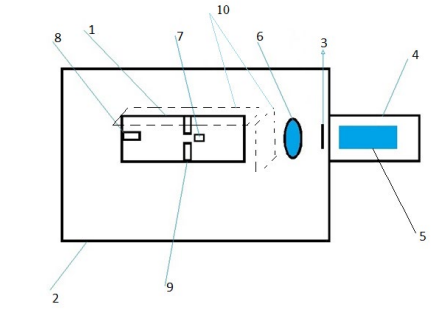
Figure 1: Schema Installation.
1- Light-hermetic, plastic chamber (SGK)
2- Lightproof camera
3- Ebonite shutter in front of the FEU-106 photocathode
4- Lightproof chamber for FEU-106
5- Photomultiplier with end photocathode-FEU-106
6- Quartz lens
7- Test sample inside (SGK)
8- LED (250nm) inside SGKv
9- Foam seal-diaphragm inside the SGK
10- Cinnabar-coated paper strips (HgS)
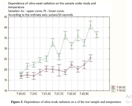
Figure 2: Dependence of ultra-weak radiation on a of the test sample and temperature.
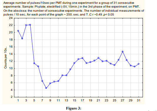
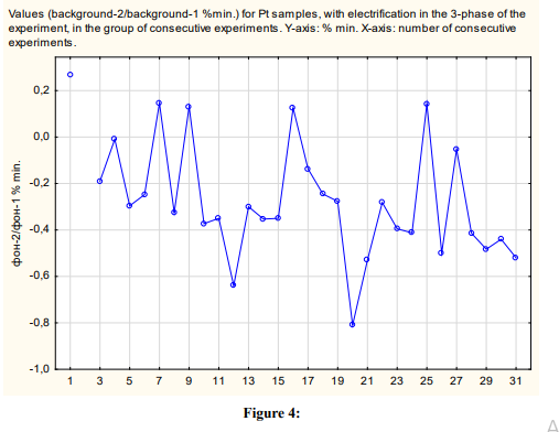
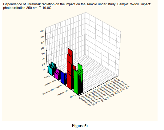

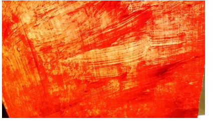
Figure 7: Diffraction lines - a Pt sample, a vertical plane, in the center of the photo, at the top left are the contours of the LED case in lateral projection (rectangle), to the right are the contours of a platinum plate in the form of an irregular W-shaped harmonic, at the top right are the contours of the company name, pushed aside on an insulating tape.
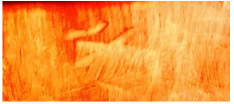
Figure 8: Diffraction lines - Pt sample, horizontal plane, in the center of the photo is an image of fragments of wiring and platinum wire inside the case (SGK) under 7-8 layers of insulating tape.
Figure 9: Diffraction lines - Al sample is a horizontal plane. In the center of the photograph is an image of individual letters (E, A) from the name of the company "ERA", pushed aside on an insulating tape.
Figure 10: Diffraction lines - Al sample, vertical plane.
Figure 11: Diffraction lines - Au sample (alloy 575 sample), horizontal plane.
Figure 12: Diffraction lines-Au (alloy, 575 sample), vertical plane.
Declarations
Ethical Approval
Institutional Review Board Statement: The study was conducted according to the guidelines of the (DIRECTIVE 2010/63/EU OF THE EUROPEAN PARLIAMENT AND OF THE COUNCIL on the protection of animals used for scientific purposes of 22.09.2010.), and approved by the Institutional Ethics Committee of the Institute of General Pathology and Pathophysiology (final protocol # 1 of 01.02.2023.)
Competing Interests
Informed Consent Statement
Informed consent was obtained from all subjects involved in the study.
Authors Contributions
Inadaptability.
Funding
This research received no external funding.
Availability of Data and Materials
Data Availability Statement
The data presented in this study are available on request from the corresponding author.
References
- Batyanov, A. P. (1988). Correlation of mitochondrial metabolism and spontaneous luminescence of incubation cells.//Biophysics 6 ,1029, t.33.-Vol.
- Beloussov, L. V., & Popp, F. A. (Eds.). (1994). Biophotonics: Non-equilibrium and Coherent Systems in Biology, Biophysics and Biotechnology. Bioinform Services Company Russia.
- J.Stauff.J. Ostrowski. Z.Naturforsch. (1967). № 226.s.734-740.
- B.Ruth. (1989). Electromagnetic Bio-Information. Edited by Fritz-Albert Popp and Ulrich Warnke, Herbert L.Konig, Walter Peschka. Urban Schwarzenberg, Munchen,Wien. Baltimore //Experimental Investigation on Ultra weak Photon Emission. p.128-143.
- A.P.Batyanov. (1989). Chemiluminescent analysis of metabolic disorders of liver mitochondria in some types of pathology. Abstract of Candidate of Dissertation Research Institute of General Pathology and Pathological Physiology. USSR Academy of Medical Sciences Moscow.
- H Frohlich,F.Kremer. (1983). Coherent excitations in biological systems: Springer.
- F.A.Popp.,K.H. Li.. W.P.Mei.,M.Galle.,R.Neurohr. (1988). Experientia Vol.44. №.7 pp.576-585.
- F.A. Popp, W. Nagl , K. H.Li , W. Scholz , O. Weingartner , R. Wolf. (1984). Cell Biophys.-Vol.6.- p.33-52.
- J.Slawinski. (1988). Experientia Vol.44 №7p.559-571.ISSN0014-4754.
- J.Pokorny. (2007). Electrodynamic activity of healthy and cancer cells. Journal of Physics:Conference Series 329 (2011)01.
- J.Pokorny. ,C. Vedruccio.,M.Cifra.,O.Kucera. (2011). Eur. Biophys. J. 40:747-759.
- C Kittel Charles Kittel. (2012). Introduction to solid state physics. Chapter 7.11. Translation from English.M. "Book on Demand".
- J.M.Ziman. (1962). Electrons and phonons. Translation from English, M. Chapter 2 pp. 66-112, 435-439.
- A.M. Gurvich. (1982). Introduction to the physical chemistry of crystal phosphors. M .: Higher school. 376s.<
- N.O.Chechik., S.M. Fainshtein, T.M. Lifshits. (1957). Electron multipliers. State publishing house of technical and theoretical literature. Moscow.
- Friedrich Liebau. (1985). Structural Chemistry of Silicates. Springer-Verlag,Berlin-Heidelberg,New-York, Tokyo
Copyright:
Copyright: ©2023 Batyanov A.P. This is an open-access article distributed under the terms of the Creative Commons Attribution License, which permits unrestricted use, distribution, and reproduction in any medium, provided the original author and source are credited.