

Advances in Bioengineering and Biomedical Science Research(ABBSR)
ISSN: 2640-4133 | DOI: 10.33140/ABBSR

Impact Factor: 1.7

Review Article - (2021) Volume 4, Issue 4
Sealing-type Three-cavities Ventilation Joint: A New Device in Painless Bronchoscopy
2Division of Hematology and Oncology, Department of Internal Medicine, School of Medicine, , University of California Davis, Sacramento, CA 95817, USA
3Fushun Second People’s Hospital, Fushun City, Liaoning Province, 113000, China
Received Date: Nov 23, 2021 / Accepted Date: Nov 26, 2021 / Published Date: Dec 03, 2021
Copyright: ©Copyright: ©2021 Xiaoshi Liu, et al. This is an open-access article distributed under the terms of the Creative Commons Attribution License, which permits unrestricted use, distribution, and reproduction in any medium, provided the original author and source are credited.
Citation: Xiaoshi Liu, Zheng Zhu, Zhenna Wang, Jingtian Han, Jiazhen He, Xiaoshi Liu (2021) Sealing-type Three-cavities Ventilation Joint: A New Device in Painless Bronchoscopy. Adv Bioeng Biomed Sci Res 4(4): 123-128.
Abstract
Background: To observe the clinical effect and safety of the sealing-type three-cavities ventilation joint in painless bronchoscopy. To compare the respiratory mechanics between I-gel laryngeal mask and tracheal tube-controlled breath during bronchoscopy.
Methods: 200 patients underwent bronchoscopy were recruited and randomly assigned to general anesthesia group (group â? , n = 100) and local anesthesia group (group â?¡, n = 100). General anesthesia group were divided into two groups, the I-gel laryngeal mask combined with sealing-type three-cavities ventilation joint group group(n=50) and the endotracheal tube combined with sealing-type three-cavities ventilation joint group(n=50). Patients in Group I were adopted by I-gel laryngeal mask or endotracheal tube with the sealing-type three-cavities ventilation joint after the induction anesthesia with remifentanil, propofol and succinylcholine. In group II, patients were anaesthetized with lidocaine and followed by 2mg/ kg propofol iv, and spontaneous respirations were retained with nasal cannula. All patients’ vital signs, endoscopic related adverse reactions and arterial blood gas analysis were recorded during the procedure.
Results: Group I showed little changes of vital signs (P <0.05), and less adverse reaction such as the intraoperative hypoxia and intraoperative body movement (P <0.05), and no significant decrease of oxygen partial pressure (P <0.05). There is no significant difference in respiratory mechanics including tidal volume and airway pressure between two subgroups in group I(P>0.05).
Conclusion: Sealing-type three-cavities ventilation joint prevents the oxygen deficit and makes it possible for us to examine patients through bronchoscope under general anesthesia without gas leakage. Moreover, sealing-type three-cavities ventilation joint provides safe and effective airway control while it does not change respiratory mechanics in endotracheal tube group compared with I-gel laryngeal mask makes endotracheal tube an alternative solution in bronchoscope. Take all these in consideration, sealing-type three-cavities ventilation joint proves to be a feasible method in bronchoscope.
Keywords
Sealing-Type Three-Cavities Ventilation Joint, Bronchoscopy, Painless
Background
Bronchoscopy, which is a common diagnostic and therapeutic method in department of respiration, provides a direct view of patients’ airway. It allows respiratory physicians to observe ab-normalities, such as bleeding, tumors, inflammations in airways as well as to remove secretions and excise tumors in airways[1]. Although bronchoscopy is widely used, some patients refuse to use bronchoscopy because of fear, anxiety and bucking. Bron¬choscopy is always followed by local or general anesthesia. Both ways of anesthesia have disadvantages. Local anesthe¬sia is often given to anesthetize the mucous membranes of the pharynx, larynx, and trachea by sedatives such as midazolam or propofol, which makes patients feel uncomfortable, and may lead a delay of diagnosis and treatment. General anesthesia is an alternative choice for bronchoscopy; however, gas leakage and intraoperative hypoxia remain a big challenge in widespread using of this technique.
To improve the bronchoscopy utilization, we aim to solve the adverse reaction such as intraoperative hypoxia and reduce the pains of patients during bronchoscopy. And to solve the air leak¬age in current general anesthesia. What is more we also want to record the accurate respiratory data during bronchoscopy in general anesthesia both in endotracheal tube group and in I-gel laryngeal mask group to see if endotracheal tube could be an alternative way in general anesthesia? To achieve these goals, we invented a novel device, sealing-type three-cavities ventila¬tion joint (Patent No.: ZL201620212426.8), as a new approach to control respiration as well as to avoid hypoxia in bronchos¬copy during general anesthesia. It optimizes general anesthesia during bronchoscopy, making bronchoscopy a painless proce¬dure, which may reduce the uncomfortable symptoms as well as endoscopic related adverse reactions of patients and stabilize patients’ vital signs. By using this device, we are also able to re-cord accurate breathing mechanics during the procedure, which demonstrated the endotracheal tube had little difference in respi-ratory mechanics compared with I-gel laryngeal mask and may be applied in general anesthesia during bronchoscopy, which is not recommended before. It is safe and efficient, which may pro¬vide a feasible choice for patients who are fear of bronchoscopy and for those who show poor compliance in bronchoscopy to get diagnosis and treatment in time.
Methods
Design
The study is designed as a randomized controlled trial (registra-tion No.: ChiCTR-INR-17014190) and is approved by the Eth-ics Committee and Medical Dept.
Patients underwent bronchoscopy from December 2017 to Jun 2018 in Fushun Mining Bureau General Hospital, without se-vere cardiopulmonary disease, severe maxillofacial deformities and neuromuscular disease, were recruited. All patients signed the informed consent before anesthesia.
Device Introduction
We drew a sketch map of sealing-type three-cavities ventilation joint as shown in Figure 1. Components include the main pipe 1, the main pipe 1 is provided with a branch pipe 2, the upper end of the main pipe 1 is provided with a gas barrier device 3, a third through hole (operating hole) 8 is opened on the top surface of the gas barrier device 3, The air-blocking cone device 4 is provided on the device 3, and the choke cone devices5 is placed below the air-blocking cone device4,the combination of the two devices can make the bronchoscope with no air-leaking in oper¬ation, so that to achieve 100% sealing in operation.
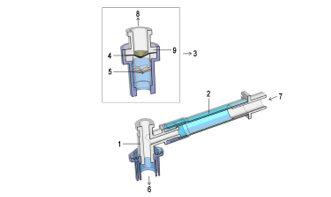
Figure 1: Components of sealing-type three-cavities ventilation
joint 1:main pipe, 2: branch pipe, 3: gas barrier device, 4: air-blocking cone device 5: choke cone devices, 6: openings on aeration de¬vice, 7, openings on anesthesia machine, 8: a third through hole (operating hole), 9:stabilizer on gas blocking piece.
Interventions
Patients were randomly divided in two groups, the general an-esthesia group (group I, N=100) and local anesthesia group (group II, N=100). Patients in group I were randomly allocated into I-gel laryngeal mask combined with sealing-type three-cav¬ities ventilation joint group (group Ia, n=50) and endotracheal tube combined with sealing-type three-cavities ventilation joint group (group Ib, n=50). All the patients in group I received rou¬tine inspections including blood examination, electrocardio-gram, chest digital radiography (DR). After12h free of fasting and 4h free of drinking, patients were noninvasively monitored vital signs in operating room before operation. Group I were narcotized by midazolam 0.02mg/kg, propofol 1mg/kg, fentanyl 0.1mg, remifentanil 20ug, succinylcholine 2mg/kg intravenous¬ly. After induction anesthesia, then connected the air passage of sealing-type three-cavities ventilation joint with the well locat-ed I-gel laryngeal mask or endotracheal tube, added ventilation end into the screwed pipe of anesthesia machine, conducted with mechanical ventilation, enforced the bronchoscope operation in the three-cavities connection juncture, pumping with propofol 4-8mg/kg/h and 1% Sevoflurane to maintain with anesthesia during whole operation, and kept watching until sobering of pa¬tients. In group II, patients were anaesthetized with lidocaine and followed by 2mg/kg propofol iv, and spontaneous respira-tions were retained with nasal cannula. The diameter of bron-choscope in each group is2.8mm.
Observations
Observation indexes including pulse wave oxygen saturation, heart rate, blood pressure during the five time point of pre-anes¬thesia (T0), after anesthesia but before bronchoscope operation (T1), bronchoscope pass by the Glottis (T2), inspection (T3), finish of Inspection (T4), body movement, bucking and laryn¬gospasm of patients were recorded. Arterial blood gas analysis was done during bronchoscopy. All patients’ satisfaction degree were investigated 1 represents for poor satisfactory whereas 10 represents for very satisfactory.
Observations
Observation indexes including pulse wave oxygen saturation, heart rate, blood pressure during the five time point of pre-anes¬thesia (T0), after anesthesia but before bronchoscope operation (T1), bronchoscope pass by the Glottis (T2), inspection (T3), finish of Inspection (T4), body movement, bucking and laryn¬gospasm of patients were recorded. Arterial blood gas analysis was done during bronchoscopy. All patients’ satisfaction degree were investigated 1 represents for poor satisfactory whereas 10 represents for very satisfactory.
Statistics
By the adopting of Statistical software analysis, measurement data was recorded by mean ± standard deviation, the compari¬son among groups was recorded by repeated measurement, the comparison of enumeration data was measured by chi-square test, and P<0.05 was considered as a statistical significance. All analyses were done using SPSS 17.0
Results
Study Population
In total, 200 patients, were included in the analysis. The mean age was (56.6±8.27) years, 109 patients were male while 91 pa¬tients were female. The difference between group I and group II in ASA classification, gender, age, height and weight were out of statistical significance (Table 1).
Table 1: Data of the patient

Vital Signs
The vital signs, including heart rate (HR), saturation of pulse oximetry (SPO2), mean arterial pressure (MAP), arterial blood oxygen partial pressure (PaO2), were observed in T0~5. Patients in group II showed an obviously fluctuation in heart rate, blood pressure and SPO2. Patients in group I showed a higher PaO2 but exhibited a slim blood pressure decline during the procedure, it might be caused by the vein injection of propofol (Table 2 and Figure 2).
Table 2: The Hemodynamic change of two groups by indu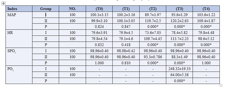
* P < 0.05
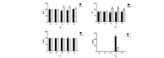
Figure 2: Vital signs variation in group I and group II
Vital signs on (a) MAP, (b) HR, (c)SPO2, (d)PaO2 in group I and group II. *, P<0.05.
Respiratory Mechanics
By the comparison of arterial blood gas analysis of two groups in group I, before and after inserting bronchoscope, the tidal volume decreased about 20-60ml but the final tidal volume and airway pressure occurrence showed no difference (Fig. 3).

Figure 3: Respiratory mechanics in group Ia and group Ib
Respiratory mechanics on tidal volume and airway pressure in group Ia and group Ib.
Adverse Reaction Comparison
Patients in group II suffered a higher adverse reaction such as body movement and cough during bronchoscopy. As patients in group, I was anesthetized and received muscle relaxant, no body movement and cough occurred in the procedure. Howev-er, 75 patients in group II occurred body movement. Aphonia and hoarseness after bronchoscopy occurred more frequently in group II, whereas patients’ satisfaction degree was higher in group I. There was no distinct difference on laryngospasm to both groups, but since laryngospasm was one of the most serious complications during bronchoscope inspection, so had paused the inspection of laryngospasm.
Patients’ Satisfaction Degree
After the procedure, patients’ satisfaction degree in group I and group II were obtained after they went back inpatient ward. Pa-tients’ satisfaction degree varied from zero to ten. Zero stands for least satisfaction and 10 represents for most satisfaction. Scores between these two groups were shown in figure 4.

Figure 4: Patients’ degree of satisfaction in group I and group II
Patients’ degree of satisfaction in group I and group II. *, P<0.05.
Discussion
Bronchoscopy is an endoscopic technique of visualizing the in-side of the airways which is widely used in the diagnosis and treatment in weasand disease. Nowadays, most patients receiv-ing bronchoscopy receive local or general anesthesia, which will influence bronchoscope operation in some aspects. Local anes-thesia may cause complications in cardiovascular, cerebrovas-cular, laryngospasm, massive hemorrhage. What is more, bron-choscope is an invasive operation, for a long time, numerous patients refused to choose this inspection due the fear, horror and avoiding strong irritation. This led to delayed diagnosis and treatment of disease. Some patients don’t receive bronchoscope until the last moment, meanwhile the late stage of the disease[2]. Researchers have attempted many kinds of anesthesia methods to deal with these problems[1], but a little of them works. Current general anesthesia applied in bronchoscopy is combined with I-gel laryngeal mask. The reason is that anesthesia drugs may cause respiratory depression and problems of weasand manage- ment. I-gel laryngeal mask is used as the newly airway manage¬ment tool, it has the features both in tracheal catheter ventilation and noninvasive of mask. It can positively control respiratory tract, with less irritation, less reaction of angiocarpy. There is no need to use bronchoscope exposing glottis[3, 4]. The curvature design in I-gel laryngeal mask is a perfect match to the curva¬ture of pharyngeal physiology dissection[5]. The ways of simple operation and can be located inside easily, which put the glottis in a better exposure[6, 7]. The front part of I-gel laryngeal mask is made up of silica gel materials, these parts do no stimulation to glottis and weasand mucous membrane, which will benefit-the depth of anesthesia and the quick woke up. Zhou Shuzhen’s research of I-gel LMA mask used in bronchoscope inspection had proved the safety of the products in ventilation and glottis control[2]. Enjoying such advantages, however, I-gel larynge¬al mask still has limitations. Leakage between I-gel laryngeal mask and anesthesia machine results in uncontrolled anesthesia depth. This may lead to anesthesia machine working improperly even lead hypoxia in patients and terminate the examiner. Such leakage also makes it impossible to obtain the accurate respira¬tory mechanics. If a patient cannot go through I-gel laryngeal mask, one will have no choice in choosing other ventilation way. Some researchers attempted to prevent such leakage by using a uni-seal ring. This device could prevent the air leakage in some degree; however, it could not prevent the leakage all the way during procedure. When placing the bronchoscope vertically or spinning it during procedure, air leakage occurs. What’s more, minor-diameter bronchoscope placement and pre-placing bron¬choscope sealing remain issues in treatment. These disadvantag¬es interrupt the bronchoscopy, force us withdraw bronchoscope and wait for the recovery of blood oxygen for next exam. These disadvantages limit the use of bronchoscope.
Our device sealing-type three-cavities ventilation joint (Patent No.: ZL201620212426.8) is a novel device and is totally differ¬ent from others. By using silica gel, our device makes it possible to block air leakage by fitting the bronchoscope to the silica gel closely in procedure. It can achieve a non-air leaking and could be parts of the outer hoop circle. Our data showed a 100% no air leaking in performance. Just like theory of one-way valve of ten¬sion pneumothorax, the way of operation will do little influence to positive pressure mechanical ventilation of anesthesia ma¬chine or to the way of anesthesia drags inhalation. By using sil¬ica gel elastic damping theory, our device can actively joint the bronchoscope, as you see, the choke blocking devices is mov¬ing in parallel with bronchoscope in rotating operation, which means one sides of the choke blocking devices will be pressed to leech on to bronchoscope, the other sides of the device will be upspring to cling to the surface of bronchoscope. The kind of operation will make the choke blocking devices clinging on to bronchoscope ignoring the ways of rotation. Besides, air-block¬ing cone devices, which is used for one-way air valve blocking, the combination of the two devices can make the bronchoscope with no air-leaking in operation, so that to achieve 100% sealing in operation.
By using sealing-type three-cavities ventilation joint to deal with bronchoscope inspection inside airways, the way can both con-trol free breathing and stabilize anesthesia drugs inhalation [9]. The higher damping and elasticity can be adaptable to all kinds of bronchoscope with different diameters ranging from 2mm to 10mm. Our results showed that Group I have a less vital signs fluctuation and bucking and quieter compared with group II, there is no more patients with decline of blood oxygen satura¬tion or drop of oxygen partial pressure. The body movement and bucking appeared in group II has increased the difficulty in op¬eration. The laryngospasm accompanied by three of the patients and led to the operation pause in group II. Patients in group I had no memory of the operation and had higher satisfaction. The widely used of our device combining I-gel laryngeal mask will reduce the suffers of patients during operation, also the conquer fear to the operation. Besides, it has more benefits to special pa¬tients with high blood pressure, coronary heart disease, dumb and deaf, hearing disorder and mentally handicapped whom cannot cooperate with the operation. Our research showed that when using a 2.8mm diameter bronchoscope to do the proce¬dure, there is no respiratory mechanics difference in group Ia and Ib. Without influence the anesthesia depth, our device could almost reach the same tidal volume, airway pressure. This is important because we could use endotracheal tube with our de¬vice when I-gel laryngeal mask is forbidden so as to expand the procedure indications. The sealing-type three-cavities ventila- tion joint could realize a totally sealing environment in placing and withdrawing the bronchoscope which provides an accurate condition to record the respiratory mechanics. These respiratory mechanics are rarely recorded due to the none sealing environ¬ment, such environment makes respiratory mechanics fluctuate drastically. Our results showed no difference between I-gel la¬ryngeal mask group and sealing-type three-cavities ventilation joint group in 2.8mm bronchoscope. Such results indicates that the sealing-type three-cavities ventilation joint could be an opti¬mal choice for bronchoscopy.
According to our research, I-gel laryngeal mask with seal-ing-type three-cavities ventilation joint can be widely and safely used in bronchoscope inspection in daily operation, which will do no influence to the breathing machine according to pre-clin¬ical application. The tidal volume is a little lower (20-60ml), and an increase in airway pressure, an inhalation of 50% oxygen concentration till the end of operation, the oxygen partial pres¬sure is 180mm Hg, CO2 pressure is 40mm Hg, other results are in normal range. With the development of technology involved in lungs disease, the bronchoscopies widely used in all kinds of surgeon operation[10]. The interventional therapy needs a long operation and accompanied by a lot of complications compared with normal inspection, I-gel laryngeal mask with Sealing-type three-cavities ventilation joint in daily inspection will reduce the chance of vital signs fluctuation and bucking, the doctors can deal with the inspection conveniently with no more concerns of breath control, no more worry of oxygen deficit, no more con¬trol of inspection and therapy, which will make more patients receive bronchoscope in early stage. The methods were proved to solve the problems caused in weasand management funda-mentally[11]. By the succeed experience of above test, and the freely use of bronchoscope in and out of weasand, we wish to widely use the technology in bronchoscope inspection therapy and children diagnosis and treatment, and offer a new way in breath control in bronchoscope inspection.
In conclusion, sealing-type three-cavities ventilation joint apply¬ing in painless bronchoscopy is safe, it controls the airway effec¬tively, and solves the problem of intraoperative hypoxia in bron¬choscopy. What is more it makes endotracheal tube a feasible choice in bronchoscopy in general anesthesia, which provides a feasible It is useful and promising as it proves to be a feasible method for patients who are afraid of bronchoscopy as well as patients who cannot cooperate with doctors?
Abbreviations
BP: blood pressure
HR: heart rate
SPO2: saturation of pulse oximetry
MAP: mean arterial pressure
PaO2: arterial blood oxygen partial pressure
LMA: laryngeal mask
T: time.
Acknowledgement
The manuscript was professionally proofread.Funding
None
Availability of Data and Materials
The datasets used and/or analyzed during the current study are available from the corresponding author on reasonable request.
Authors Contributions
Xiaoshi Liu and Zixuan Wang: conception, design, experimen-tal work. Zheng Zhu: Drafting article. Zhenna Wang: Analyzing data. Jiazhen He: Sample collection. Jingtian Han: Sample col-lection and guide technique.
Ethics Approval and Consent to Participate
The study is approved by the Ethics Committee and Medical Dept. Patients were informed about the research and agreed to join in.
Consent for Publication
Not applicable.
Competing interests
The authors declare that they have no competing interests
References
- Pathak V, Welsby I, Mahmood K, Wahidi M, MacIntyre N (2014) Shofer S.Ventilation and anesthetic approaches for rigid bronchoscopy[J].Annals of the American Thoracic Society 11: 628-634.
- Fadaizadeh L, Hosseini MS (2013) Laryngeal mask airway in interventional bronchoscopy for tracheal lesions[J]. Acta anaesthesiologica Taiwanica : official journal of the Taiwan Society of Anesthesiologists 51: 137.
- Fadaizadeh L, Hosseini MS, Dabir S (2013) Role of laryngeal mask airway in interventional bronchoscopy procedures for upper tracheal stenosis: case series[J]. Middle East journal of anaesthesiology 22: 223-227.
- Martin Flores M, Cortright CC, Koba SJ (2015) Removal of an Airway Foreign Body via Flexible Endoscopy Through a Laryngeal Mask Airway[J]. Journal of the American Ani-mal Hospital Association 51: 325-328.
- Belena JM, Nunez M, Anta D, Carnero M, Gracia JL, et al. (2013) Comparison of Laryngeal Mask Airway Supreme and Laryngeal Mask Airway Proseal with respect to oropha-ryngeal leak pressure during laparoscopic cholecystectomy: a randomised controlled trial[J]. European journal of anaesthesiology 30: 119-123.
- Belena JM, Gracia JL, Ayala JL, Nunez M, Lorenzo JA, et al. (2011) The Laryngeal Mask Airway Supreme for positive pressure ventilation during laparoscopic cholecystecto-my[J]. Journal of clinical anesthesia 23: 456-460.
- Chandrakar S, Sreevastava DK, Bhasin S, Dhar M (2017) Comparison of laryngeal tube suction II and proseal LMA in pediatric patients, undergoing elective surgery[J]. Saudi journal of anaesthesia 11: 432-436.
- Kimijima T, Edanaga M, Yamakage M (2018) Superior sealing effect of a three-dimensional printed modified supraglottic airway compared with the i-gel in a three-dimensional printed airway model[J]. Journal of anesthesia.
- Arevalo Ludena J, Arcas Bellas JJ, Alvarez Rementeria R, Flandes J, Moris L, et al. (2016) comparison of the I-Gel supraglottic device with endotracheal intubation for bron-choscopic lung volume reduction coil treatment[J]. Journal of clinical anesthesia 31: 137-141.
- Kang F, Li J, Chai X, Yu J, Zhang H, et al. (2015) Comparison of the I-gel laryngeal mask airway with the LMA-supreme for airway management in patients undergoing elective lumbar vertebral surgery[J]. Journal of neurosurgical anesthesiology 27: 37-41.
- Jose RJ, Shaefi S, Navani N (2014) Anesthesia for bron-choscopy[J]. Current opinion in anaesthesiology 27: 453-457.