

Advances in Bioengineering and Biomedical Science Research(ABBSR)
ISSN: 2640-4133 | DOI: 10.33140/ABBSR

Impact Factor: 1.7

Research Article - (2021) Volume 4, Issue 2
Probiotic Bifidobacterium Lactis Combined with Lactobacillus Plantarum Effectively Reduced Weight and Intestinal Microbiota
2Graduate Institute of Biomedical Engineering, National Chung Hsing University, Taichung, Taiwan
3Research & Design Center, TCI CO., Ltd., Taipei, Taiwan
4Guizhou yangsen big health industry CO., Ltd., Guizhou, China
5nstitute of Food Safety and Risk Management, National Taiwan Ocean University, Keelung, Taiwan
Received Date: May 12, 2021 / Accepted Date: May 17, 2021 / Published Date: May 28, 2021
Copyright: ©Copyright: ©2021 Chi Fu Chiang, et al. This is an open-access article distributed under the terms of the Creative Commons Attribution License, which permits unrestricted use, distribution, and reproduction in any medium, provided the original author and source are credited.
Citation: Yung Kai Lin, Tao Ping, Yung Hsiang Lin, Wei Chun Hu, Chi Fu Chiang (2021) Probiotic Bifidobacterium lactis Combined with Lactobacillus Plantarum Effectively Reduced Weight and Intestinal Microbiota. Adv Bioeng Biomed Sci Res 4(2): 54-58.
Abstract
Probiotics intake can ameliorate body weight and fatty liver development. The fruit and vegetable also performed antiinflammation effects. The fermented vegetable solid drink (FVSD) was combined fruit and vegetable with probiotics and further examining the anti-obesity efficacy in this study. The FVSD promote lipolytic activity effect was examined on OP9 cells and the anti-inflammation effect was performed via Q-PCR analysis on C2BBe1. To investigate the weight management potential and gut microbiota influence. The obese subjects were recruited, and then performed anthropometric measurement and next-generation sequencing (NGS) after FVSD intervention. In the results, the lipolytic activity effects were significantly increased and the LPS induced inflammation response was significantly reversed by FVSD co-treatment. After FVSD administration, obese subjects were significantly ameliorated body weight, body fat weight, and body fat at 4 weeks. And the waist-to-hip ratio was improved at 8 weeks. Both aspartate aminotransferase (AST), alanine aminotransferase (ALT) were significantly improved with anti-fatty liver potential. The NGS analysis suggested FVSD intervention could increase Christensenellaceae and Parabacteroides abundance of subjects’ gut microbiota. In conclusion, FVSD performed a great anti-obese effect in vitro and in vivo.
Keywords
Bifidobacterium, Lactobacillus, Probiotics, Weight reduction.Introduction
Obesity is a global epidemic and is considered to cause serious chronic diseases such as hypertension, cardiovascular disease, di-abetes, cancer [1]. The current strategy is to fight obesity diet con¬trol, exercise, medication and surgery, but diet and exercise do not continue to strictly, and enforce the adverse side effects of drugs also limits its therapeutic use [2]. Fortunately, most recently about some interesting research on obesity is helping to validate this new method of medical disease control.
Obesity can lead to a change in the composition of the intestinal microbiota [3]. The main types of bacteria in the intestinal microbi¬ota are Firmicutes, Bacteroidetes, Proteobacteria, Actinobacteria, and Verrucomicrobia [4]. The intestinal microbiota can regulate the metabolism of the host, including weight control. In animal and human studies, it has been found that the composition of the intestinal microbiota related to obesity [5]. The study showed that through gene sequence analysis, it was found that intestinal bac- teria such as Bacteroidetes and Firmicutes were colonized [6]. Weight loss makes the ratio of Bacteroidetes to Firmicutes up regulated in humans [7]. Thus, changes in the intestinal microbiota, may be a novel strategy for the treatment of obesity.
Recent studies had demonstrated that probiotic strains play an im¬portant role in modulating immune responses and exert anti-obesi¬ty effects [8]. Bifidobacterium was one of the most abundant pro¬biotic bacteria in the intestine of mammals [9]. Studies had shown that Bifidobacterium can inhibit the absorption of cholesterol in the small intestine, and Bifidobacteria are believed to improve me-tabolism-related diseases, including reducing insulin resistance, fat accumulation, and fatty liver [10, 11]. Lactobacillus probiotic has many functions, can reduce blood fat, protect the cardiovas¬cular system and reduce obesity [12]. Lactobacillus rhamnosus, Lactobacillus brevis, Lactobacillus plantarum and Lactobacil¬lus paracasei had lipid-lowering effects, which can reduce body weight and increase metabolism [13]. In addition, Lactobacteria and Bifidobacteria suppressed weight gain of mice fed a high fat diet [14]. Therefore, specific strains of Lactobacteria and Bifido-bacteria may be potential therapeutic candidates for anti-obesity.
In this study, we used fruit and vegetable powder with TCI604 (Bifidobacterium lactis) and TCI507 (Lactobacillus plantarum) to make a solid drink (FVSD). We used solid beverages to examine whether it increased fat metabolism and reduced intestinal inflammation. At the same time, we recruited obese subjects and taking solid beverages for 8 weeks to examine whether it decreased weight, and regulated intestinal microbiota.
Materials and Methods
Lipolytic Activity Assay
8 × 10⁴ OP9 cells were seeded with 500 µl pre-adipocyte expan-sion medium in 24 wells. And incubated at 37 â?? for 7 days and replace with a fresh differentiation medium every other day. After 7 days, observe lipid droplet formation using microscopy to make sure the cells are fully differentiated. Add 0.25% of FVSD and incubate for another 7-10 days and change the medium every other day. And proceed to the glycerol content analysis (Cayman: Item No.10010755). Then collect cell culture supernatant from each well, and then transfer 25 μl of cell culture supernatant from each well and standard into a new 96-well plate. Add 100 μl of recon¬stituted free glycerol assay reagent per well. After 15 minutes at room temperature and further measuring at 540 nm.
Inflammation Related Genes mRNA Expression Analysis
1.5 × 10⁵ C2BBe1 cells in 2 ml of the media with 0.25% of FVSD were seeded in each well of 6-well plates and incubated for 24 hours. And change the medium with or without lipopolysaccharide (LPS) and FVSD for examining inflammation-related gene expres¬sion. Then, we collected the cells and used the RNA extraction kit for RNA collection. Finally, we adjusted the RNA concentration to 75 ng/μL for mRNA expression analysis. The IL-1β, IL-8, IL-18, and TNF-α mRNA expression level was analyzed by qPCR.
Clinical Trial
This clinical study was approved by the ethics committee of the Antai Medical Care Corporation Antai Tian-sheng Memorial Hos-pital (IRB No. 20-039-A), and the study protocol was registered with the ClinicalTrials.gov (NCT04501601). All methods were performed following the relevant guidelines and regulations. 25 adult subjects were recruited, and informed consent was obtained from all subjects before the study.
Subjects consumed daily solid drink for 8 weeks, and recorded weight, body fat, waist-to-hip ratio at 0, 4, and 8 weeks. In blood analysis, liver function indicators were analyzed at 0, 4, and 8 weeks, the fecal samples were obtained from subjects at weeks 0, 4, and 8. and intestinal microbiota was also analyzed through next-generation sequencing (NGS). Inclusion criteria included: i) age over 20 and below 60 years old; ii) body mass index (BMI) ≥ 24 or body fat > 25% (male) and body fat > 30% (female); Ex-clusion criteria included: i) Women who are breastfeeding, preg-nant and menopause. ii) History of serious diseases associated with heart, liver, kidney, endocrine systems or other organs; iii) Type 2 diabetes or performed other weight loss methods within six months, including diet control, exercise, drugs; iv) drug consump¬tion, alcohol addiction, gastrointestinal diseases.
Test Sample
The main ingredients of solid beverages were fruit and vegetable powder, Bifidobacterum lactis (TCI604) and Lactiplantibacillus plantarum (TCI507), water. Each subject was required to consume daily solid drink before lunch for 8 weeks. The test sample is a pack of 13 grams, which is melted with 180 ml of 45°C water before consumption.
Statistical Analysis
All values were expressed as mean ±SD between sample populations differences statistical result was determined by an unpaired two-tailed Student’s t-test. Statistical significance was considered at P value < 0.5.
Results
FVSD increased the Lipolytic Activity and Decreased Inflammation in Vitro
First, in order to examine whether FVSD increased lipolytic activity, using the mouse embryonic stem cells derived stromal cells, OP9. The OP9 was differentiated into adipocytes, and treated with FVSD, then examined the glycerol content. Triglyceride was digested by lipases, and converted into fatty acid and glycerol [15]. We found that solid drinks could increase glycerol by about 121.5% (Figure 1). This result revealed that FVSD increased lipolytic activity. Second, in order to explore whether FVSD decreased intestinal inflammation, we used the human-derived colorectal cells, C2BBel, and using lipopolysaccharides (LPS) to mimic inflamed environment, and treated with FVSD, then examined inflammation related gene expression. We found that IL-1β, IL-8, IL-18, and tu¬mor necrosis factor-alpha (TNF-α) were increased in LPS stimula¬tion. Conversely, as LPS combined with FVSD treatment, the IL-1β, IL-8, IL-18, and TNF-α were decreased by 31.4, 2.1, 3.3, and 10.6 fold respectively (Figure 2). This result suggested that FVSD treatment could ameliorate the intestinal inflammation response.
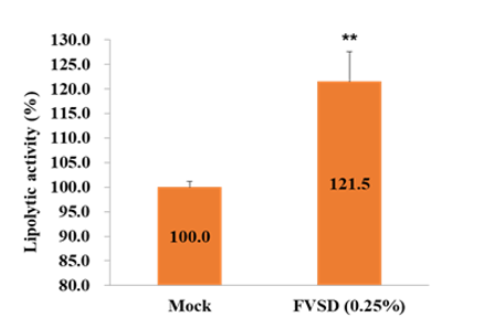
Figure 1: FVSD treatment can increase the lipolytic activity. Glycerol content was measured on OP9 cells after FVSD treatment. (n = 3; mean value ± S.D.) (*p < 0.05; **, p < 0.01; ***, p < 0.001).
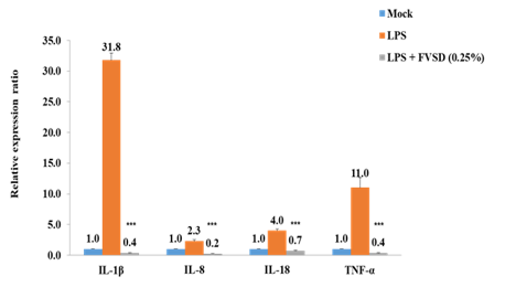
Figure 2: FVSD treatment can reverse LPS-induced inflammation related genes induction on C2BBel. (n = 3; mean value ± S.D.) (*p < 0.05; **, p < 0.01; ***, p < 0.001).
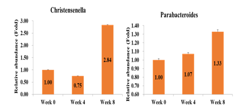
Figure 3: FVSD can ameliorate obesity through regulating the in-testinal microbiota of obese subjects. Christensenella and Parabac teroides abundance were increased after FVSD administration by NGS analysis. (n = 25; mean value ± S.D.)
FVSD had Anti-Obesity Effect in Obesity Subjects
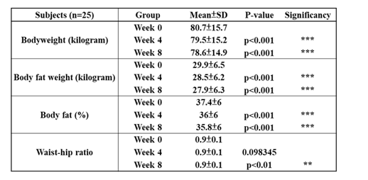
To further explore the weight management efficacy of FVSD in a clinical trial. A total of 25 obesity subjects were recruited and administrated FVSD daily for 8 weeks and examined at weeks 0, 4, 8 weeks. Table 1 showed the results of anthropometric measure-ments before and after the study. After 4 weeks of FVSD inter-vention, the result of body weight, body fat weight, and body fat performed significantly improvements in comparison with week 0. Follow up to the week 8 result, the body weight, body fat weight, body fat was significantly improved. Besides, the waist-hip ratio (WHR) also performed significantly improved at week 8 interven-tion.
Table 1: Results of anthropometric measurements. (N= 25; mean value ± SD). *p < 0.05; **, p < 0.01; ***, p < 0.001 (sig-nificant difference between week 0 and week 4 or week 8 after FVSD administration)
FVSD Improved Fatty Liver in Obese Subjects
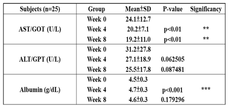
To examine whether the FVSD administration decreased fatty liver, obesity subjects were examined aspartate aminotransferase (AST), alanine aminotransferase (ALT) and albumin. ALT and AST was known as indicator enzymes of the presence of liver dis¬ease. Elevated AST or ALT was associated with fatty liver [16]. When liver cells were damaged, the albumin content decreased [17]. The table 2 showed the results of biochemical analysis before and after FVSD intervention. As a result, both AST and ALT were performing a decreasing trend. In static results, the AST was decreased significantly, but ALT was not, suggesting FVSD im¬proved fatty liver in obese subjects.
Table 2: Results of biochemical analyses. (N= 25; mean val¬ue ± SD). *p < 0.05; **, p < 0.01; ***, p < 0.001 (significant difference between week 0 and week 4 or week 8 after FVSD administration)
FVSD Improved Intestinal Microbiota in Obese Subjects
To investigate whether FVSD regulated intestinal microbiota in obese subjects, we examined microbiota from subjects’ feces by next generation sequencing (NGS). The Christensenellaceae and Parabacteroides on decreasing weight gain, hyperglycemia, and hepatic steatosis in high-fat diet (HFD)-fed mice [18]. In figure 3, both of Christensenellaceae and Parabacteroides related abundance were increased after FVSD intervention. The Christensenellaceae was increased by 2.84 fold and the Parabacteroides was increased by 1.29 fold. The results suggested that FVSD could ameliorate obesity through regulating the intestinal microbiota of subjects.
Discussion
In this study, we found that FVSD increased the lipolytic activity and decreased inflammation in vitro, as well as, FVSD decreased body weight, improved fatty liver, and regulated intestinal micro-biota in clinical trial. FVSD contained two major bacteria, includ-ing TCI604 (Bifidobacterium lactis) and TCI507 (Lactobacillus plantarum). Studies showed that Lactobacteria and Bifidobacteria can inhibit weight gain in high fat diet induced mice [19]. Bifidobacterium longum exhibited a more significant effect in lowering serum total cholesterol [20]. Lactobacteria can inhibit cholesterol synthase, thereby reducing the production of cholesterol, and Bifidobacteria promote the elimination of cholesterol in feces [21]. Consistent with our results, FVSD can increase the fatty acid breakdown. Probiotic treated-mice improved glucose-tolerance, and decreased inflammatory cytokines [22]. Lactobacteria was shown to adhere to intestinal epithelial cell line and had anti-in¬flammation in vitro. Bifidobacteria and Lactobacteria can produce superoxide dismutase (SOD), reduced inflammatory cytokines IL-1β, TNF-α, and improved intestinal conditions [23]. Bifidobacteria interfere with pro-inflammatory signals upstream of the pathway of NF-κB activation for LPS and TNF-α [24]. Consistent with our results, FVSD decreased the inflammation-related gene induction by LPS.
Probiotic supplement resulted in signifcant reductions in body weight, BMI, waist circumference and waist-to-height ratio [25]. L. plantarum and L. gasseri reduced the body weight, and choles-terol level. The Lactobacteria was showed lowering effects on ab-dominal adiposity, body weight, suggesting its beneficial influence on metabolic disorders [26]. Multi-strain probiotic contained Lactobacteria and Bifidobacteria can reduce BMI, body weight and WHR in overweight/obese adults [27]. Consistent with our results, FVSD decreased body weight, body fat weight, and WHR. Some probiotics can inhibit TNFα and enhance adiponectin to improve the intestinal microbiota, leading to regulation of blood sugar, lipid metabolism and protection of the liver [28]. Bifidobacterium, Lactobacillus, had shown beneficial effects in rodent models of nonalcoholic fatty liver disease (NAFLD). Some studies indicated that activating the Nrf2/ARE pathway had a hepato-protective effect [29]. L. plantarum caused the activation of Nrf2 in liver, thus alleviating oxidized oil induced hepatic injury in mice [30]. Short-term oral supplementation with B. bifidum and L. plantarum can decrease AST, ALT activity, and regulated bowel flora [31]. Consistent with our results, FVSD decreased AST, ALT expression.
The gut microbiota appeared to play a role in the pathogenesis of obesity and associated diseases. The relative abundance of Christensenellaceae in the human gut is inversely related to host BMI [32]. Parabacteroides reduced obesity was associated with increased adipose tissue thermogenesis, and reduced inflammation and insulin resistance in HFD-fed mice [8]. In the oral Bifidobacterium treatment group, Christensenellaceae continued to increase [33]. The intake of Bifidobacterium, Lactobacillus can increase Christensenellaceae or Parabacteroides [33]. Consistent with our results, FVSD increased Christensenellaceae or Parabacteroides. In addition, we also found that FVSD decreased Eggerthella, Clos-tridium, Acinetobacter (data not shown).The possible mechanism was that branched chain fatty acids (BCFA), especially isobutyric acid and isovaleric acid, affected human fat cells by inhibiting li-pogenesis and glucose metabolism [33].
Conclusion
This study showed that solid drinks containing probiotics Bifido¬bacterium, Lactobacillus supplementation could increase the lip¬olysis process and reduce intestinal inflammation. In clinical trials, it was found that obese subjects had a significant reduction in body weight, body fat, and waist-to-hip ratio after taking it for 8 weeks. And it had been observed that it can improve liver function indica¬tors and reduced the fatty liver. It was further discovered that after 8 weeks of taking it, it can increase the beneficial bacteria in the intestines and improved fat metabolism, such as increasing Christensenellaceae, Parabacteroide. Although the detailed mechanism was not yet clear, more studies were needed to confirm, but this study proposed a new weight reduction strategy, using the combi¬nation of probiotics to regulate the intestinal flora and achieved the anti-obesity effects [34, 35].
Acknowledgement
Thank TCI gene group for their full technical and funding sup¬ports.References
- Jaacks LM, Vandevijvere S, Pan A, McGowan CJ, Wallace C, et al. (2019) The obesity transition: stages of the global epidemic. Lancet Diabetes Endocrinol 7: 231-240.
- Sivamaruthi BS, Kesika P, Suganthy N, Chayasut C (2019) A Review on Role of Microbiome in Obesity and Antiobesity Properties of Probiotic Supplements. Biomed Res Int 2019: 3291367.
- Rinninella E, Cintoni M, Raoul P, Lopetuso LR, Scaldaferri F, et al. (2019) Food Components and Dietary Habits: Keys for a Healthy Gut Microbiota Composition. Nutrients 11: 2393.
- Stanislawski MA, Dabelea D, Lange LA, Wagner BD, Lozu-pone CA (2019) Gut microbiota phenotypes of obesity. NPJ Biofilms Microbiomes 5: 18.
- Fontane L, Benaiges D, Goday A, Llaurado G, Pedro Botet J (2018) Influence of the microbiota and probiotics in obesity. Clin Investig Arterioscler 30: 271-279.
- Ubeda C, Bucci V, Caballero S, Djukovic A, Toussaint NC, et al. (2013) Intestinal microbiota containing Barnesiella species cures ancomycin-resistant Enterococcus faecium colonization. Infect Immun 81: 965-973.
- Jandhyala SM, Talukdar R, Subramanyam C, Vuyyuru H, Sasikala M, et al. (2015) Role of the normal gut microbiota. World J Gastroenterol 21: 8787-8803.
- Wu TR, Lin CS, Chang CJ, Lin TL, Martel J, et al. (2019) Gut commensal Parabacteroides goldsteinii plays a predominant role in the anti-obesity effects of polysaccharides isolated from Hirsutella sinensis. Gut 68: 248-262.
- Wang J, Tang H, Zhang C, Zhao Y, Derrien M, et al. (2015) Modulation of gut microbiota during probiotic-mediated attenuation of metabolic syndrome in high fat diet-fed mice. ISME J 9: 1-15.
- Ruscica M, Pavanello C, Gandini S, Macchi C, Botta M, et al. (2019) Nutraceutical approach for the management of cardiovascular risk - a combination containing the probiotic Bifidobacterium longum BB536 and red yeast rice extract: results from a randomized, double-blind, placebo-controlled study. Nutr J 18: 13.
- Chen J, Wang R, Li XF, Wang RL (2012) Bifidobacterium ad-olescentis supplementation ameliorates visceral fat accumulation and insulin sensitivity in an experimental model of the metabolic syndrome. Br J Nutr 107: 1429-1434.
- Liu YW, Liong MT, Tsai YC (2018) New perspectives of Lactobacillus plantarum as a probiotic: The gut-heart-brain axis. J Microbiol 56: 601-613.
- Lee E, Jung SR, Lee SY, Lee NK, Paik HD, et al. (2018) Lactobacillus plantarum Strain Ln4 Attenuates Diet-Induced Obesity, Insulin Resistance, and Changes in Hepatic mRNA Levels Associated with Glucose and Lipid Metabolism. Nutrients 10: 643.
- Li Z, Jin H, Oh SY, Ji GE (2016) Anti-obese effects of two Lactobacilli and two Bifidobacteria on ICR mice fed on a high fat diet. Biochem Biophys Res Commun 480: 222-227.
- Rozhkova TA, Titov VN, Amelyushkina VA, Kaba SI, Kukhartchuk VV (2015) [the Lipolysis in Phylogenetically Early Lipoproteins of Low Density and More Later Lipoproteins of Very Low Density: Function and Diagnostic Value of Apoe and Apoc-Iii]. Klin Lab Diagn 60: 4-14.
- Kwo PY, Cohen SM, Lim JK (2017) ACG Clinical Guideline: Evaluation of Abnormal Liver Chemistries. Am J Gastroenterol 112: 18-35.
- Seidkhani Nahal A, Allameh A, Soleimani M (2019) Antioxidant and reactive oxygen species scavenging properties of cellular albumin in HepG2 cells is mediated by the glutathione redox system. Biotechnol Appl Biochem 66: 163-171.
- Wang, K, Liao M, Zhou N, Bao L, Ma K, et al. (2019) Parabacteroides distasonis Alleviates Obesity and Metabolic Dysfunctions via Production of Succinate and Secondary Bile Acids. Cell Rep 26: 222-235.
- Yin YN, Yu QF, Fu N, Liu XW, Lu FG (2010) Effects of four Bifidobacteria on obesity in high-fat diet induced rats. World J Gastroenterol 16: 3394-3401.
- An HM, Park SY, Lee DK, Kim JR, Cha MK, et al. (2011) Antiobesity and lipid-lowering effects of Bifidobacterium spp. in high fat diet-induced obese rats. Lipids in Health and Disease 10: 116.
- Beena A, Prasad V (1997) Effect of yogurt and bifidus yogurt fortified with skim milk powder, condensed whey and lactose-hydrolysed condensed whey on serum cholesterol and triacylglycerol levels in rats. J Dairy Res 64: 453-457.
- Cani PD, Neyrinck AM, Fava F, Knauf C, Burcelin RG, et al. (2007) Selective increases of bifidobacteria in gut microflo-raimprove high-fat-diet-induced diabetes in mice through amechanism associated with endotoxaemia. Diabetologia 50: 2374-2383.
- Han W, Fioramonti J (2008) Anti-inflammatory properties of lactic acid bacteria producing superoxide dismutase. Am J Physiol Gastrointest Liver Physiol 294: 353-354.
- Riedel CU, Foata F, Philippe D, Adolfsson O, Eikmanns BJ, et al. (2006) Anti-inflammatory effects of bifidobacteria by inhibition of LPS-induced NF-kappaB activation. World J Gastroenterol 12: 3729-3735.
- Michael DR, Jack AA, Masetti G, Davies TS, Loxley KE, et al. (2020) A randomised controlled study shows supplementation of overweight and obese adults with lactobacilli and bifidobacteria reduces bodyweight and improves well-being. Scientific Reports 10: 4183.
- Kadooka Y, Sato M, Imaizumi K, Ogawa A, Ikuyama K, et al. (2010) Regulation of abdominal adiposity by probiotics (Lactobacillus gasseri SBT2055) in adults with obese tendencies in a randomized controlled trial. Eur J Clin Nutr 64: 636-643.
- Sudha MR, Ahire JJ, Jayanthi N, Tripathi A, Nanal S (2019) Effect of multi-strain probiotic (UB0316) in weight management in overweight/obese adults: a 12-week double blind, randomised, placebo-controlled study. Benef Microbes 10: 855-866.
- Wang W, Shi LP, Shi L, Xu L (2018) Efficacy of probiotics on the treatment of non-alcoholic fatty liver disease. Zhonghua Nei Ke Za Zhi 57: 101-106.
- Long M, Liu Y, Cao Y, Wang N, Dang M, et al. (2016) Proan-thocyanidins Attenuation of Chronic Lead-Induced Liver Oxidative Damage in Kunming Mice via the Nrf2/ARE Pathway. Nutrients 8: 656.
- Lin X, Xia Y, Wang G, Yang Y, Xiong Z, et al. (2018) Lactic Acid Bacteria With Antioxidant Activities Alleviating Oxidized Oil Induced Hepatic Injury in Mice. Front Microbiol 9: 2684.
- Kirpich IA, Solovieva NV, Leikhter SN, Shidakova NA, Leb-edeva OV, et al. (2008) Probiotics restore bowel flora and improve liver enzymes in human alcohol-induced liver injury: a pilot study. Alcohol 42: 675-682.
- Waters JL, Ley RE (2019) The human gut bacteria Christensenellaceae are widespread, heritable, and associated with health. BMC Biology 17: 83.
- Hibberd AA, Yde CC, Ziegler ML, Honore AH, Saarinen MT, et al. (2019). Probiotic or synbiotic alters the gut microbiota and metabolism in a randomised controlled trial of weight management in overweight adults. Benef Microbes 10: 121-135.
- Chang CS, Ruan JW, Kao CY (2019) An overview of microbiome based strategies on anti-obesity. Kaohsiung J Med Sci 35: 7-16.
- Wang T, Yan H, Lu Y, Li X, Wang X, et al. (2020) Anti-obesity effect of Lactobacillus rhamnosus LS-8 and Lactobacillus crustorum MN047 on high-fat and high-fructose diet mice base on inflammatory response alleviation and gut microbiota regulation. Eur J Nutr 59: 2709-2728.