

Archives of Epidemiology & Public Health Research(AEPHR)
ISSN: 2833-4353 | DOI: 10.33140/AEPHR

Impact Factor: 1.98


Research Article - (2026) Volume 5, Issue 2
Prevalence and Genetic Diversity of Human Immunodeficiency Virus Type 1 (HIV-1) Among Blood Donors in the Service Area of the Santos/Sp Blood Center
2Bachelor’s degree in Medicine from the Centro Universitário Lusíada (1988), Master’s degree in Medicine (Preventive Medicine) from the University of São Paulo (1996), and Ph.D. in Infectious Diseases from the Federal University of São Paulo (2001). Infectious disease specialist at GUILHERME ÁLVARO HOSPITAL, assistant professor at São Judas Tadeu University, professor at Santa Cecília University, and associate professor at Lusíada University Center, Brazil
3Ph.D. in Infectious Diseases from the School of Medicine, at the University of São Paulo and completed a postdoctoral fellowship at the Federal University of São Paulo. She is currently a professor at the São Paulo Institute for Health Research and Education (IPESSP), Brazil
4Bachelor’s degree in Biological Sciences from the Catholic University of Santos, UNISANTOS (1990), a Master’s degree in Health Sciences from Lusíada University Center-UNILUS (2004), and a Ph.D. in Infectious Diseases from the Federal University of São Paulo—UNIFESP (2009) in the field of Infectious Diseases (Retrovirology Laboratory), Brazil
5Bachelor’s degree in Biological Sciences from the Federal University of Viçosa, (1997), Master’s degree (1999), Ph.D. (2003), and postdoctoral fellowship (2006) in Basic Sciences (Infectious and Parasitic Diseases) from the Federal University of São Paulo, Brazil
Received Date: Aug 04, 2026 / Accepted Date: Sep 08, 2026 / Published Date: Sep 17, 2026
Copyright: ©Â©2026 Rosane Rezende de Souza Giuliani, et al. This is an open-access article distributed under the terms of the Creative Commons Attribution License, which permits unrestricted use, distribution, and reproduction in any medium, provided the original author and source are credited.
Citation: de Souza Giuliani, R. R., Caseiro, M. M., Vasconcelos, S. C., Gagliani, L. H., de Sá Filho, D. J. (2026). Prevalence and Genetic Diversity of Human Immunodeficiency Virus Type 1 (HIV-1) Among Blood Donors in the Service Area of the Santos/Sp Blood Center. Arch Epidemiol Pub Health Res, 5(2), 01-17.
Abstract
Introduction: The HIV-1 epidemic in the Baixada Santista region is complex due to the presence of different subtypes, recombinants, and mutations conferring resistance to antiretrovirals. Therefore, the study of samples from newly diagnosed individuals allows us to characterize the viruses currently circulating in the local transmission network.
Objectives: To determine the prevalence and genetic diversity of HIV-1 among blood donors in the Baixada Santista region, whose serological testing is performed at the Hemonúcleo de Santos.
Study Population and Methods: To calculate prevalence, we assessed the number of HIV-positive samples, confirmed by Western blot, relative to the number of donations during the period from January2009 a to May 2011. To analyze viral genetic diversity, we collected serum and/or plasma samples from the donors. Viral RNA was extracted, and reverse transcription was performed to generate cDNA. This material was used for PCR amplification of the HIV-1 pol gene to analyze subtypes, recombinants, and the presence of primary antiretroviral resistance mutations using bioinformatics tools.
Results: During the analyzed period, a total of 65,428 donations were recorded. Of these, 49 samples were identified as HIV-positive, with a prevalence of 0.07% during this period. Of the total 49 HIV-positive donors, it was only possible to characterize the viral genotype of 9 samples due to the inadequacy of the stored biological material for molecular biology techniques. Of these 9 sequenced samples, 7 belong to subtype B, 1 to subtype F, and 1 sample was identified as a B/F recombinant. Regarding primary resistance mutations, 6 samples exhibited isolated accessory mutations, such as: K20R, A71V, and M36I in the protease gene, and A62V, V118I, and T215I in the reverse transcriptase gene. With the exception of the T215I mutation, which causes a low level of resistance to zidovudine (AZT) and stavudine (D4T), these mutations alone do not cause complete resistance to any antiretroviral drug.
Final Considerations: The identified prevalence of HIV-positive donors was 0.07%. Despite the small number of sequenced samples, subtype B was found to predominate, and mutations associated with HIV-1 resistance to antiretrovirals were frequently detected. This indicates the need for more comprehensive studies to investigate primary HIV-1 resistance in the Baixada Santista region.
Keywords
HIV-1, Genetic Diversity, HIV-1 Subtypes, Antiretroviral Resistance, Blood Donors, Molecular Epidemiology, Baixada Santista
Introduction
Ever since the transmission of AIDS through blood transfusions, blood components, and blood products was well established, the Ministry of Health has made it a priority to increase control over the entire “blood cycle” and ensure the safety of transfusions in our country.It is well known that, today, blood therapy is the most heavily regulated medical specialty in Brazil. New Resolutions (RDCs) and Standards are continually published and must be strictly adhered to by all blood therapy services, which are periodically inspected by municipal and state health surveillance agencies.
Currently, blood therapy activities in Brazil are regulated by Ministry of Health Ordinance No. 1353, published on June 13, 2011, which establishes the Technical Regulations for Blood Therapy Procedures. These regulations must be followed in conjunction with compliance with the health requirements for the operation of blood therapy services defined by ANVISA (National Health Surveillance Agency).
The so-called Hemorede of the State of São Paulo is one of the best-organized in the country. It currently consists of 6 blood centers and approximately 23 regional blood units.
The Santos Hematology and Blood Therapy Center (Santos Blood Center) is part of the São Paulo State Blood Network (HEMOREDE). It is located in the city of Santos and its mission is to ensure transfusion safety and access to care for benign hematological diseases and oncohematological diseases in the region known as the Baixada Santista Metropolitan Region, which comprises nine municipalities(Peruíbe, Itanhaém, Mongaguá, Praia Grande, São Vicente, Santos, Cubatão, Guarujá, and Bertioga) and has an estimated population of approximately 1,600,000 inhabitants, representing 3.8% of the state’s population.
Because it is home to the largest port in Latin America (the Port of Santos), one of the country’s largest petrochemical hubs (Cubatão), and a region with a high volume of tourists due to its coastal location, the Baixada Santista has always presented a very unique profile regarding AIDS, which is why significant studies of international renown have been and continue to be produced by local researchers.
The Baixada Santista currently has fifteen blood services, only two of which are private; all others are public or nonprofit facilities, and the blood collected there undergoes mandatory serological screening-as required by law-at the serology department of the Santos Blood Center. Tests are conducted to detect syphilis, Chagas disease, HTLV-1 and -2, hepatitis C, hepatitis B (HBsAg and anti-HBc), and two tests for HIV-1.
Any blood donor who tests positive for any of the parameters tested is counseled by a medical professional from the Service and referred to a referral center for follow-up and treatment, when necessary.
In accordance with Technical Standards, donor serum samples are aliquoted and stored in the Blood Center’s own serum bank.
The profile of donors in the region was found to be similar to that of other regions in Brazil: the majority of donors are male, the age group of most donors is between 18 and 29 years old, and repeat donors account for only about 20% of the total.
The high degree of viral diversity of the Human Immunodeficiency Virus (HIV-1), caused primarily by the reverse transcription stage, favors the emergence of variants, which may result from three interrelated factors: the number of mutations produced in each replication cycle, the number of replication cycles over time, and the selective advantage or disadvantage of the properties acquired by viral variants. The study of HIV variability is essential for understanding the infection and, above all, the response to antiretroviral (ARV) drugs, as the virus is capable of adapting to the host’s immune response and drug therapy.
The success of antiretroviral treatment has increased the life expectancy and quality of life of infected individuals. However, this success can be compromised by the development of resistance to ARVs, leading to treatment failure.
Currently, many patients taking potent combinations of antiretrovirals (also known as HAART—highly active antiretroviral therapy) do not maintain plasma viral load at undetectable levels. As a consequence of basal replication, mutant viral strains resistant to antiretrovirals emerge [1,2]. Patients with antiretroviral-resistant viruses typically progress to treatment failure in subsequent regimens, particularly because cross-resistance is very common.
There are several factors related to the development of mutations in HIV-1. A nd poor treatment adherence (“compliance”) is the most common of these. Pharmacological causes, such as changes in absorption, rapid elimination, insufficient penetration into certain anatomical reservoirs of HIV, and drug interactions are also involved. Viral factors or those related to the host’s immune system may also be involved [3]. The presence of mutations may lead to genotypic or phenotypic viral resistance. This resistance can be primary or secondary. Primary resistance is present even before the use of medications, while secondary resistance-and the consequent emergence of mutations-results from the selective pressure exerted by antiretroviral therapy (ART) [4].
Studies have shown that the level of transmitted resistance is directly related to the replicative capacity of the resistant strains involved and increases as access to treatment increases [5-7].
Determining the prevalence of primary resistance is of great importance in monitoring the molecular epidemiology of HIV-1 and can serve as a guide for initial treatment regimens, pre- and post-exposure prophylaxis, and the prevention of vertical transmission. It aids in the development of preventive measures such as vaccines and microbicides and, ultimately, helps in assessing the validity of efforts to perform resistance testing prior to initial treatment [8].
The Etiological Agent
The Retrovirus Family
Retroviruses comprise a large family of viruses, primarily found in vertebrates, but which can also occasionally be found in insects and mollusks. They have a genome ranging from 7 to 12 Kb, containing RNA as genetic material, and use the enzyme reverse transcriptase (RT) for transcription into DNA. The DNA is subsequently integrated into the host cell’s genome via proviral DNA. As a result of this integration, the host’s permanent cells lead to a persistent infection.
This family of viruses is associated with a variety of diseases, such as slow- or fast-progressing cancers, neurological disorders, and immunodeficiencies, which may also be associated with persistent viremia without disease. In addition, retroviruses or parts of them can be found as endogenous genetic elements in humans and other hosts.
Until the early 1980s, retroviruses were of interest only as models for studying cancer in animals; they were not yet considered to be of interest as human pathogens. The discovery of human T-cell leukemia viruses (HTLVs) in 1980 and 1982 by and, subsequently, the virus causing Acquired Immunodeficiency Syndrome (AIDS) by generated enormous scientific interest in this class of viruses [9-11]. Retroviruses have evolved over long periods of time within their hosts and among primate lentiviruses. Certainly, several transfers between humans and primates have occurred in the course of this evolution, as well as recombinations among related retroviruses [12-15].
Recently, a retrovirus was described that was repeatedly isolated from a patient with multiple sclerosis and that showed homology to a human endogenous retrovirus, ERV-9 [16]. Although its role as an etiological agent in this disease remains to be proven, it is likely that the involvement of a retrovirus in other human diseases may yet be discovered.
Another important aspect of retrovirus research concerns their potential as tools for gene transfer [17]. Because these viruses have the ability to integrate in their proviral form into the host cell’s genome, retroviruses are capable of transferring permanent genes to the target cell and thus function as vectors for a variety of gene therapies.
Despite a variety of host species and different clinical manifestations, retroviruses are similar in their structure and genomic organization
The virions are enveloped, measure approximately 80 to 100 nm in diameter, and bud from the cytoplasmic membrane of cells. A specific viral enzyme, reverse transcriptase, which transcribes viral RNA into DNA, gave this family of viruses its name. However, reverse transcription is also found in other viruses (e.g., hepatitis B virus) and in cellular transposons; thus, it is not a property exclusive to retroviruses.
Classification
Retroviruses are classified into six distinct groups based on the genetic relationship of their reverse transcriptase genes see Table 1 [18,19].

Table 1: Classification of Retroviruses (modified from Unger R.E., 2000)
Spumaviruses, which were once thought to belong to the retrovirus family, have recently been shown to have DNA rather than RNA as their genetic material. In addition, they share several properties with hepadnaviruses (e.g., hepatitis B virus) [20]. These viruses also reverse-transcribe their RNA within the cell and are released as DNA-containing particles; however, morphologically, spumaviruses fit well within the retrovirus family. Retroviruses can also be classified according to the shape of their virions (Types A–D; Figure 2). These particles were first observed as immature intracellular forms of murine mammary tumor virus (MMTV), and the term still refers to strict intracellular structures consisting of immature forms of Type B and Type D viruses. These particles are virtually indistinguishable; furthermore, they are found in cell lines derived from a large number of rodent species and are products of a large number of endogenous provirus-like particles.
Type-C particles constitute the majority of the mammalian and avian viruses studied. Complete intracellular forms are rarely observed, but we can visualize crescent-shaped structures protruding from the surface. Like Type-B particles, newly released forms have an empty nucleocapsid and must mature to produce a central, electron-dense spherical structure.
Unlike Type-B particles, Type-C particles have barely visible surface projections. Type-D particles resemble those of type B; they have a complete intracellular nucleocapsid and an acentric core, but with prominent surface projections. Bovine leukemia virus and HTLV resemble type C in their budding pattern but differ in the appearance of their envelope.
Lentiviruses, including human immunodeficiency virus (HIV), caprine encephalitis-arthritis virus (CEAV), equine infectious anemia virus (EIAV), sheep maedi visna virus (MMV), and feline immunodeficiency virus (FIV), as well as bovine immunodeficiency virus (BIV), also bud similarly to type-C viruses, but the mature virion has a cone-shaped nucleocapsid.
Spumaviruses behave similarly to type-B viruses and preform their nucleocapsid within the cell, which condenses into its mature form after being released as a virion.
Structure of HIV
When observed under an electron microscope, HIV-1 exhibits the typical characteristics of a lentivirus; these infectious particles (virions) are spherical, with an average diameter of 100 nm, and feature a central region—the core or nucleocapsid—of cylindrical shape, surrounded by an envelope known as the viral capsid, which is nothing more than a lipoglycoprotein envelope derived from the host cell’s membrane. Inside the nucleocapsid lies the viral genome, consisting of two identical molecules of single-stranded, non-segmented, positive-sense ribonucleic acid (RNA). The inner portion of the viral membrane is surrounded by a meristalized core p17 (MA) protein, which provides the matrix for the viral structure and is vital for the integrity of the virion.
This protein appears to be required for the incorporation of Env proteins into the mature virion. Associated with the core are the Vif and Nef proteins [21,22].
It is estimated that 7 to 20 Vif molecules are produced for each virion. The products of the viral accessory gene vpr (Vpx in the case of HIV-2) can also be found within the virion and, more likely, outside the core; more recently, it has been demonstrated that Tat must be located within the virion; all of these viral proteins within the viral particle suggest that they play an important role in the initial events of HIV infection.
Characteristically, 72 trimeric or tetrameric projections of envelope glycoproteins are observed on the surface of the virus. Recent electron microscopy and X-ray crystallography data on the ectodomain of gp41 suggest that the envelope proteins are organized as a trimer.
The envelope structures are derived from the 160-kDa precursor, gp160, which is cleaved inside the cell—likely within the Golgi complex—into gp120, an outer envelope surface protein, and gp41, a transmembrane protein.
These proteins are then transported to the cell surface, where part of the central and N-terminal regions of gp41 are expressed on the outer surface of the virion. The central region of the transmembrane protein binds to the outer gp120 in a noncovalent manner, primarily to two hydrophobic regions at the N- and C-termini of gp120. Studies suggest that the protrusions surrounding the envelope region, as well as other regions of the envelope-including V1/V2 and the C2 and C3 domains of gp120—help stabilize this association. In general, the virion contains about 100 times more Gag p24 protein than gp120 in its envelope, and 10 times more p24 than in the polymerase molecule [23].
The gp120 on the virion’s surface contains the binding sites for cellular receptors and the primary neutralization domains. However, other external portions of the virion, such as gp41 and parts of gp17, are also susceptible to neutralizing antibodies. These antibodies against p17 may indicate that this core protein protrudes from the virion surface onto the cell surface.
These antibodies, however, may represent a cross-reaction with anti-gp41 antibodies [24].
Genomic Organization
The retrovirus genome ranges from 7,000 to 12,300 nucleotides; the HIV-1 genome is approximately 9.8 Kb in size and is unique among viruses in its physical organization, mode of synthesis, and replication pathway. It is a virus with a diploid genome consisting of identical RNA molecules linked by a 5’ terminal region. The RNA genome is reverse-transcribed to form a DNA provirus, which in turn serves as a template for transcription into RNA by cellular enzymes.
Like all other retroviruses, HIV-1 has three genes that encode structural proteins (Gag, Pol, and Env) and several regulatory and accessory genes (Vif, Vpr, Tat, Ver, Nef, and Vpu), the latter of which is present only in HIV-1 and SIVcpz (Figure 1).
The first transcript of HIV-1 is an mRNA that is translated to form the Gag and Pol proteins. Through proteolytic cleavage, the Gag p55 precursor gives rise to the small proteins p24, p17, p9, and p6.

Figure 1: Genome Organization and Representation of the Major HIV-1 Proteins
The Pol precursor is cleaved into products consisting of the viral enzymes reverse transcriptase (RT), protease, and integrase. The Gag and Gag-Pol products are synthesized in a 20:1 ratio. The coordination of these mRNA splicing and unsplicing events appears to be determined by the rev gene, which is itself the product of multiple mRNA splicing events.
An important regulatory protein in these events is Tat, a transactivation protein that plays the largest role in regulating HIV replication.
Another viral regulatory protein, Rev (Regulator of viral protein expression), interacts with the “cis-acting RNA loop”-a structure known as the Rev response element—located in the viral envelope mRNA. This interaction involves cellular proteins and Rev multimers and allows the mRNA to be released from the nucleus into the cytoplasm, where it gives rise to all the proteins necessary for the production of viral progeny. In this way, Rev appears to affect the function of spliceosomes. Tat and Rev are RNA-binding proteins that interact with cellular factors to optimize intracellular activities.
Another protein, Nef (Negative Factor), appears to have a variety of potential functions, including cell activation and perhaps downregulation of viral expression. Its activity, however, is pleiotropic and appears to require interaction with other viral proteins involved in signal transduction and cell activation. How this effect influences HIV replication is not yet fully understood. In addition, the products of the accessory genes—Vif, Vpr, and Vpu/ Vpx—interfere with events such as binding and release, as well as with the production of infectious virus during the infectious cycle. Some studies suggest that the accessory genes Vpr, Vpu, and Nef may be more important for HIV-1 replication in macrophages than in CD4+ lymphocytes.
Several other characteristics described in various reviews reveal multiple interrelationships in the regulation of this virus’s gene expression, leading to high or low HIV-1 replication during the establishment of the latent state [25,26]. The entire coding process is based on three main enzymes, transcribed from the pol gene, which function at different stages during the HIV-1 replication cycle.
These enzymes are thus the primary targets used in antiretroviral therapy; they are: RNA-dependent DNA polymerase (which is nothing more than ribonuclease H), known as reverse transcriptase (RT), which is a heterodimer composed of two protein subunits, p66 and p51. It possesses two catalytic activities: the first acts early in the replication process to form a double-stranded DNA copy (cDNA), or proviral DNA; the second is ribonuclease H (RNase H) activity, which degrades the RNA component of RNA-DNA hybrid molecules. When the proviral DNA is transported into the host cell nucleus, it is incorporated into the chromosomal DNA via another product of the Pol gene, namely integrase; finally, a last enzyme called protease (p10) acts during the maturation of viral particles, whether on the surface of the host cell or in the released virion, by cleaving the Gag and Gag-Pol polyproteins, transforming them into functional proteins [27].
Origin, Evolution, and Diversity
Various lines of evidence have been used to support zoonotic transmission of lentiviruses from other primates to humans, among them are the similarity in viral genomic organization; second, the phylogenetic relationship; third, the prevalence of infection in the natural host; fourth, the geographic overlap between primates and humans; and fifth, the plausibility of transmission routes [28].
For HIV-2, a virus called the Sooty mangabey simian immunodeficiency virus (SIVsm)-which is genomically indistinguishable and closely related phylogenetically-has been found in large numbers of sooty mangabeys whose natural habitat coincides with the epicenter of the HIV-2 epidemic [29-31].
Very close contact between sooty mangabeys and humans occurs in this region, where the former are hunted for food or kept as pets [32]. It is believed that at least six independent insertions or transmissions must have occurred from SIVsm to humans [31,32]. However, the origin of HIV-1 was much less certain [28].
HIV-1 is closer, both in sequence and genomic organization, to the viruses found in chimpanzees (SIVcpz) (Figure 2), but an apparent low prevalence of SIVcpz infection in wild animals, combined with the presence of these chimpanzees in geographic regions of Africa where AIDS was not initially recognized, has cast doubt on the chimpanzee as a host and reservoir for HIV-1 [33,34]. Undoubtedly, another, as yet unidentified, primate species has been suggested as a possible natural host for SIVcpz and HIV-1 [35].
Definitive evidence that the origin of HIV-1 infection in humans occurred through cross-species transmission from chimpanzees was described by in which these authors demonstrate that all HIV-1 strains are phylogenetically related to SIVcpz strains that infect the chimpanzee Pan troglodytes, a primate whose habitat overlaps with the areas where the HIV-1 M, N, and O groups are endemic [14,34,36-38].
These same authors have shown, based on the evolutionary history of mitochondrial DNA in chimpanzee species, that chimpanzees are a natural reservoir of SIVcpz, dating back to the divergence between the two host species, *Pan troglodytes troglodytes* and *Pan troglodytes schweinfurthii*, which must have occurred hundreds of years ago; they also show that in West Equatorial Africa, chimpanzees are captured for food, thus representing a plausible route for the zoonotic transmission of SIVcpz to humans; Meanwhile, the phylogenetic position of HIV-1 subtypes M, N, and O within the HIV-1/SIVcpz radiation suggests that these three subtypes were introduced via zoonotic transmission at separate times from SIVcpz in Pan troglodytes to humans; finally, they show that another possible explanation for the origin of subtypes M, N, and O—as resulting from diversification within the human population or acquisition of the virus from another primate species—would be inconsistent with the phylogenetic data or implausible.
Recently, following extensive research published in a book (The River), the author attempted to link the introduction of HIV-1 infection into humans to the polio vaccination trials conducted in Africa-the “Chat” vaccine, which was tested between 1957 and 1960 on more than 1 million Africans; however, several claims made by HOOPER would need to be proven: 1) to show that chimpanzee kidneys were used to produce the “Chat” vaccine; 2) that these kidneys were contaminated with the SIVcpz virus; 3) that no cases of AIDS (HIV-1) occurred prior to the vaccine trials; 4) to prove that the first cases of HIV-1, group M, occurred in the locations where the “Chat” vaccines were tested-Congo and Burundi; 5) that the outbreaks caused by subtypes O and N coincided with French vaccine trials conducted in Gabon and Cameroon; and finally, to prove that the HIV-2 epidemic coincides with the areas of Guinea-Bissau where vaccinations were carried out by the Portuguese in the 1960s [39].
Virtually all of this evidence has been refuted, and some authors have demonstrated, using a new method of temporal molecular analysis called “Site Stripping for Clock Detection” (SSCD)-which allows for the selection of nucleotide sites exhibiting the same rate of variation across different lineages-that the origin of Group M radiation occurred in the 1930s and that the emergence of the HIV-1 ancestor related to SIVcpz occurred around the 17th century [14,40,41].
HIV-1 Subtypes
Strains of the Human Immunodeficiency Virus type 1 (HIV-1) circulating globally exhibit an extraordinary degree of genetic diversity, which can influence aspects of their biology such as infectivity, transmissibility, and immunogenicity. Sequences derived from these HIV-1 strains have historically been classified into groups and subtypes based on their phylogenetic relationships. However, the increasing complexity of new HIV-1 sequences has highlighted the need to reevaluate the current nomenclature system. Below, we describe the most recent classification used, according to [42].
Classification of HIV-1 Subtypes
The first attempt to classify HIV-1 sequences involved subdividing them into European/North American and African strains; as a result, isolates from Europe and North America formed distinct clusters on the phylogenetic tree, while strains from Africa were grouped into different lineages [43,44].
However, when different sequences from other geographic regions became available, it became clear that this classification system was also limited. Phylogenetic analysis of envelope region sequences revealed the existence of multiple phylogenetic clusters (subtypes or “clades”), which were approximately equidistant from one another. These “clades” were designated as subtypes A through F, with the “European/North American” prototype strain classified as subtype B [45]. Subsequently, five of these six envelope-based subtypes/clades (A, B, C, D, and F, but not subtype E) were inferred and identified phylogenetically from the gag region [46].
In subsequent years, four additional subtypes-G through J (excluding I)—were characterized based on phylogenetic comparisons of partial sequences [47-50]. More recently, subtype F was reported to be divided into sub-subtypes F1, F2, and F3 based on phylogenetic comparisons of the gag and env regions, however, following subsequent whole-genome analyses, sub-subtype F3 was renamed subtype K [51,52]. Collectively, all HIV-1 subtypes form a cluster known as group M (main), which distinguishes it from the distantly related HIV-1 group O (outlier), and the recently discovered group N (non-M/non-O) “news,” or “new”, Figure 2 [53,54]. The vast majority of HIV-1 strains clustered in phylogenetic trees fall within the same subtype, regardless of which region of their genome was analyzed. However, it was soon recognized that some HIV-1 strains exhibit discrepancies in the phylogenetic tree inferred from different parts of their genome [55].
This finding, together with the fact that all these viruses originated from geographic regions where the same divergent sequence of co-circulating subtypes was present, strongly suggests that these viruses were the products of recombination.

Figure 2: Evolutionary Relationship Among non-recombinant HIV-1 Strains from HIV-1/SIVcpz Lineages, based on the Neighbor-Joining Method Using the Complete Sequences of these Strains
This propensity for recombination in HIV was not unexpected, given the results of previous research on retroviruses and specific studies on HIV have definitively established that recombination is a relatively common event occurring between different HIV strains [43,56-61].
This recombination most obviously occurs between members of different subtypes, but it is also likely to occur between members of the same subtype, although currently available methods have failed to identify intrasubtype recombination.
One of the most interesting and epidemiologically important examples of recombinant strains is the so-called subtype E, which is most prevalent in Thailand and neighboring countries in Southeast Asia.
Evidence that subtype E may represent recombinants was initially inferred from phylogenetic studies of gag and env, and subsequently from a complete analysis of the subtype E genome [43,62-64].
In the extracellular regions of gp120 and gp41, subtype E is known to form a distinct cluster, a finding that led to its initial classification as an independent subtype [45]. In contrast, in regions such as gag and pol, all subtype E strains fall within a radiation of subtype A.
Thus, subtype E appears to comprise a recombinant lineage between subtype A and subtype E; however, since a (non- recombinant) subtype E lineage has not been identified, this has led to some debate regarding the recombinant status of subtype E. Recent advances in PCR technology have made it possible to routinely generate complete genomic sequences from total HIV-1 RNA [65-67].
This fact has influenced the nomenclature of HIV-1; it is now clear that recombination frequently occurs throughout the HIV genome. It has also become apparent that subtyping in the region of the accessory genes can be difficult.
For example, all known strains of subtype G are relatively more closely related to subtype A in the vif/vpr region. While this may indicate an anomaly or a recombinant ancestor of subtype G, these issues remain to be resolved [68,69].
Recent studies have also shown that new subtypes cannot be determined based solely on subgenomic sequences. For example, an HIV isolate previously classified as subtype I based on its C2V3 segment sequence was found to be a complex recombinant between subtypes A and G, with regions not found in any currently described subtype [48,70,71].
However, different noncontiguous regions of a subtype of unknown origin, initially called “subtype I,” were later shown to be closely related to subtype H or K [72].
This case illustrates the need for complete genomic sequencing to designate a new subtype. Finally, some intersubtype recombinants of HIV-1 are spreading epidemically [73-75]. These HIV-1 variants are being termed “circulating recombinant forms” (CRFs) to indicate that these representative strains are contributing to the global epidemic [69].
In summary, four categories should be used to refer to the lineages of the HIV-1 Major (M) group: groups, subtypes, sub-subtypes, and CRFs.
• Groups: refer to the distinct HIV-1 lineages, M, N, and O; Group M was initially named M for “Main,” O for “Outlier,” and N for “News”—a new lineage that is neither M nor O [76-78].
• Subtypes: These refer to the divisions of the “clades” within group M; currently, 9 subtypes are identified (A, A2, B, C, D, F1, F2, G, H, J, and K) see Figure 3 [44].
• Sub-subtype: Refers to distinct lineages that are closely related to a particular subtype but are not genetically distinct enough to warrant classification as a new subtype.
• CRF (Circulating Recombinant Forms): These describe a recombinant lineage that plays an important role in the HIV-1 pandemic. Members of the CRF must share an identical mosaic structure-that is, they must be descended from the same recombination event-see Table 4 and Figure 5.
Objectives
To determine the prevalence of HIV-1 among blood donors undergoing serological screening at the Hemonúcleo de Santos in São Paulo and to characterize the genetic diversity of HIV-1 present in the analyzed samples.
Case Series and Methods
The study followed the following flowchart 1 of activities:

HIV-1 Prevalence Among Hemonúcleo Donors
To determine the prevalence of HIV-1 among blood donors in the Hemonúcleo de Santos region, we surveyed the number of donations between January2009 a nd May 2011. All samples underwent screening tests in accordance with Hemonúcleo’s standard protocol: Chagas disease, anti-HCV; anti-HBc and HBsAg, syphilis, anti-HTLV I/II, and anti-HIV I/II.
Samples that tested positive for HIV-1 underwent a confirmatory Western blot test. To calculate prevalence, the number of confirmed HIV-positive samples was assessed in relation to the number of donations.
Survey of Samples from HIV-Positive Donors
A survey was conducted of samples from donors with confirmed HIV-1-positive serology during the period from January2009 a to May 2011. The aim was to select the largest possible number of HIV-positive samples stored at the Blood Center for viral genotyping. Hemonúcleo stores only serum from the HIV-positive pool, and this material is not stored in a -70â?» °C freezer, which hinders the preservation of viral RNA for molecular biology tests. As an alternative method for obtaining this biological material, we verified the adherence of HIV-positive donors to the public referral serviceem AIDS. Foi and checked whether the HIV-positive donor is listed in SISCEL (Laboratory Test Control System of the National Network for CD4+/CD8+ Lymphocyte Counting and Viral Load) of the Ministry of Health.
The viral load test is performed at the Molecular Biology Laboratory of the Santos City Government; this is the testing site in the Baixada Santista region where plasma aliquots from patients seeking the service are stored.
Thus, samples were retrieved from the Hemonúcleo serum bank or from plasma collected during the initial sample collection for the viral load test.
RNA Extraction and Reverse Transcription
HIV-1 RNA extraction and reverse transcription to generate cDNA from each sample were performed using commercially available reagents from the QIAamp Viral RNA Mini Kit (Qiagen), following the manufacturer’s instructions. The extraction product was eluted in 50 µL and properly stored at-70°C in pre-labeled tubes for the Polymerase Chain Reaction (PCR).
Polymerase Chain Reaction
The region of the HIV-1 genome used for this study was the pol gene, which contains the genes for the viral protease and reverse transcriptase enzymes. A two-step PCR (nested PCR) was used for amplification. In the first step, the K1 and K2 primer pair was used. The product from this reaction was used for the second step, in which the primers DP10 and F2 were used, yielding a fragment of approximately 1,200 base pairs (bp).
The cycling conditions were94°C s for 10 min, followed by 35 cycles at94°C s for 30 s,55°C s for 30 s,72°C s for 1.30 min, and a final extension at72°C s for 5 min. All amplification reactions were performed in an Applied Biosystems thermocycler. The PCR amplification reactions used the following amounts of reagents per 1 mL of final reaction mixture: 100μ L buffer (10X), 16μ L dNTP (25 mM), 50μ L MgCl2 (50 mM), 6μ L of each primer (200 pmoles/μ L), 5μ L Taq DNA polymerase (Invitrogen, USA), and sufficient ultrapure water to make up to 1 mL.
Agarose gel electrophoresis
The amplification products were analyzed on a 1% agarose gel in 0.5% TBE (89 M Tris-borate and 2 mM EDTA, pH 8.0). The run was performed at 110 V for an average time of 40 min. A 100-bp standard (Invitrogen) was applied alongside the samples on the gel to compare the molecular weights of the amplified DNA. The gel was stained with ethidium bromide to visualize the fragments under ultraviolet light.
Sequencing of PCR Products
The PCR products were purified using the QIAquick PCR kit (Qiagen). After purification, the fragments underwent sequencing using the ABI PRISM DyeTerminator Cycle Sequencing Ready Reaction Kit – with Ampli Taq DNA Polymerase (ABI). The products of this reaction were analyzed using an ABI 3100 automated sequencer (Applied Biosystems).
Analysis of Genetic Diversity
The subtypes were identified using the Clustal X program, and the recombination pattern was identified using the Bootscan program. The reference sequences used were: subtype B (U39362.B; 55529.B), subtype F (AF005494.F; 594621Ith.F), subtype C (AF286228.C; 56860Ith.C), and subtype A (AB098330.A). The subtypes and the recombinant sample, as well as antiretroviral resistance mutations, were also analyzed using the HIV Drug Resistance Database (Stanford).
Results
In the data collection conducted at the Santos Blood Center from January2009 a May 2011, a total of 65,428 donations were recorded. Of these, 49 were identified as HIV-positive using the confirmatory Western blot test. The prevalence during this period was 0.07%.
Stratified by year, the following were identified: 16 HIV-positive donors out of a total of 27,597 donations in 2009; 21 HIV-positive donors among 26,393 donations in 2010; and 12 HIV-positive donors among 11,438 donations from January through May2011. A prevalence identified in 2009 was 0.06%; in 2010 it was 0.08%, and during the analyzed period (2011 a ) the prevalence was 0.1% (Table X).
The 49 HIV-positive donors are residents of municipalities in the Baixada Santista region: Santos, São Vicente, Praia Grande, Guarujá, Itanhaém, Cubatão, and Mongaguá, with a mean age of 34 years; 79.6% are male. Five individuals were identified as co-infected with the hepatitis B virus, one with the hepatitis C virus, and four with syphilis.
Of the 49 positive donations, 42 samples were found in the Hemonúcleo serum bank. Viral RNA extraction, reverse transcription, and PCR for the HIV-1 pol gene were performed on these 42 serum samples. However, only 3 tested positive by PCR. This finding indicates that the storage of biological material at Hemonúcleo is not adequate to maintain the integrity of viral RNA for molecular biology techniques.
The adherence of the 49 HIV-positive donors to the public AIDS referral service for obtaining stored plasma following viral load testing was assessed. Of the 49 donors, only 12 were registered in SISCEL. This demonstrates the low adherence of newly diagnosed donors to the Ministry of Health’s monitoring system. Of the 16 HIV-positive donors diagnosed in 2009, five adhered (5/16; 31%); of the 21 HIV-positive donors from 2010, seven adhered (7/21; 33.3%). Of the HIV-positive donors from 2011, none had enrolled in the program as of the most recent survey conducted in June of this year.
HIV-positive donors who sought care through the public health system began treatment an average of 4 months after diagnosis, with a mean viral load of 4.5 log10 copies/mL, a mean CD4+ cell count of 490 cells/μ L, and a mean CD8+ cell count of 1,053 cells/μ L. Of the 12 patients, we obtained stored plasma from 9 samples; of these, 6 tested positive by PCR. We repeated the RNA extraction, reverse transcription, and PCR procedures on the negative samples, and the negative results persisted.
Of the total of 49 donors, it was only possible to characterize the HIV-1 genotype of 9 samples: 3 from the Hemonúcleo serum bank and 6 from plasma samples obtained for viral load testing. Of these 9 sequenced samples, 7 belong to subtype B (Samples: Pol01, Pol03, Pol04, Pol08, Pol13, Pol17, and Pol23), 1 to subtype F (Sample: Pol05), and 1 was characterized as a B/F recombinant (Pol09).
Figure 3 illustrates the phylogenetic reconstruction performed, showing the clustering of the pure subtypes B and F with the reference sequences. The recombinant sample Pol09 was characterized as having a subtype B protease and a subtype F reverse transcriptase (Figure X).
None of the samples showed complete genotypic resistance to protease inhibitors or reverse transcriptase inhibitors. The T215I mutation, which confers a low level of resistance to nucleoside reverse transcriptase inhibitors—zidovudine (AZT) and stavudine (D4T)—was detected in only one subtype B sample. Other accessory mutations were identified in isolation, such as K20R, A71V, and M36I in the protease gene, and A62V and V118I in the reverse transcriptase gene; however, these mutations only cause resistance when associated with other major mutations (Table 2).
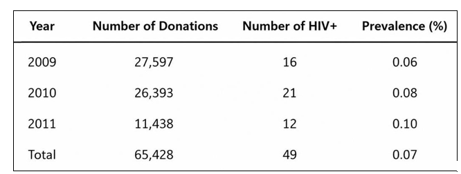
Table 2: Prevalence of HIV-1 Among Donors at Hemonúcleo in Santos, São Paulo
The table shows the year, the number of donations, the number of HIV-positive donors confirmed by Western blot, and the prevalence oem porcentagem. No the period from January2009 a May2011 a , the prevalence was 0.07%.

Figure 3: Phylogenetic tree showing the clustering of the seven HIV-1 sequences belonging to subtype B (Pol01, Pol03, Pol04, Pol08, Pol13, Pol17, Pol23) and one sequence belonging to subtype F (Pol05) with the corresponding reference sequences. The reference sequences used were: subtype B (U39362.B; 55529.B), subtype F (AF005494.F; 594621Ith.F), subtype C (AF286228.C; 56860Ith.C), and subtype A (AB098330.A)
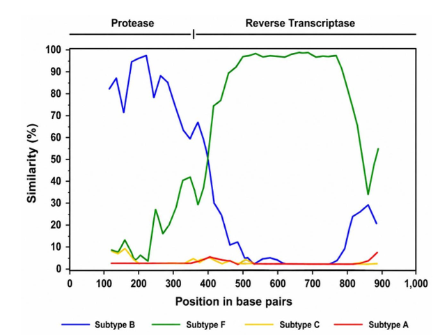
Figure 4: Recombination pattern between HIV-1 subtypes B and F identified in sample Pol09. The similarity of the sample was compared with the patterns of subtype B (blue), F (green), C (yellow), and A (red). We can see on the horizontal axis that the protease gene region was characterized as subtype B and the reverse transcriptase region was characterized as subtype F
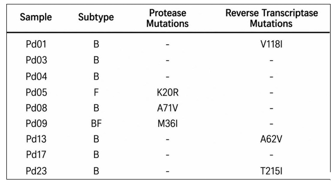
Table 3: Resistance Mutations Identified in Samples from HIV-1-positive Donors at the Santos Blood Center
The table lists the samples analyzed, the subtypes identified, and the mutations in the protease and reverse transcriptase genes. Six samples exhibited accessory mutations associated with resistance. Sample Pol23, belonging to subtype B, exhibited the T215I mutation, which confers a low level of resistance to nucleoside reverse transcriptase inhibitors: zidovudine (AZT) and stavudine (D4T).
Discussion
Several studies in recent years have focused on characterizing HIV-1 isolates circulating in the Baixada Santista region [79-83]. These studies demonstrate the complexity of the local epidemic, due to the presence of different subtypes, recombinants, and mutations conferring resistance to antiretrovirals. Our study focused on investigating the prevalence and genetic diversity of HIV-1 among blood donors at the Santos Blood Center, with the aim of characterizing the viruses currently circulating in the local transmission network.
Studies conducted in our region, based on analysis of the HIV-1 a pol gene, describe a predominance of subtype B (65%), followed by B/F recombinants (29%), subtype F (5%), and, less frequently, subtype C (~1%) [81]. When more than one gene is analyzed, such as pol and env, the frequency of recombinants increases to approximately 50% [82].
In the present study, despite the small number of sequenced samples, subtype B was predominant, with the presence of subtype F and the B/F recombinant. Initially, our sample set consisted of 49 samples; however, likely due to inadequate storage of serum for molecular biology techniques, it was not possible to amplify the viral genomic material. The absence of subtype C and the low frequency of recombinants are likely due to the small number of samples analyzed.
In the Baixada Santista region, two circulating recombinant forms of HIV-1 were identified, designated CRF28_BF and CRF29_BF, with a reported frequency of approximately 24% [81,83].
These CRFs share a common recombination pattern in the pol gene, with the protease belonging to subtype F and the reverse transcriptase belonging to subtype B. However, unique recombinant forms (URFs) have also been described in various articles, with a frequency of approximately 5% [81]. The recombinant sample identified in our analysis does not have the recombinant structure characteristic of CRFs, as the protease belongs to subtype B and the reverse transcriptase to subtype F (Figure X), and can be characterized as yet another URF identified in this location. This finding supports the hypothesis that dual HIV-1 infection events are common in samples from the Baixada Santista region [80,81].
Regarding primary resistance mutations, 6 out of 9 samples exhibited isolated accessory mutations, such as: K20R, A71V, and M36I in the protease gene, and A62V, V118I, and T215I in the reverse transcriptase gene (Table X). These mutations alone do not cause complete resistance to any antiretroviral drug, but they indicate that viruses from patients treated with antiretrovirals are being transmitted in the region. Report a frequency of 18.2% of complete antiretroviral resistance identified in samples from the Baixada Santista region [80]. Also report a high frequency of primary resistance mutations in samples from Santos (36%) [82].
Despite the small number of samples analyzed, our data support the hypothesis that samples from the Baixada Santista region may exhibit a high frequency of resistance mutations, even if they are only accessory mutations. Found a 31.2% rate of primary resistance and observed an association between primary resistance to antiretrovirals and virologic failure during clinical follow-up [79]. The authors suggest that, from a cost-benefit perspective, it would be advantageous to perform resistance testing before initiating antiretroviral therapy. Taken together, these studies reinforce the need for comprehensive discussions regarding HIV-1 resistance in the Baixada Santista region.
In the data collection conducted at the Hemonúcleo de Santos from January2009 a May2011 a , the identified prevalence of HIV-positive donors was 0.07%. Table X shows the prevalence rates stratified by year; these values are similar to the 0.04% rate found at the Pró-Sangue Foundation/São Paulo Blood Center by [84]. We emphasize that these studies used a group selected through interviews, thereby excluding donors at risk of infection.
Final Considerations
In the data collection conducted at the Santos Blood Center from January2009 a May2011 a , the identified prevalence of HIV-positive donors was 0.07%.
Despite our small sample size, a predominance of HIV-1 subtype B was identified, along with the presence of subtype F and the B/F recombinant. Six of the nine samples analyzed exhibited accessory mutations associated with antiretroviral resistance.
Although HIV-positive donors’ adherence to the public health system was not an initial objective of this study, our data reveal the need for improvements in counseling newly diagnosed individuals regarding clinical follow-up [85,86].
References
- Larder, B. A., Darby, G., & Richman, D. D. (1989). HIV with reduced sensitivity to zidovudine (AZT) isolated during prolonged therapy. Science, 243(4899), 1731-1734.
- Aleman, S., Söderbärg, K., Visco-Comandini, U., Sitbon, G., & Sönnerborg, A. (2002). Drug resistance at low viraemia in HIV-1-infected patients with antiretroviral combination therapy. Aids, 16(7), 1039-1044.
- Accetturi, C. A., Pardini, R., Pinto, G. H. N., Turcato Jr, G., & Lewi, D. S., et al. (2000). Effects ofCCR5Genetic Polymorphism and HIV-1 Subtype in Antiretroviral Response in Brazilian HIV-1-Infected Patients. JAIDS Journal of Acquired Immune Deficiency Syndromes, 24(4), 399-400.
- Shafer, R. W. (2004). Genotypic Testing for HIV-1 Drug Resistance., HIV Insite.
- Blower, S. M., Aschenbach, A. N., Gershengorn, H. B., & Kahn, J. O. (2001). Predicting the unpredictable: transmission of drug-resistant HIV. Nature medicine, 7(9), 1016-1020.
- Blower, S., & Farmer, P. (2003). Predicting the public health impact of antiretrovirals: preventing HIV in developing countries. AIDScience, 3(11), eado3591.
- Blower, S., Ma, L., Farmer, P., & Koenig, S. (2003). Predicting the impact of antiretrovirals in resource-poor settings: preventing HIV infections whilst controlling drug resistance. Current Drug Targets-Infectious Disorders, 3(4), 345-353.
- World Health Organization (2006). WHO case definitions of HIV for surveillance and revised clinical staging and immunological classification of HIV-related disease in adults and children. Geneva: World Health Organization.
- Poiesz, B. J., Ruscetti, F. W., Gazdar, A. F., Bunn, P. A., & Minna, J. D., et al. (1980). Detection and isolation of type C retrovirus particles from fresh and cultured lymphocytes of a patient with cutaneous T-cell lymphoma. Proceedings of the National Academy of Sciences, 77(12), 7415-7419.
- Yoshida, M., Miyoshi, I., & Hinuma, Y. (1982). Isolation and characterization of retrovirus from cell lines of human adult T-cell leukemia and its implication in the disease. Proceedings of the National Academy of Sciences, 79(6), 2031-2035.
- Barré-Sinoussi, F., Chermann, J. C., Rey, F., Nugeyre, M. T., & Chamaret, S., et al. (1983). Isolation of a T-lymphotropic retrovirus from a patient at risk for acquired immune deficiency syndrome (AIDS). Science, 220(4599), 868-871.
- Myers, G., Korber, B. T., Foley, B. T., Jeang, K.-T., & Mellors,J. W. et al. (1997.). Human Retroviruses and AIDS 1996: A Compilation and Analysis of Nucleic Acid and Amino Acid Sequences. Los Alamos, NM: Los Alamos National Laboratory.
- Eigen, M., & Winkler-Oswatitsch, R. (1990). [32] Statistical geometry on sequence space.
- Gao, F., Bailes, E., Robertson, D. L., Chen, Y., & Rodenburg,C. M., et al. (1999). Origin of HIV-1 in the chimpanzee Pan troglodytes troglodytes. Nature, 397(6718), 436-441.
- Dietrich, U., Adamski, M., Kreutz, R., Seipp, A., Kühnel, H., & Rübsamen-Waigmann, H. (1989). A highly divergent HIV-2-related isolate. Nature, 342(6252), 948-950.
- Perron, H., Garson, J. A., Bedin, F., Beseme, F., & Paranhos-Baccala, G., et al. (1997). Molecular identification of a novel retrovirus repeatedly isolated from patients with multiple sclerosis. Proceedings of the National Academy of Sciences, 94(14), 7583-7588.
- Miller, M. D., Farnet, C. M., & Bushman, F. D. (1997). Human immunodeficiency virus type 1 preintegration complexes: studies of organization and composition. Journal of virology, 71(7), 5382-5390.
- Doolittle, R. F., Feng, D. F., McClure, M. A., & Johnson, M.S. (1990). Retrovirus phylogeny and evolution. Retroviruses: Strategies of Replication, 1-18.
- McClure, M. A. (1993). 19 Evolutionary History of Reverse Transcriptase. Cold Spring Harbor Monograph Archive, 23, 425-444.
- Dettenhofer, M., & Yu, X. F. (1999). Highly purified human immunodeficiency virus type 1 reveals a virtual absence of Vif in virions. Journal of virology, 73(2), 1460-1467.
- Camaur, D., & Trono, D. (1996). Characterization of human immunodeficiency virus type 1 Vif particle incorporation. Journal of virology, 70(9), 6106-6111.
- Welker, R., Kottler, H., Kalbitzer, H. R., & Kräusslich, H. G. (1996). Human immunodeficiency virus type 1 Nef protein is incorporated into virus particles and specifically cleaved by the viral proteinase. Virology, 219(1), 228-236.
- Layne, S. P., Merges, M. J., Dembo, M., Spouge, J. L., et al. (1992). Factors underlying spontaneous inactivation and susceptibility to neutralization of human immunodeficiency virus. Virology, 189(2), 695-714.
- Levy, J. A. (1998). HIV and the Pathogenesis sof AIDS. 2. ed.Washington, DC: ASM Press.
- Peterlin, B. M. (1995). Molecular biology of HIV. In The retroviridae (pp. 185-238). Boston, MA: Springer US.
- Ou, S. H. I., & Gaynor, R. B. (1995). Intracellular factors involved in gene expression of human retroviruses. In The Retroviridae (pp. 97-184). Boston, MA: Springer US.
- Turner, B. G., & Summers, M. F. (1999). Structural biology of HIV. Journal of molecular biology, 285(1), 1-32.
- Sharp, P. M., Robertson, D. L., & Hahn, B. H. (1995). Cross-species transmission and recombination of'AIDS'viruses. Philosophical Transactions: Biological Sciences, 41-47.
- Hirsch, V. M., Olmsted, R. A., Murphey-Corb, M., Purcell,R. H., & Johnson, P. R. (1989). An African primate lentivirus (SIVsmclosely related to HIV-2. Nature, 339(6223), 389-392.
- Gao, F., Yue, L., White, A. T., Pappas, P. G., & Barchue, J., et al. (1992). Human infection by genetically diverse SIVSM-related HIV-2 in west Africa. Nature, 358(6386), 495-499.
- Chen, Z., Luckay, A., Sodora, D. L., Telfer, P., & Reed, P., et al. (1997). Human immunodeficiency virus type 2 (HIV-2) seroprevalence and characterization of a distinct HIV-2 genetic subtype from the natural range of simian immunodeficiency virus-infected sooty mangabeys. Journal of virology, 71(5), 3953-3960.
- Chen, Z., Telfier, P., Gettie, A., Reed, P., Zhang, L., Ho, D. D., & Marx, P. A. (1996). Genetic characterization of new West African simian immunodeficiency virus SIVsm: geographic clustering of household-derived SIV strains with human immunodeficiency virus type 2 subtypes and genetically diverse viruses from a single feral sooty mangabey troop. Journal of virology, 70(6), 3617-3627.
- Gilden, R. V., Arthur, L. O., Robey, W. G., Kelliher, J. C., &Graham, C. E., et al. (1986). HTLV-III antibody in a breeding chimpanzee not experimentally exposed to the virus.
- Teleki, G. (1989). Population status of wild chimpanzees (Pan troglodytes) and threats to survival. In Understanding chimpanzees (pp. 312-353). Harvard University Press.
- Huet, T., Cheynier, R., Meyerhans, A., Roelants, G., & Wain-Hobson, S. (1990). Genetic organization of a chimpanzee lentivirus related to HIV-1. Nature, 345(6273), 356-359.
- Sharp, P. M., Bailes, E., Stevenson, M., Emerman, M., & Hahn, B. H. (1996). Gene acquisition in HIV and SIV. Nature, 383(6601), 586.
- Simon, F., Mauclère, P., Roques, P., Loussert-Ajaka, I., & Müller-Trutwin, M. C., et al. (1998). Identification of a new human immunodeficiency virus type 1 distinct from group M and group O. Nature medicine, 4(9), 1032-1037.
- Zhu, T., Korber, B. T., Nahmias, A. J., Hooper, E., & Sharp, P. M., et al. (1998). An African HIV-1 sequence from 1959 and implications for the origin of the epidemic. Nature, 391(6667), 594-597.
- Hooper, E. (2000). The River: A Journey to the Source of HIV and AIDS. London: Penguin Books.
- Vandamme, A. M., Bertazzoni, U., & Salemi, M. (2000). Evolutionary strategies of human T-cell lymphotropic virus type II. Gene, 261(1), 171-180.
- Salemi, M., Strimmer, K., Hall, W. W., Duffy, M., & Delaporte, E., et al. (2001). Dating the common ancestor of SIVcpz and HIV-1 group M and the origin of HIV-1 subtypes by using a new method to uncover clock-like molecular evolution. The FASEB Journal, 15(2), 276-278.
- Kuiken, C. L., Foley, B., Hahn, B., Marx, P. A., & McCutchan, F., et al. (1999). Human Retroviruses and AIDS 1999: A Compilation and Analysis of Nucleic Acid and Amino Acid Sequences. Los Alamos, NM: Los Alamos National Laboratory.
- Li, W. H., Tanimura, M., & Sharp, P. M. (1988). Rates and dates of divergence between AIDS virus nucleotide sequences. Molecular biology and evolution, 5(4), 313-330.
- Myers, G., Joseph, S., Berzofsky, J. A., Rabson, A. B., & Smith, T. F., et al. (1988).. Human Retroviruses and AIDS 1988: A Compilation and Analysis of Nucleic Acid and Amino Acid Sequences. Los Alamos, NM: Los Alamos National Laboratory.
- Myers, G., MacInnes, K., & Korber, B. (1992). The emergence of simian/human immunodeficiency viruses. AIDS research and human retroviruses, 8(3), 373-386.
- Louwagie, J., McCutchan, F. E., Peeters, M., Brennan, T. P., & Sanders-Buell, E., et al. (1993). Phylogenetic analysis of gag genes from 70 international HIV-1 isolates provides evidence for multiple genotypes. Aids, 7(6), 769-780.
- Janssens, W., Heyndrickx, L., Fransen, K., Motte, J., & Peeters, M., et al. (1994). Genetic and phylogenetic analysis of env subtypes G and H in Central Africa. AIDS research and human retroviruses, 10(7), 877-879.
- Kostrikis, L. G., Bagdades, E., Cao, Y., Zhang, L., & Dimitriou, D., et al. (1995). Genetic analysis of human immunodeficiency virus type 1 strains from patients in Cyprus: identification of a new subtype designated subtype I. Journal of Virology, 69(10), 6122-6130.
- Leitner, T., Alaeus, A., Marquina, S., Lilja, E., & Lidman, K., et al. (1995). Yet another subtype of HIV type 1?. AIDS research and human retroviruses, 11(8), 995-997.
- Louwagie, J., Janssens, W., Mascola, J., Heyndrickx, L., & Hegerich, P., et al. (1995). Genetic diversity of the envelope glycoprotein from human immunodeficiency virus type 1 isolates of African origin. Journal of Virology, 69(1), 263-271.
- Triques, K., Bourgeois, A., Saragosti, S., Vidal, N., & Mpoudi-Ngole, E., et al. (1999). High diversity of HIV-1 subtype F strains in Central Africa. Virology, 259(1), 99-109.
- Triques, K., Bourgeois, A., Vidal, N., Mpoudi-Ngole, E., & Mulanga-Kabeya, C., et al. (2000). Near-full-length genome sequencing of divergent African HIV type 1 subtype F viruses leads to the identification of a new HIV type 1 subtype designated K. AIDS research and human retroviruses, 16(2), 139-151.
- Gürtler, L. G., Hauser, P. H., Eberle, J., Von Brunn, A., & Knapp, S., et al. (1994). A new subtype of human immunodeficiency virus type 1 (MVP-5180) from Cameroon. Journal of virology, 68(3), 1581-1585.
- Simon, F., Mauclère, P., Roques, P., Loussert-Ajaka, I., & Müller-Trutwin, M. C., et al. (1998). Identification of a new human immunodeficiency virus type 1 distinct from group M and group O. Nature medicine, 4(9), 1032-1037.
- Robertson, D. L., Sharp, P. M., McCutchan, F. E., & Hahn,B. H. (1995). Recombination in HIV-1. Nature, 374(6518), 124-126.
- Coffin, J. M. (1979). Structure, replication, and recombination of retrovirus genomes: some unifying hypotheses. Journal of General Virology, 42(1), 1-26.
- Hu, W. S., & Temin, H. M. (1990). Genetic consequences of packaging two RNA genomes in one retroviral particle: pseudodiploidy and high rate of genetic recombination. Proceedings of the National Academy of Sciences, 87(4), 1556-1560.
- Sabino, E. C., Shpaer, E. G., Morgado, M. G., Korber,B. T., & Diaz, R. S., et al. (1994). Identification of human immunodeficiency virus type 1 envelope genes recombinant between subtypes B and F in two epidemiologically linked individuals from Brazil. Journal of Virology, 68(10), 6340-6346.
- Diaz, R. S., Sabino, E. C., Mayer, A., Mosley, J. W., & Busch,M. P. (1995). Dual human immunodeficiency virus type 1 infection and recombination in a dually exposed transfusion recipient. The Transfusion Safety Study Group. Journal of Virology, 69(6), 3273-3281.
- Robertson, D. L., Gao, F., & Saksena, N. (1998). Recombination of HIV Genomes.
- Quiñones-Mateu, M. E., & Arts, E. J. (1999). Recombination in HIV-1: update and implications. Aids Rev, 1(2), 89-100.
- McCUTCHAN, F. E., Hegerich, P. A., Brennan, T. P., Phanuphak, P., & Singharaj, P., et al. (1992). Genetic variants of HIV-1 in Thailand. AIDS research and human retroviruses, 8(11), 1887-1895.
- Carr, J. K., Salminen, M. O., Koch, C., Gotte, D., & Artenstein,A. W., et al. (1996). Full-length sequence and mosaic structure of a human immunodeficiency virus type 1 isolate from Thailand. Journal of virology, 70(9), 5935-5943.
- Gao, F., Robertson, D. L., Morrison, S. G., Hui, H., & Craig, S., et al. (1996). The heterosexual human immunodeficiency virus type 1 epidemic in Thailand is caused by an intersubtype (A/E) recombinant of African origin. Journal of virology, 70(10), 7013-7029.
- Salminen, M. O., Koch, C., Sanders-Buell, E., Ehrenberg, P. K., & Michael, N. L., Carr, et al. (1995). Recovery of virtually full-length HIV-1 provirus of diverse subtypes from primary virus cultures using the polymerase chain reaction. Virology, 213(1), 80-86.
- Salminen, M. O., Carr, J. K., Burke, D. S., & McCUTCHAN,F. E. (1995). Identification of breakpoints in intergenotypic recombinants of HIV type 1 by bootscanning. AIDS research and human retroviruses, 11(11), 1423-1425.
- Salminen, M. O., Johansson, B. O., Sönnerborg, A., Ayehunie, S., & Gotte, D., et al. (1996). Full-length sequence of an Ethiopian human immunodeficiency virus type 1 (HIV-1) isolate of genetic subtype C. AIDS research and human retroviruses, 12(14), 1329-1339.
- Carr, J. K., Foley, B. T., Leitner, T., Salminen, M., & Korber, B., et al. (1998). Reference sequences representing the principal genetic diversity of HIV-1 in the pandemic. Human retroviruses and AIDS, 111(10).
- Gao, F., Robertson, D. L., Carruthers, C. D., Morrison, S.G., & Jian, B., et al. (1998). A comprehensive panel of near-full-length clones and reference sequences for non-subtype B isolates of human immunodeficiency virus type 1. Journal of virology, 72(7), 5680-5698.
- Gao, F., Robertson, D. L., Carruthers, C. D., Li, Y., & Bailes, E., et al. (1998). An isolate of human immunodeficiency virus type 1 originally classified as subtype I represents a complex mosaic comprising three different group M subtypes (A, G, and I). Journal of virology, 72(12), 10234-10241.
- Nasioulas, G., Paraskevis, D., Magiorkinis, E., Theodoridou, M., & Hatzakis, A. (1999). Molecular analysis of the full-length genome of HIV type 1 subtype I: evidence of A/G/I recombination. AIDS Research and Human Retroviruses, 15(8), 745-758.
- SALMINEN, M. O. Unpublished observations, 1999.
- McCutchan, F. E., Carr, J. K., Bajani, M., Sanders-Buell, E., & Harry, T. O., et al. (1999). Subtype G and multiple forms of A/G intersubtype recombinant human immunodeficiency virus type 1 in Nigeria. Virology, 254(2), 226-234.
- Montavon, C., Bibollet-Ruche, F., Robertson, D., Koumare, B., & Mulanga, C., et al. (1999). Sequence Note The Identification of a Complex A/G/I/J Recombinant HIV Type 1 Virus in Various West African Countries. AIDS research and human retroviruses, 15(18), 1707-1712.
- Nasioulas, G., Paraskevis, D., Magiorkinis, E., Theodoridou, M., & Hatzakis, A. (1999). Molecular analysis of the full-length genome of HIV type 1 subtype I: evidence of A/G/I recombination. AIDS Research and Human Retroviruses, 15(8), 745-758.
- De Leys, R., Vanderborght, B., Vanden Haesevelde, M., Heyndrickx, L., & Van Geel, A., et al. (1990). Isolation and partial characterization of an unusual human immunodeficiency retrovirus from two persons of west-central African origin. Journal of virology, 64(3), 1207-1216.
- Charneau, P., Borman, A. M., Quillent, C., Guétard, D., & Chamaret, S., et al. (1994). Isolation and envelope sequence of a highly divergent HIV-1 isolate: definition of a new HIV-1 group. Virology, 205(1), 247-253.
- Simon, F., Mauclère, P., Roques, P., Loussert-Ajaka, I., & Müller-Trutwin, M. C., et al. (1998). Identification of a new human immunodeficiency virus type 1 distinct from group M and group O. Nature medicine, 4(9), 1032-1037.
- Gagliani, L. H., Alkmim Maia, W. T., Sá-Filho, D., Janini, L. M., & Sucupira, M. C., et al. (2011). The association between primary antiretroviral resistance and HAART virologic failure in a developing set. AIDS research and human retroviruses, 27(3), 251-256.
- de Sa-Filho, D. J., Ambar, R. F., Duarte, N. B., Matias, R. B.R., Candido, V., Gagliani, L. H., & Caseiro, M. M. (2009). HIV type 1 diversity from newly diagnosed patients in Santos metropolitan area/Brazil. AIDS research and human retroviruses, 25(9), 925-929.
- de Sa-Filho, D. J., Soares, M. D. S., Candido, V., Gagliani, L. H., Cavaliere, E., Diaz, R. S., & Caseiro, M. M. (2008). HIV type 1 pol gene diversity and antiretroviral drug resistance mutations in Santos, Brazil. AIDS research and human retroviruses, 24(3), 347-353.
- Sucupira, M. C. A., Caseiro, M. M., Alves, K., Tescarollo, G., & Janini, L. M., et al. (2007). High levels of primary antiretroviral resistance genotypic mutations and B/F recombinants in Santos, Brazil. AIDS patient care and STDs, 21(2), 116-128.
- Filho, D. J. D. S., Sucupira, M. C. A., Casiero, M. M., Sabino,E. C., & Diaz, R. S., et al. (2006). Identification of two HIV type 1 circulating recombinant forms in Brazil. AIDS research and human retroviruses, 22(1), 1-13.
- Salles, N. A., Sabino, E. C., Barreto, C. C., Barreto, A. M., & Otani, M. M., et al. (2003). The discarding of bloodunits and the prevalence of infectious diseases in donors at the Pro-Blood Foundation/Blood Center of Sao Paulo, Sao Paulo, Brazil. Revista Panamericana de Salud Publica= Pan American Journal of Public Health, 13(2-3), 111-116.
- Gagliani, L. H., Alkmim Maia, W. T., Sá-Filho, D., Janini, L. M., Sucupira, & M. C., et al. (2011). The association between primary antiretroviral resistance and HAART virologic failure in a developing set. AIDS research and human retroviruses, 27(3), 251-256.
- HIV, A. T. (2003). Economics of aids and Access to hiv/aids Care in Developing Countries. Issues and Challenges.