


Research Article - (2026) Volume 9, Issue 3
Multi-System Secondary Autonomic Dysfunction Recovery: A Three-Hit Injury Model and Six Clinical Flags of Autonomic Network Repair

Received Date: Jun 01, 2026 / Accepted Date: Jun 22, 2026 / Published Date: Jul 02, 2026
Copyright: ©2026 Bruce H. Knox. This is an open-access article distributed under the terms of the Creative Commons Attribution License, which permits unrestricted use, distribution, and reproduction in any medium, provided the original author and source are credited.
Citation: Knox, B. H. (2026). Multi-System Secondary Autonomic Dysfunction Recovery: A Three-Hit Injury Model and Six Clinical Flags of Autonomic Network Repair. Adv Neur Sci, 9(3), 01-10.
Abstract
Autonomic dysfunction may arise through primary neurodegenerative disease or secondary injury affecting autonomic regulatory networks. Distinguishing between these mechanisms is clinically important because secondary autonomic dysfunction may retain substantial capacity for recovery. This paper integrates a patient-investigator hypothesis describing a three-hit model of severe secondary autonomic dysfunction with a practical framework for recognising recovery. The first hit consisted of Chikungunya virus (CHIKV) infection in 2008, likely producing chronic autonomic vulnerability through small-fibre and autonomic nervous system injury. The second and third hits occurred in October 2021 during a left ventricular outflow tract premature ventricular contraction ablation complicated by ventricular perforation, cardiac tamponade, emergency thoracotomy, and cardiopulmonary bypass repair. Following these events, widespread autonomic collapse developed, affecting cardiovascular, thermoregulatory, gastrointestinal, genitourinary, neuroendocrine, cognitive, motivational, and sexual domains.
Although the syndrome initially resembled Pure Autonomic Failure (PAF), subsequent progressive recovery across multiple autonomic systems argues strongly against a primary neurodegenerative process. Six clinical recovery flags emerged during longitudinal observation: return of sweating and thermoregulation; improved exercise capacity and heart-rate responsiveness; improved cognitive clarity; restoration of motivation and anticipation; return of sexual function and ideation; and increased facial hair growth requiring daily shaving. Collectively, these observations support a model in which severe secondary dysautonomia represents autonomic network failure, while recovery reflects gradual autonomic network repair.
Keywords
Dysautonomia, Secondary Autonomic Dysfunction, Chikungunya Virus, Baroreflex Failure, Neuroplasticity, Autonomic Recovery, Network Failure, Thermoregulation, Sexual Function, Neuroendocrine Recovery
Introduction
The autonomic nervous system (ANS) regulates cardiovascular stability, thermoregulation, gastrointestinal function, genitourinary function, endocrine coordination, emotional processing, and physiological homeostasis [1-5]. Dysfunction may arise through progressive neurodegenerative disease or through acquired injury [6-10].
Primary autonomic disorders such as Pure Autonomic Failure (PAF) and Multiple System Atrophy (MSA) are generally characterised by progressive deterioration without expectation of significant recovery [11-15]. By contrast, secondary autonomic dysfunction arises from identifiable insults including infection, trauma, surgery, inflammation, autoimmune disease, metabolic disturbance, or critical illness [6-10]. Because neural pathways may remain structurally intact to varying degrees, secondary disorders may retain the capacity for substantial recovery [29-39,66-70]. A central challenge for both clinicians and patients is determining whether recovery is occurring. Much of the dysautonomia literature focuses on diagnosis and symptom management rather than the identification of recovery markers [6–10,66–70]. Consequently, patients often struggle to interpret subtle improvements that may represent genuine physiological repair. This paper proposes that severe secondary autonomic dysfunction is best understood as a disorder of autonomic network failure and that recovery can be recognised through a constellation of clinical indicators reflecting restoration of network function [1–5,29–39,66–70].
The Three-Hit Model
Hit One: Chikungunya Virus Infection
In 2008, the patient contracted Chikungunya virus infection during a documented epidemic in Indonesia. Acute illness was severe and required hospitalisation upon return to New Zealand. Increasing evidence demonstrates that CHIKV may affect autonomic and peripheral nervous system structures through inflammatory and immune-mediated mechanisms. Documented sequelae include neurological complications, chronic inflammatory activation, prolonged fatigue syndromes, and persistent neurological symptoms [16–20]. Rather than producing immediate catastrophic autonomic failure, the infection may have established a state of reduced autonomic reserve and increased vulnerability to future physiological stressors [6–10,16–20].
Hit Two: Cardiac Perforation and Tamponade
On 15 October 2021, an elective catheter ablation procedure for frequent premature ventricular contractions was complicated by ventricular perforation and cardiac tamponade. Cardiac tamponade represents one of the most profound acute physiological stressors encountered in cardiovascular medicine. Rapid accumulation of blood within the pericardial space compromises cardiac filling, reduces cardiac output, and initiates intense sympathetic activation. The event exposed autonomic regulatory systems to acute haemodynamic collapse, massive catecholamine release, systemic inflammatory activation, and global physiological stress [21–25].
Hit Three: Emergency Thoracotomy and Cardiopulmonary Bypass
Emergency open-heart surgery immediately followed the tamponade event. This intervention required thoracotomy, cardiopulmonary bypass, pericardial repair, and prolonged intensive care recovery. Cardiopulmonary bypass is recognised to induce widespread inflammatory activation, endothelial dysfunction, neurohumoral disturbance, and autonomic imbalance [26–30]. The combined effects of tamponade and bypass likely transformed a vulnerable autonomic system into one exhibiting overt failure.
Development of Multi-System Autonomic Network Failure
Between 2022 and 2023, dysfunction developed across virtually every major autonomic domain. The breadth of involvement supports a network-based interpretation rather than isolated organ pathology [1–10].
Cardiovascular Dysfunction
Manifestations included severe orthostatic hypotension, supine hypertension, baroreflex instability, blood-pressure volatility, autonomic storms, and profound exercise intolerance [21–25,66–70].
Thermoregulatory Failure
Loss of sweating and severe heat intolerance emerged as prominent features. The development of anhidrosis suggested widespread sympathetic cholinergic dysfunction [54–57].
Gastrointestinal Failure
Digestive dysfunction involved gastroparesis, severe reflux, delayed gastric emptying, exocrine pancreatic insufficiency, and functional gallbladder failure. The simultaneous involvement of multiple digestive organs suggested impairment of autonomic coordination rather than isolated organ pathology [40–44].
Genitourinary Dysfunction
Symptoms included urinary retention, impaired bladder emptying, erectile dysfunction, loss of libido, and loss of sexual ideation. Sexual and genitourinary function require coordinated autonomic, endocrine, vascular, and emotional integration [45–48].
Neuroendocrine and Motivational Dysfunction
The patient experienced profound reductions in motivation, anticipation, emotional engagement, reward processing, cognitive clarity, and future orientation. These manifestations suggested dysfunction within hypothalamic-autonomic-limbic networks extending beyond traditional cardiovascular concepts of dysautonomia [1–5,49–53].
Why Pure Autonomic Failure Was Initially Suspected
The clinical picture overlapped substantially with recognised presentations of Pure Autonomic Failure. Features included orthostatic hypotension, supine hypertension, thermoregulatory failure, genitourinary dysfunction, and widespread autonomic impairment [11–15]. At the height of illness, differentiation between severe secondary autonomic dysfunction and primary autonomic neurodegeneration was challenging.
Evidence Supporting Secondary Rather Than Primary Disease
Over time, progressive recovery occurred across multiple systems. Recovery was observed in sweating, heat tolerance, heart-rate responsiveness, exercise capacity, cognitive clarity, sexual desire, emotional engagement, gastrointestinal function, and orthostatic tolerance. Autonomic storms resolved completely. Such widespread improvement is atypical of primary synucleinopathies such as PAF and MSA. Instead, the pattern is more consistent with recovery following severe secondary injury [11–15,31–39,66–70].
Mechanisms of Autonomic Recovery
Resolution of Neuroinflammation
As inflammatory burden diminished, neuronal signalling likely improved. This proposed mechanism is consistent with the broader literature on inflammatory and immune-mediated autonomic disorders and post-infective neurological sequelae [6–10,16–20].
Neural Plasticity
Autonomic circuits retain adaptive capacity. Alternative pathways may develop, damaged pathways may regain function, and partially injured neurons may recover [31–34].
Baroreflex Recalibration
Improvement in blood-pressure regulation suggests progressive repair and recalibration of baroreflex pathways [21–25].
Peripheral Nerve Recovery
Small-fibre autonomic nerves possess regenerative potential, particularly when injury is inflammatory rather than degenerative [35–39].
Physiological Reconditioning
Recovery was likely supported through activity pacing, rehabilitation, hydration strategies, compression therapy, medication support, and nutritional optimization [66–70].
Six Clinical Flags of Autonomic Network Repair
Flag One: Return of Sweating and Thermoregulation
The return of sweating represents one of the most objective indicators of autonomic recovery. Patients may notice sweating during exercise, sweating in warm weather, improved heat tolerance, reduced overheating, and improved recovery following exertion. This flag reflects restoration of sympathetic cholinergic signaling [54–57].
Flag Two: Improved Exercise Capacity and Heart-Rate Responsiveness
Patients may begin walking further, climbing stairs more easily, and tolerating sustained activity with fewer post-exertional crashes. Improved exercise tolerance reflects recovery across cardiovascular regulation, baroreflex function, vascular tone, skeletal muscle perfusion, and metabolic efficiency [21–25,66–70].
Flag Three: Improved Cognitive Clarity and Mental Endurance
Patients frequently report better concentration, improved memory, faster thinking, improved reading endurance, and longer conversational engagement. These improvements likely reflect improved cerebral perfusion and reduced autonomic burden [1–5,21–25].
Flag Four: Return of Motivation, Anticipation, and Emotional Engagement
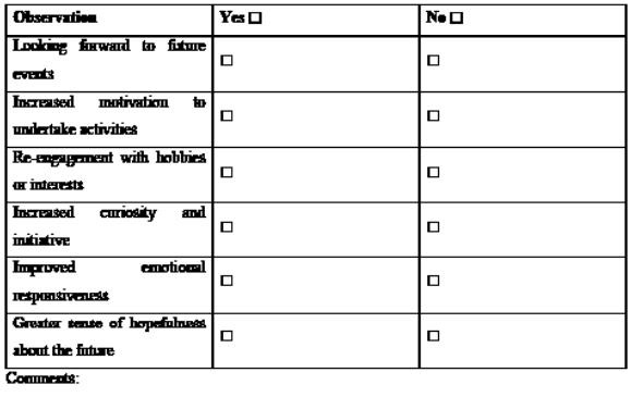
Patients may begin planning future activities, anticipating events, re-engaging with hobbies, demonstrating curiosity, and experiencing renewed hopefulness. The return of anticipation may represent one of the earliest signs of recovery within deeper autonomic-limbic system [49–53].
Flag Five: Return of Sexual Function and Sexual Ideation
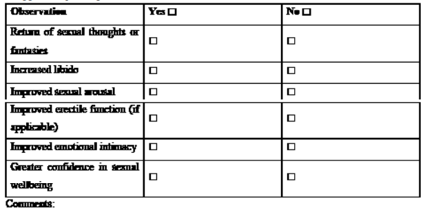
Recovery may be reflected by return of libido, return of sexual thoughts, improved erectile function, improved arousal, and increased emotional intimacy. Because sexual function requires coordinated autonomic, endocrine, vascular, and emotional integration, it serves as a sensitive indicator of network recovery [45–53].
Flag Six: Increased Facial Hair Growth and Return to Daily Shaving
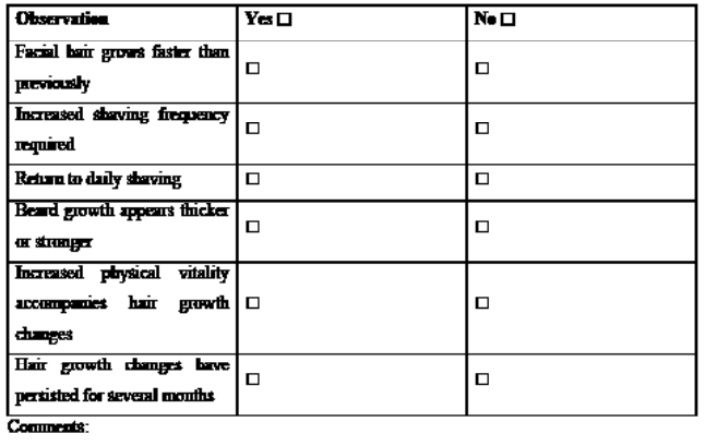
Facial hair growth represents a visible peripheral marker of androgen signalling and neuroendocrine function. Patients may observe faster facial hair growth, transition from shaving every several days to daily shaving, thicker beard growth, and return of other androgen-dependent characteristics. This may reflect restoration of hypothalamic-pituitary-gonadal axis activity following prolonged autonomic and inflammatory stress [58–65].
A Framework for Identifying Multi-System Secondary Dysautonomia Recovery
Taken individually, each recovery flag may appear subjective. Viewed collectively, however, they demonstrate recovery across multiple interconnected physiological networks:

Table 1:The simultaneous emergence of these indicators, mirroring their earlier collapse into dysfunction, argues strongly for restoration of network function rather than isolated organ recovery [1–5,31–39,66–70].
Implications for Clinical Practice
This integrated model has important implications. First, clinicians should recognise that recovery in severe secondary dysautonomia may occur gradually over years rather than months. Second, recovery may become apparent clinically before conventional autonomic testing demonstrates substantial change. Third, observation of recovery across multiple domains may assist in distinguishing secondary autonomic dysfunction from primary neurodegenerative disorders. Finally, these findings support a broader conceptual shift from organ-centred interpretations toward a network-based understanding of autonomic illness and recovery [1–15,66–70].
Conclusion
This patient-investigator hypothesis suggests that severe secondary autonomic dysfunction developed through a three-hit sequence: Chikungunya virus infection, cardiac tamponade, and emergency surgery with cardiopulmonary bypass. The resulting syndrome caused widespread autonomic network failure involving cardiovascular, thermoregulatory, gastrointestinal, genitourinary, neuroendocrine, cognitive, emotional, and sexual systems. Although the condition initially resembled Pure Autonomic Failure, gradual improvement across multiple domains argues against a primary neurodegenerative disorder and instead supports severe secondary autonomic network dysfunction. Six clinical signs emerged as especially meaningful indicators of recovery: return of sweating and temperature regulation, improved exercise tolerance and heart-rate responsiveness, better cognitive clarity and mental endurance, return of motivation, anticipation, and emotional engagement, return of sexual function and sexual thoughts, and increased facial hair growth and a return to daily shaving.
Together, these findings support a model in which autonomic recovery reflects restoration of interconnected physiological networks rather than isolated organ recovery. Recognising these markers may help clinicians and patients identify meaningful progress during prolonged autonomic rehabilitation and distinguish recovering secondary dysautonomia from progressive neurodegenerative autonomic disease. The difference between secondary autonomic dysfunction and neurodegenerative autonomic dysfunction is more than a diagnostic label. It has major implications for prognosis, treatment, patient expectations, and how recovery is interpreted. At initial presentation, the two conditions may look very similar. Both can involve orthostatic hypotension, impaired cardiovascular regulation, gastrointestinal dysmotility, bladder dysfunction, sexual dysfunction, thermoregulatory abnormalities, fatigue, and cognitive impairment. Because the overlap can be extensive, the early clinical picture may not reliably distinguish between them [6–15].
The key diagnostic challenge is therefore not only to identify autonomic failure, but also to determine its trajectory. In secondary autonomic dysfunction, the autonomic nervous system is understood to have been injured by a defined insult such as infection, inflammation, surgery, trauma, autoimmune disease, metabolic disturbance, or cardiovascular catastrophe [6–10,16–30]. Although the resulting dysfunction may be severe and long-lasting, the biological expectation is often stabilisation and at least partial recovery [31–39,66–70]. By contrast, neurodegenerative autonomic dysfunction follows a different biological course. In these conditions, autonomic failure is part of an ongoing degenerative process marked by progressive neuronal loss. Disorders such as Pure Autonomic Failure, Parkinson disease, Dementia with Lewy Bodies, and Multiple System Atrophy are not caused by a temporary injury, but by continuing neurodegeneration [11–15].
Ultimately, longitudinal observation is one of the most powerful diagnostic tools available. The central question is not simply whether autonomic failure is present, but whether the autonomic nervous system shows signs of recovery or signs of ongoing degeneration. The answer shapes diagnosis, prognosis, treatment strategy, and future expectations for both clinician and patient. The core message of this review is simple: autonomic dysfunction is not a single disease entity. It is a clinical syndrome with different biological pathways. Determining whether the course is one of recovery or degeneration is the key diagnostic challenge and a major determinant of long-term outcome.
Article Declarations
Article Type: Hypothesis and narrative review with patient-investigator clinical observation. Ethics Statement: This manuscript is a patient-investigator hypothesis and narrative review based on the author’s own longitudinal clinical experience and published literature. No external human participant recruitment, intervention, or identifiable third-party data are reported.
Consent Statement: The patient-investigator is the author and consents to publication of the clinical narrative contained in this manuscript.
Funding: No external funding was received.
Conflicts of Interest: The author declares no competing interests. Data Availability: No datasets were generated or analysed. All literature cited is publicly identifiable through the reference list.
References
- Benarroch, E. E. (1993, October). The central autonomic network: functional organization, dysfunction, and perspective. In Mayo clinic proceedings (Vol. 68, No. 10, pp. 988-1001). Elsevier.
- Benarroch, E. E. (2012). Central autonomic control. In Primer on the autonomic nervous system (pp. 9-12). Academic press.
- Low, P. A., & Benarroch, E. E. (Eds.). (2008). Clinical autonomic disorders. IMO Publishing.
- Goldstein, D. S. (2000). The autonomic nervous system in health and disease. Informa Health Care.
- Wehrwein, E. A., Orer, H. S., & Barman, S. M. (2016). Overview of the anatomy, physiology, and pharmacology of the autonomic nervous system. Comprehensive physiology, 6(3), 1239-1278.
- Freeman, R. (2005). Autonomic peripheral neuropathy. The Lancet, 365(9466), 1259-1270.
- Vernino, S. (2020). Autoimmune autonomic disorders. CONTINUUM: Lifelong Learning in Neurology, 26(1), 44-57.
- Kaufmann H, Norcliffe-Kaufmann L, Palma JA (2017).Autonomic disorders. Handb Clin Neurol. 140:245-265.
- Cheshire, W. P. (2013). Highlights in clinical autonomic neuroscience: Autonomic correlates of social cognition. Autonomic Neuroscience, 174(1-2), 5-7.
- Gibbons CH, Freeman R (2015). Clinical implications of delayed diagnosis of autonomic dysfunction. Auton Neurosci.193:1-3.
- Kaufmann H, Norcliffe-Kaufmann L, Palma JA (2017). Pure autonomic failure. Handb Clin Neurol.,140:243-257.
- Palma JA, Kaufmann H (2017). Neurogenic orthostatic hypotension and autonomic failure. Neurol Clin. 35(1):39-54.
- Fanciulli, A., & Wenning, G. K. (2015). Multiple-system atrophy. New England Journal of Medicine, 372(3), 249-263.
- Goldstein, D. S., Holmes, C., Sharabi, Y., & Wu, T. (2015). Survival in synucleinopathies: a prospective cohort study. Neurology, 85(18), 1554-1561.
- Kaufmann, H., & Biaggioni, I. (2003). Autonomic failure in neurodegenerative disorders. In Seminars in neurology (Vol. 23, No. 04, pp. 351-364). Copyright© 2003 by Thieme Medical Publishers, Inc., 333 Seventh Avenue, New York, NY 10001, USA. Tel.:+ 1 (212) 584-4662.
- Simon, F., Javelle, E., Oliver, M., Leparc-Goffart, I., & Marimoutou, C. (2011). Chikungunya virus infection. Current infectious disease reports, 13(3), 218-228.
- Mehta, R., Gerardin, P., de Brito, C. A. A., Soares, C. N., Ferreira, M. L. B., & Solomon, T. (2018). The neurological complications of chikungunya virus: A systematic review. Reviews in medical virology, 28(3), e1978.
- Leparc-Goffartd, P. G., Réniaa, L., de Lamballeried, X., & Nga,L. F. (2015). Caribbean and La Réunion Chikungunya virus isolates differ in their capacity to induce pro-inflammatory Th1 and NK cell 2 responses and acute joint pathology 3.
- Burt, F. J., Rolph, M. S., Rulli, N. E., Mahalingam, S., & Heise, M. T. (2012). Chikungunya: a re-emerging virus. The Lancet, 379(9816), 662-671.
- Economopoulou A, Dominguez M, Helynck B, et al (2009). Atypical chikungunya virus infections. Emerg Infect Dis.15(8):1345-1347.
- Biaggioni I, Robertson D (1987). Endogenous restoration of blood pressure in autonomic failure. Hypertension. 10(3):289-294.
- Jahshan, S., Dayan, L., & Jacob, G. Running Title: NO-cGMP and cerebrovascular CO2-reactivity 14.
- Goldstein, D. S., Robertson, D., Esler, M., Straus, S. E., & Eisenhofer, G. (2002). Dysautonomias: clinical disorders of the autonomic nervous system. Annals of internal medicine, 137(9), 753-763.
- Palma, J. A., & Benarroch, E. E. (2014). Neural control of the heart: recent concepts and clinical correlations. Neurology, 83(3), 261-271.
- Biaggioni I (2018). Mechanisms of orthostatic hypotension. JClin Hypertens.20(1):16-20.
- Raja, S. G., & Dreyfus, G. D. (2005). Modulation of systemic inflammatory response after cardiac surgery. Asian Cardiovascular and Thoracic Annals, 13(4), 382-395.
- Bronicki, R. A., & Hall, M. (2016). Cardiopulmonary bypass-induced inflammatory response: pathophysiology and treatment. Pediatric Critical Care Medicine, 17(8), S272-S278.
- Wan, S., LeClerc, J. L., & Vincent, J. L. (1997). Inflammatory response to cardiopulmonary bypass: mechanisms involved and possible therapeutic strategies. Chest, 112(3), 676-692.
- Paparella, D., Yau, T. M., & Young, E. (2002). Cardiopulmonary bypass induced inflammation: pathophysiology and treatment. An update. European Journal of Cardio-Thoracic Surgery, 21(2), 232-244.
- Ascione, R., Lloyd, C. T., Underwood, M. J., Lotto, A. A., Pitsis, A. A., & Angelini, G. D. (2000). Inflammatory response after coronary revascularization with or without cardiopulmonary bypass. The Annals of thoracic surgery, 69(4), 1198-1204.
- Merzenich, M. M., Van Vleet, T. M., & Nahum, M. (2014). Brain plasticity-based therapeutics. Frontiers in human neuroscience, 8, 385.
- Cramer, S. C., Sur, M., Dobkin, B. H., O'Brien, C., Sanger, T. D., Trojanowski, J. Q., ... & Vinogradov, S. (2011). Harnessing neuroplasticity for clinical applications. Brain, 134(6), 1591-1609.
- Kleim, J. A., & Jones, T. A. (2008). Principles of experience-dependent neural plasticity: implications for rehabilitation after brain damage. Journal of speech, language, and hearing research, 51(1), S225-S239.
- Benarroch EE. Neuroplasticity and autonomic function.Neurology. 2006,67(5):S20-S23.
- Oaklander, A. L., & Nolano, M. (2019). Scientific advances in and clinical approaches to small-fiber polyneuropathy: a review. JAMA neurology, 76(10), 1240-1251.
- Devigili, G., Cazzato, D., & Lauria, G. (2020). Clinical diagnosis and management of small fiber neuropathy: an update on best practice. Expert review of neurotherapeutics, 20(9), 967-980.
- Levine TD (2020). Small fiber neuropathy. Neurol Clin. 38(3):695-711.
- Lauria G, Hsieh ST, Johansson O, et al (2010). European Federation guidelines on small fibre neuropathy. Eur J Neurol.17(7):903-912.
- Camilleri, M. (2006). Integrated upper gastrointestinal response to food intake. Gastroenterology, 131(2), 640-658.
- 40. Camilleri M, Bharucha AE, Farrugia G. Epidemiology and mechanisms of gastroparesis. Gastroenterology. 2011,140(5):1225-1235.
- Grover, M., Farrugia, G., & Stanghellini, V. (2019). Gastroparesis: a turning point in understanding and treatment. Gut, 68(12), 2238-2250.
- Pasricha PJ, Yates KP, Sarosiek I, et al (2015). Outcomes and pathophysiology of gastroparesis. Gastroenterology. 149(7):1762-1774.
- Furness, J. B. (2008). The enteric nervous system. John Wiley & Sons.
- Giuliano, F., & Rampin, O. (2004). Neural control of erection.Physiology & behavior, 83(2), 189-201.
- Burnett, A. L. (2006). Erectile dysfunction. The Journal of urology, 175(3S), S25-S31.
- Menezes, A., Artham, S., Lavie, C. J., Milani, R. V., & O'Keefe, J. (2011). Erectile dysfunction and cardiovascular disease. Postgraduate Medicine, 123(3), 7-16.
- Freeman R (2002). Sexual dysfunction and autonomic disorders. Clin Auton Res. 12(Suppl 1):I56-I61.
- Benarroch EE. The central autonomic network and behavior. Neurology. 1997,48(6):1450-1455.
- Porges, S. W., & Carter, C. S. (2017). Polyvagal theory and the social engagement system. Complementary and integrative treatments in psychiatric practice, 221.
- Damasio, A. (1999). Thefeeling ofwhat happens. Body and emotion in the makingofconsciousness. New York: Harcou Brace.
- Rolls, E. T. (2014). Emotion and decision making explained.OUP Oxford.
- Berridge, K. C., & Kringelbach, M. L. (2015). Pleasure systems in the brain. Neuron, 86(3), 646-664.
- Cheshire Jr, W. P., & Fealey, R. D. (2008). Drug-induced hyperhidrosis and hypohidrosis: incidence, prevention and management. Drug safety, 31(2), 109-126.
- Low, P. A. (2004). Evaluation of sudomotor function. Clinical neurophysiology, 115(7), 1506-1513.
- Fealey RD. Thermoregulatory sweating and autonomic disorders. Clin Auton Res. 2001,11(6):349-355.
- Illigens, B. M., & Gibbons, C. H. (2009). Sweat testing to evaluate autonomic function. Clinical Autonomic Research, 19(2), 79-87.
- Randall, V. A. (2008). Androgens and hair growth.Dermatologic therapy, 21(5), 314-328.
- Kaufman, K. D. (2002). Androgens and alopecia. Molecular and cellular endocrinology, 198(1-2), 89-95.
- Chrousos, G. P. (2009). Stress and disorders of the stress system. Nature reviews endocrinology, 5(7), 374-381.
- Charmandari, E., Tsigos, C., & Chrousos, G. (2005). Endocrinology of the stress response. Annu. Rev. Physiol., 67(1), 259-284.
- Tilbrook AJ, Turner AI, Clarke IJ. Stress and reproduction.Physiol Rev. 2000,80(1):1-44.
- Kalyani, R. R., Gavini, S., & Dobs, A. S. (2007). Male hypogonadism in systemic disease. Endocrinology and metabolism clinics of North America, 36(2), 333-348.
- Handelsman DJ. Testosterone and male ageing. Nat Rev Endocrinol. 2013,9(7):414-424.
- Corona, G., Vignozzi, L., Sforza, A., Mannucci, E., & Maggi,M. (2015). Obesity and late-onset hypogonadism. Molecular and cellular endocrinology, 418, 120-133.
- Novak, P. (2011). Quantitative autonomic testing. Journal of visualized experiments: JoVE, (53), 2502.
- Gibbons CH, Cheshire WP, Fife TD. Clinical autonomic testing. Neurol Clin Pract. 2021,11(5):385-396.
- Freeman, R., Wieling, W., Axelrod, F. B., Benditt, D. G., Benarroch, E., Biaggioni, I., ... & Van Dijk, J. G. (2011). Consensus statement on the definition of orthostatic hypotension, neurally mediated syncope and the postural tachycardia syndrome. Autonomic Neuroscience, 161(1-2), 46-48.
- Vernino S, Bourne KM, Stiles LE, Grubb BP, Fedorowski A. Postural Orthostatic Tachycardia Syndrome and related disorders. Mayo Clin Proc. 2021,96(7):1861-1878.
- Benarroch EE. The autonomic nervous system and recovery after injury. Neurology. 2012,79(18):1833-1838.
- Goldstein DS. Concepts of autonomic recovery and resilience. Clin Auton Res. 2020,30(2):95-103.
Appendix
A Six Clinical Flags of Recovery in Multi-System Secondary Autonomic Dysfunction
Patient and Clinician Recovery Assessment Checklist
Purpose: This checklist is designed to assist patients and clinicians in identifying potential signs of autonomic network recovery. It is not intended as a diagnostic tool in isolation but as a structured observational aid. Recovery is suggested when multiple flags demonstrate sustained improvement over time.
Assessment Date:
Patient Name:
Clinician (if applicable):
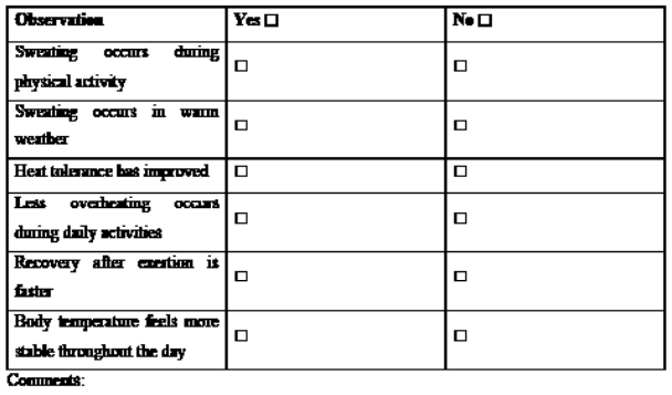
FLAG 1: Return of Sweating and Thermoregulation
Clinical significance: Suggests improving sympathetic cholinergic function and restoration of temperature regulation pathways.
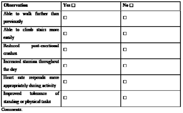
FLAG 2: Improved Exercise Capacity and Heart-Rate Responsiveness
Clinical significance: Suggests improving cardiovascular autonomic regulation, baroreflex function, vascular responsiveness, and exercise tolerance.
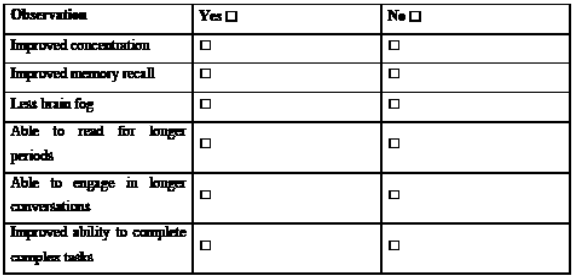
FLAG 3: Improved Cognitive Clarity and Mental Endurance
Clinical significance: Suggests improving cerebral perfusion, reduced autonomic burden, and enhanced cognitive resilience.
FLAG 4: Return of Motivation, Anticipation, and Emotional Engagement
Clinical significance: Suggests recovery within autonomic-limbic-hypothalamic pathways affecting motivation, reward processing, and emotional regulation.
FLAG 5: Return of Sexual Function and Sexual Ideation
Clinical significance: Suggests improving coordination between autonomic, endocrine, vascular, and emotional systems.
FLAG 6: Increased Facial Hair Growth and Return to Daily Shaving
Clinical significance: May reflect restoration of neuroendocrine function, androgen signalling, sleep quality, metabolic stability, and reduced chronic stress burden.
Overall Recovery Assessment
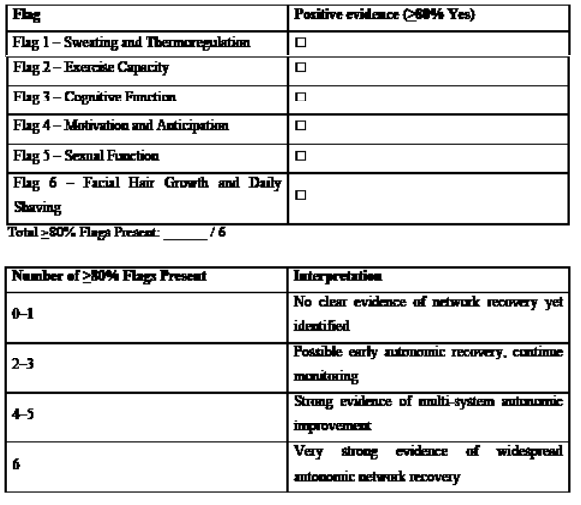
Important Clinical Note: No single flag should be considered diagnostic in isolation. The value of this assessment lies in recognising recovery occurring simultaneously across multiple autonomic domains. The emergence of several flags together supports the concept of autonomic network repair and may assist in distinguishing recovering secondary autonomic dysfunction from progressive primary autonomic neurodegenerative disease.
Reviewer Signature:
Date: