
Advances in Theoretical & Computational Physics(ATCP)
ISSN: 2639-0108 | DOI: 10.33140/ATCP
Impact Factor: 2.6

Research Article - (2025) Volume 8, Issue 3
Modeling of the Cervical Spine as a System with Several Degrees of Freedom Subjected to a Non-Periodic Excitation Force
2University of Kinshasa, Polytechnic Faculty, Department of Mechanical Engineering, at Kinshasa, Democratic, Republic of the Congo
3University of Kinshasa, Faculty of at Kinshasa, Democratic Republic of the Congo, Department of Internal Medicine; at Kinshasa, Republic of the Congo
Received Date: Feb 05, 2025 / Accepted Date: Jun 02, 2025 / Published Date: Jul 09, 2025
Copyright: ©2025 Georges Meya Kiala, et al. This is an open-access article distributed under the terms of the Creative Commons Attribution License, which permits unrestricted use, distribution, and reproduction in any medium, provided the original author and source are credited.
Citation: Kiala, G. M., Mossi, F. B., Temo, V. S., Mabenza, B. M., Bikuku, H, N., et al. (2025). Modeling of the Cervical Spine as a System with Several Degrees of Freedom Subjected to a Non-Periodic Excitation Force. Adv Theo Comp Phy, 8(3), 01-09.
Abstract
Background and Rationale: Previous cervical traction studies have investigated the impact of traction force intensity and duration of traction force on outcomes of cervical traction therapy.
Objective: The present study aims to demonstrate the impact of the intensity of the traction force and the time of the rise of this force on the measurement of separation during cervical spine traction.
Method: the human cervical spine was modeled as a system with eight degrees of freedom, an undamped system sub- jected, first of all, to a force of rectangular impulse then to a force of progressive intensity (sloping). The equations of motion describing this model were written and then solved using modal analysis. The intensity of the traction force and the time rise of the force were gradually modified in order to verify their impact on the evolution of the increase in intervertebral spaces.
Result: the numerical results showed that the intervertebral spaces calculated using different increments of the rise time (1’’, 5’’, 10’’, 15’’ and more) do not differ much and this remains proportional to the gradual increase in the tensile force applied (100N, 150N, 200N). A gradual increase in the intervertebral spaces has also been observed, these spaces being all the higher as the tensile force is greater.
Conclusion: This study confirms the need to monitor the speed of deployment of the traction force (rise time) as well as the progressive evolution of the widening of the intervertebral spaces, proportional to the traction force applied. However, it emphasizes that the time variation of the climb has almost no influence on the gain in intervertebral space. Hence the need on the one hand to monitor the speed of execution of the cervical traction to avoid damage to the anato- mo- physiological structures and on the other hand the downward revision of the duration of a cervical traction session.
Keywords
Cervical Spine Modeling, Intervertebral Space Assessment, Cervical Traction Force
Introduction
The modeling of the cervical spine is a design and use of an anticipated mathematical model, allowing a simplified under- standable representation, a form of prediction of the functioning of the cervical spine, thanks to these biomechanical properties. Thanks to this modeling it will be possible to parameterize, to calibrate or adjust the functioning of the spine in order to draw practical conclusions applicable in humans, during treatment of cervico-spondyloarthrosis by cervical traction.
Cervical spondyloarthrosis clinically manifesting itself in vari- ous forms, the most common of which is radiculopathy on ver- tebral disco impingement or cervico-brachial neuralgia (CBN) is one of three clinical syndromes of cervical spondyloarthrosis (CS) [1]. CBN is caused by compression or irritation of nerve roots [2,3]. The World Health Organization recommends that degenerative joint conditions should be managed by non-inva- sive, non-pharmacological therapeutic measures. This can be done manually or mechanically by applying a traction force to stretch the muscles, lengthen the soft tissues and create the space between the cervical vertebrae (figure1,2), [4 - 5]. The littera- ture suggests that for a good clinician, the traction force applied should neither be excessive nor insufficient. Thus, an appropri- ate pulling force must be fixed in advance.
The problem justifying this study is in the fact that previous re- search has shown that a traction force of 11 to 16 kg and a duration of 20 to 25 minutes was necessary to obtain a measurable change in the structures of the cervical spine and a good muscle relaxation [6,7]. However, there is not yet clear evidence on the impact of loading time rise (the evolution of the increase in intervertebral spaces) and on the results. Therefore, the present study aimed to evaluate the influence of the intensity of the traction force and the time of the rise on the progressive evolution of the intervertebral spaces, for a controlled, effective and safe cervical traction.
This study will provide the necessary information on the evaluation of the evolution of the spacing of the intervertebral spaces during a brutal loading, or rectangular impulse and during a pro- gressive loading following a slope or progressive impulse. was put on the impact of the time of the rise or the loading on the gain of intervertebral spaces observed. Finally, a comparison will be made in the discussion part with the experiment carried out in vitro in goats in previous study.
Methodology
The methodology adopted in this study and described below, is a combination of main two parts such as sustained traction of the human cervical spine, modeling and evaluation of intervertebral space separation measure. First, in the modeling part, the human cervical spine was modeled as an eight degrees of freedom, un- damped system and the sustained pulling force was first modeled as a rectangular impulse (The pulling force is applied suddenly at a certain level), then following a slope (The pulling force is applied gradually). In the evaluation of intervertebral space mea- surement, the equations of motion of the system under arbitrary excitation were written and solved using modal analysis.
Modeling Traction of The Human Cervical Rachis
Structure of the Human Cervical Spine
As shown in the images (figure1), the human cervical spine is made up of seven vertebrae (C1-C7) connected by soft tissues which are the intervertebral discs, ligaments, neck muscles and facet joints. The intervertebral discs act as a shock absorber during axial loading and carry the load from one vertebra to the other; the articular facets only support the load of one vertebra on the other; the ligaments and muscles of the neck stabilize the cervical spine.

Figure 1: Cervical spine: Vertebrae and ligaments

Figure 2: Facet joints and muscles
Physical Model of the Human Cervical Spine
To facilitate the analysis, a few assumptions were made on the human cervical spine model:
• The vertebrae are considered as rigid and dimensionless, identical bodies whose masses in grams per unit of vertebra are 6.3. [8].
• Discs and ligaments are treated as in parallel combination of linear parts, springs providing longitudinal stiffness.
Using the previous assumptions, the eight degrees of freedom model is established as shown in (Figure 3), where kij(i = OC,1,2,3,4,5,6,7;j=1,2,3,4,5,6,7,T1)represent different stiffnesses.

Figure 3: Axial traction of the cervical spine (model)
Stiffness values are given in Table 1 and Table 2 for, respectively, various intervertebral ligaments and discs.
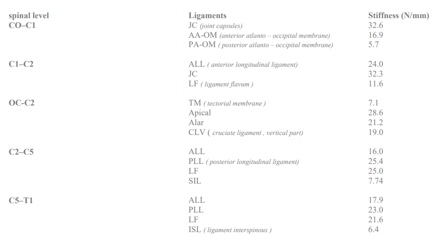
Table 1: Ligament stiffness [9]
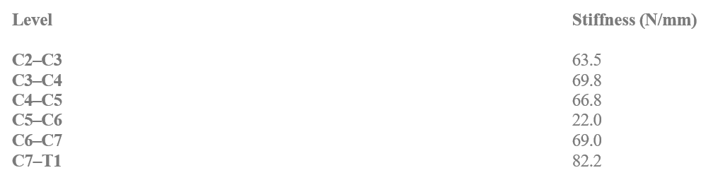
Table 2: Disc stiffness [9]
Modeling of The Axial Tensile Force
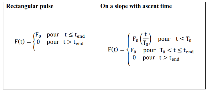
To model the pulling force, we first consider a rectangular pulse of magnitudeF0 (Figure 4), which represents a pulling force suddenly applied to the occiput.
We then apply a force gradually following a linear transition in time T0(rise time) to a constant load of amplitude F0, as shown in Figure 5, which represents a pulling force applied gradually to the occiput (OC).

Figure 4: Rectangular Pulse

Figure 5: Inclination of the slope with time of the rise
ASSESSMENT OF INCREASE IN INTERVERTEBRAL SPACE MEASUREMENT
Equations of Motion/Governing Equations
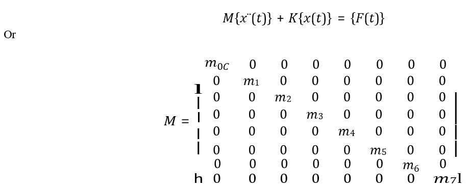
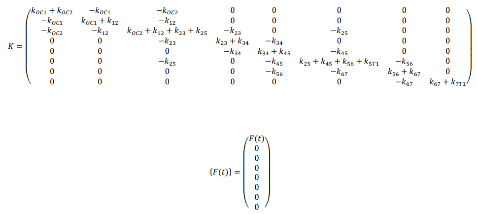
The cervical spine was modeled as an eight-degree-of-freedom system as shown in Figure 3 , so the motion of the system can be completely described by the coordinatesxOC (t), x1 (t), x2 (t), x3 (t), x4 (t), x5 (t), x6 (t), x7 (t)which define the vertical displacements of the vertebra represented by the massesmOC, m1, m2, m3, m4, m5, m6, m7at any time tfrom equilibrium positions.
Applying Lagrange’s principle, principle of derivative of the equations of motion to the model leads to the system of equa- tions which can be written in general matrix–vector form as:
Resolution
To solve the problem of governance equation, we applied the method of modal analysis exploiting the properties of orthogo- nality properties, vibration modes to transform the eight equa- tions of motion from a physical system where the equations are coupled to a modal coordinate system where the equations of motion are decoupled. Each of the decoupled equations can be solved independently. The flowchart displayed in the figure be- low summarizes the modal analysis method (figure 6):
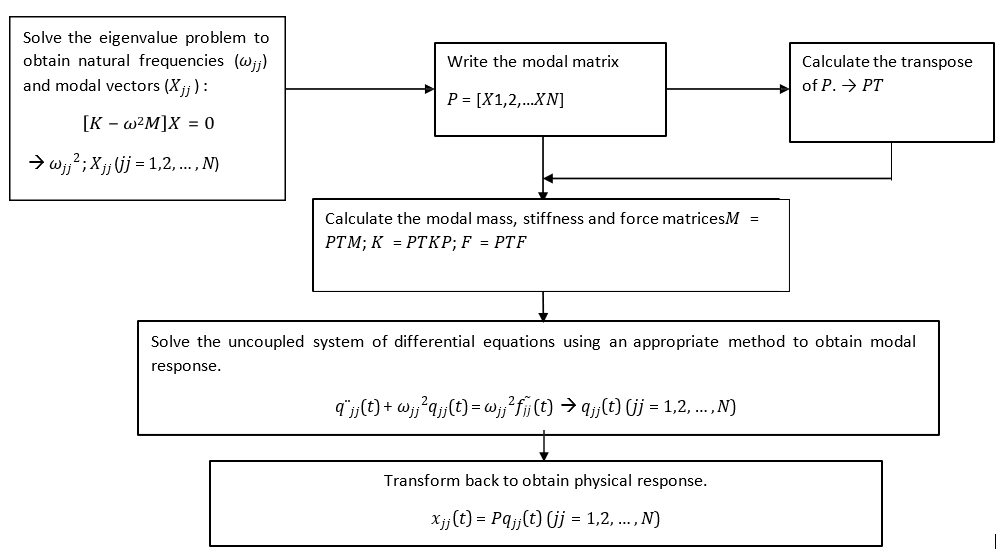
Figure 6: The Flowchart of the Modal Analysis Method
Results
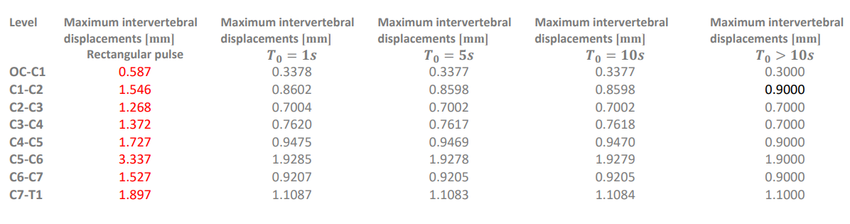
Table 3: Displacements (in mm) of the intervertebral spaces for an initial excitation force of 100N (F0=100N) applied without linear transiton (rectangular puls) and with linear transition.(1s,5s,10s, and >10s)
The application of the rectangular mode (application without linear transition over time), displacements (in mm) result in large intervertebral spaces ( first column).Concerning the mode with linear transition for the 100N excitation forces the displace- ments observed (in mm) are of small dimensions (3rd, 4th and 5th) column. Compared to the time rize of the (1s, 5s, 10s and more than 10s) we observed no impact on the evolution of the intervertebral spaces.
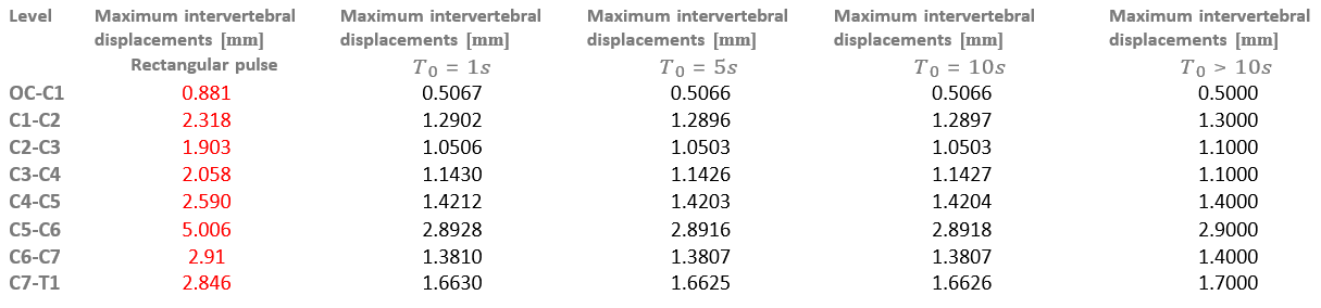
Table 4: Displacements (in mm) of the intervertebral spaces for an initial excitation force of 100N (F0=150N) applied without linear transiton (rectangular puls) and with linear transition (1s,5s,10s, and >10s)
The application of the rectangular mode (application without linear transition over time), displacements (in mm) result in large intervertebral spaces (first column). Concerning the mode with linear transition for the 150N excitation forces the displace- ments observed (in mm) are of small dimensions (3rd, 4th and 5th) column. Compared to the time rize of the (1s, 5s, 10s and more than 10s) we observed no impact on the evolution of the intervertebral spaces.
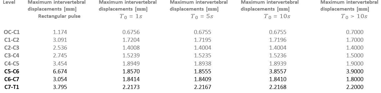
Table 5: Displacements (in mm) of the intervertebral spaces for an initial excitation force of 100N (F0=200N) applied abrupt- ly (Rectangular impulse case) and for a tensile force applied with a linear transition in time  0 (rise time: 1S , 5S , 10S and > 10S).
The application of the rectangular mode (application without linear transition over time), displacements (in mm) result in large intervertebral spaces (first column) Concerning the mode with linear transition (for the same initial excitation forces) the displacements observed (in mm) are of small dimensions (3rd, 4th and 5th) column. Compared to the time rize of the (1s, 5s, 10s and more than 10s) we observed no impact on the evolution of the intervertebral spaces.

Figure 7: Variation of intervertebral cervical spaces during cervical traction with an excitation intensity of 150 N, following a linear transition over time: 1, 5, 10 and > 10 minutes
For an excitation force of 150 N, the evolution of the rise of the intervertebral spaces during traction (blue, red, green and purple) is not at all influenced by the time of rise (1, 5, 10 and > 10 seconds).
Discussion
The influence of the intensity of the pulling force on the inter- vertebral space is studied for F0 = 100 N ,150 N and 200 N and the influence of the time rise is studied first for a rectangular impulse, then for a pulse with linear transition (T 0 = 1, 5 S , 10 And > 10 S ). Tables 3, 4 and 5 show the simulation results:
1 Each intervertebral space increases proportionally with F 0, this offers the possibility of a gradual readjustment of the traction force for a progressive tolerance in case of intolerance. This result was observed during in vitro experimentation in goats [10].
2 Contrary to E. Viel, in his publication dating from 1978 (Ann. Kinésithérapie 1978.5.27-39) which mentioned the possibility of subjecting the cervical spine to moderate traction of the order of 5 to 10 kg for a duration of 1 at 2 o’clock in the context of continuous traction, we must today specify, on the basis of this result, that this traction was only effective during the first 5 minutes. The rest of the time was only used to save the gain and not to exert traction. When the tensile force is applied gradually and for the same force intensity, there is no noticeable impact of the rise time on the intervertebral space (figure7). The same observation was made during the above-mentioned experiment. The advantage that derives from this result is that the duration of a cervical traction session can be reduced, because beyond certain duration the intervertebral spaces remain static.
When the traction force is applied without linear transition, as is the case of the rectangular impulse, the intervertebral spaces reach higher values. Such an elevation carries obvious risks to the soft tissues. Sudden maneuvers are therefore to be avoided. The 5th and 6th intervertebral spaces show increasing values compared to the overlying spaces. This observation, also made by E. Viel quoted above, is justified by the low rigidity of the C5-C6 intervertebral disc (22 N/mm),(table 2) compared to other discs above and below. However, this larger increase in the intervertebral spaces of the lower cervical segment benefits the strengthening of the therapeutic power of cervical traction, be- cause this segment is the privileged site of the disc degeneration responsible of disco-radicular impingement. According to our first study, the lower cervical level is more loaded than the upper levels [3].
Considering the result observed during the variation of the time of the ascent whatever the intensity of the traction load, we can argue that the duration of a cervical traction session can be re- vised downwards, because this it does not influence the variation of the intervertebral spaces significantly. The in vitro experiment in goats confirmed this study when it specified that beyond 5 minutes no increase in gain on the intervertebral spaces was ob- served [10].
Conclusion
This numerical model developed to investigate the impact of traction force intensity and rise time on the intervertebral spaces during sustained traction therapy on the human cervical spine, achieved its goal: The spaces calculated using different times of the climbs are quite the same. However, when the traction force is applied suddenly following a rectangular impulse, the intervertebral spaces reach higher values, which can be uncom- fortable or cause injury. This study helps to understand how loading conditions during traction therapy affect spaces. This understanding is vital for patient safety.
References
- Woods, B. I., & Hilibrand, A. S. (2015). Cervical radicu- lopathy. Journal of Spinal Disorders and Techniques, 28(5), E251-E259.
- Yang, F., Li, W. X., Liu, Z., & Liu, L. (2016). Balance chi- ropractic therapy for cervical spondylotic radiculopathy: study protocol for a randomized controlled trial. Trials, 17(1), 1-6.
- Carette, S., & Fehlings, M. G. (2005). Cervical radiculop- athy. New England Journal of Medicine, 353(4), 392-399.
- Madson, T. J., & Hollman, J. H. (2017). Cervical traction for managing neck pain: a survey of physical therapists in the United States. journal of orthopaedic & sports physical therapy, 47(3), 200-208.
- Alshami, A. M., & Bamhair, D. A. (2021). Effect of manual therapy with exercise in patients with chronic cervical ra- diculopathy: a randomized clinical trial. Trials, 22(1), 1-12.
- Chung, C. T., Tsai, S. W., Chen, C. J., Wu, T. C., Wang, D., Lan, H. C. H., & Wu, S. K. (2009). Comparison of the intervertebral disc spaces between axial and anterior lean cervical traction. European Spine Journal, 18, 1669-1676.
- Lenrow, D. A., & Ostrowski, J. (2008). Rehabilitation Meth- ods in Cervical Radicular Pain. In Interventional Spine (pp. 635-644). WB Saunders.
- Lowrance, E. W., & Latimer, H. B. (1967). Weights and variability of components of the human vertebral column. The Anatomical Record, 159(1), 83-88.
- Yoganandan, N., Kumaresan, S., & Pintar, F. A. (2001). Biomechanics of the cervical spine Part 2. Cervical spine soft tissue responses and biomechanical modeling. Clinical biomechanics, 16(1), 1-27.
- Kiala, G. M., Mosi, F. B., Temo, V. S., Mabenza, B. M.,Bikuku, H. N., Thsibamba, P. K., ... & Muamba, J. M. M. (2023). Deformation Threshold of Cervical Spine Struc- tures Subjected to Tensile Stress: In Vitro Experiment on Goats. Int J Phys Med Rehabil. 11:673.