

Advances in Bioengineering and Biomedical Science Research(ABBSR)
ISSN: 2640-4133 | DOI: 10.33140/ABBSR

Impact Factor: 1.7

Case Report - (2022) Volume 5, Issue 2
Minimally Invasive Endoscopic Treatment of Gastric Leiomyosarcoma: A Rare Case Report and Review of Literature
Received Date: Jan 31, 2022 / Accepted Date: Feb 04, 2022 / Published Date: May 10, 2022
Copyright: ©Copyright: ©2022 Gui-Xiang Kong. This is an open-access article distributed under the terms of the Creative Commons Attribution License, which permits unrestricted use, distribution, and reproduction in any medium, provided the original author and source are credited.
Citation: Gui-Xiang Kong, Xiao-Lu Su, Xiao-Jun Huang (2022) Minimally Invasive Endoscopic Treatment of Gastric Leiomyosarcoma: A Rare Case Report and Review of Literature. Adv Bioeng Biomed Sci Res 5(2): 108- 112.
Abstract
Background Since the c-kit was described in 1998, gastrointestinal LMS remains a relatively rare disease in the post-GIST era. Previous studies have reported that gastric LMS accounts for only 1% of gastric tumors. This report provides valuable endoscopic and endoscopic ultrasound data for rare diseases such as primary gastric leiomyosarcoma, which can add experience for clinical diagnosis to the current literature. Particularly, this case proved that the early-stage lesions can be treated with minimally invasive endoscopic treatment with high safety, good tolerance, and satisfactory clinical results.
Case Presentation A 63-year-old female patient visited our hospital mainly because of “epigastric pain and discomfort for 2 months.” After hospitalization, the relevant examinations were performed, and laboratory examination showed no obvious abnormalities. Abdominal contrast-enhanced CT found slight thickening of the gastric body wall. Ultrasonic gastroscopy revealed a round protuberant mass that originated from the muscularis mucosa on the great curvature of the gastric body, with a largest diameter of 16 mm. After full communication with the patient and exclusion of contraindications, the patient successfully underwent diagnostic endoscopic submucosal dissection (ESD) surgery. The postoperative pathological findings, immunohistochemical staining panel and molecular detection all improved that the mass was leiomyosarcoma. After 18 months of follow-up, the patient was in good condition, and there was no relapse on gastroscopy and abdominal enhanced CT.
Conclusion Gastrointestinal leiomyosarcoma can be successfully resected by minimally invasive endoscopic treatment with high safety, good tolerance, and satisfactory clinical results. This shows that ESD is an effective method for the treatment of these diseases and has promising prospects for clinical application.
Keywords
Gastric Leiomyosarcoma; Endoscopy; Minimally Invasive Treatment
Introduction
Gastric leiomyosarcoma (LMS) is a rare gastrointestinal tu-mor, with limited studies reporting on its clinical imaging and endoscopy findings. Thus, subepithelial tumors, such as LMS and gastrointestinal stromal tumors (GISTs), are difficult to differentiate. Especially for endoscopic manifestations, them are subepithelial tumors with solid round appearance, normal surface covering mucosa, tough or hard texture, and originated from muscularis mucosae to muscularis propria. Large tumors may present with ulcers and other manifestations. In general, only deep biopsy or endoscopic ultrasound-guided fine-needle aspiration (EUS-FNA) tissue can be differentiated by immuno-histochemical detection. As a result, pathological immunohis-tochemistry has become the key to differential diagnosis and endoscopic ultrasonography (EUS) can be of great value in its diagnosis. In particular, minimally invasive endoscopic therapy has a high safety profile and satisfactory effect; based on the case described herein, it is promising for clinical application. Herein, we provide the endoscopic image findings and describe current practices in endoscopic diagnosis and minimally invasive treat¬ment both at home and abroad.
Case Report
A 63-year-old female patient visited our hospital with chief com¬plaints of “epigastric pain and discomfort for 2 months.” The patient underwent esophagogastroduodenoscopy (EGD) for screening and a mass was detected at the gastric body. Upon ex¬amination, we noted that she had a poor diet and had lost about 2 kg of body weight. The patient had a 15-year history of hypertension that was well-controlled with oral drugs.
At hospitalization, the patient vital signs were stable. There was mild tenderness in her left upper abdomen. Laboratory tests showed no abnormalities. Abdominal contrast-enhanced CT found slight thickening of the gastric body wall. Repeat EGD revealed a polyp-like mass protruding into the gastric lumen was seen, indicating roughness and unevenness with ulcer formation, with mucosal aggregation, and when touched with biopsy for-ceps, the mass was found to be hard and fixed(Fig. 1A).EUS revealed a round protuberant mass that originated from the muscularis mucosa on the greater curvature of the gastric body, with a maximum diameter of 16 mm (Fig. 2).Considering the tumor was mesenchymal tissue origin, and after pursuing the medical history, it was found that the lesion changed significantly within a short period of time, with potential malignancy. In addition, the lesion could be completely resected. At the same time, the low positive rate of ordinary endoscopic biopsy was also considered, and the cost of EUS to take biopsy was expensive but not fully confirmed, therefore, after full communication with the patient and exclusion of contraindications, the patient successfully underwent diagnostic endoscopic submucosal dissection (ESD) surgery (Fig. 1B-D).
Figure 1: Endoscopic features: A protruding tumor is in the gastric lumen with the ulceration on the surface and it has a positive elevation sign after intraoperative after submucosal injection. Eventually, the tumor is completely removed by ESD surgery
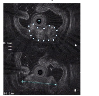
Figure 2: Features under endoscopic ultrasonography: The lesion originates from the second muscularis mucosa layer, and has an uneven internal echo and cystic space.The white dots are the outline of the tumor.
The postoperative pathological examination of the surgical specimens (Fig. 3) show pleomorphic, spindled, eosinophilic, and mitotic figures in the tumor cells, with approximately 10 mito-ses per high-power fields, and abundant blood vessels around the tumor. The immunohistochemical staining panel (Fig. 3) included vimentin (+), desmin (+), epithelial membrane anti¬gen (-), CD117 (-), DOG-1 (-), S100 (-), smooth muscle actin (-), synapsin (-), CD56 (-), p63 (-), P40(-), thyroid transcription factor-1(-), CK5/6 (-), C-reactive protein (-), transducer-like enhancer of split 1(-), HMB45(-), SOX10(-), PMS-2(+), MSH- 2(+), MSH-6(+), and Ki67 (40%).Vascular endothelial cells were immunostained with anti-CD34 antibody. Based on these results, LMS was considered as the diagnosis; however, GIST was not excluded. Subsequently, molecular detection showed that exons 9, 11, 13, and 17 of the c-kit gene and exons 12 and 18 of the PDGFRA gene were of the wild type. Since this was a rare case, the specimens were taken to two other authorita¬tive pathological institutions for consultation, and a diagnosis of LMS was finally established. The patient recovered well and was discharged from the hospital.

Figure 3: Postoperative pathological findings A: hematoxylin and eosin (HE) stain; B: 40x HE staining; C: 400x HE staining; D: tumor cells are desmin positive; E: tumor cells are vimentin positive; F: tumor cells are CD117 negative; G: tumor cells are DOG-1 negative; H: tumor cells are CD34 negative
After 20 months of follow-up, the patient remains in good con-dition, and follow-up gastroscopy and enhanced CT of the abdo¬men indicated no relapse.
Discussion
Since c-kit staining was first described in 1998, which led to the recognition of GISTs; ever since, gastrointestinal LMS has been a relatively rarely diagnosed disease. It has been reported that gastrointestinal LMS accounts for less than 0.5% of all mesen-chymal tumors. The most commonly site in LMS is the colon, while the least site is the stomach [1]. Yamamoto et al. reported 55 cases of gastrointestinal LMS, and only four cases occurred in the stomach, indicating that the stomach is the least common site. In March 2018, Japan published the first report in English, describing the case of a patient with gastric LMS treated by ESD, thereby indicating the safety and efficacy of the procedure [2, 3]. In March 2020, Rajat et al., after a comprehensive search of multiple databases and additional sources, reported the cases of 16 patients with gastric LMS [4]. The mean age of LMS onset in these patients was 52 years, and the ratio of males to females was equal. The most common symptom in these patients was gastrointestinal bleeding, followed by abdominal pain, weight loss, and gastroesophageal reflux, with the average tumor size being 4.80 cm. The tumors occur most frequently in the body of the stomach, especially in the upper part of the gastric body, fol¬lowed by the antrum and fundus, and appear as either a solitary lesion or multiple lesions. These lesions rarely affect gastrointes¬tinal function in their early stage and are often treated in the mid¬dle and late stages, wherein the patient typically presents with hemorrhage, anemia, and/or an epigastric mass. In June 2021, Toru Takagi et al. summarized that only two cases were origi¬nated from the muscularis mucosae (table1) [5]. However, due to the lack of known clinical manifestations, the tumors can be easily misdiagnosed as a peptic ulcer, gastric cancer, subepithe¬lial tumors-such as stromal tumors and other diseases. The lack of understanding of the disease is the main cause of overlooked diagnosis and misdiagnosis, while timely diagnosis is the main factor affecting the survival rate of patients with gastric LMS.
Table 1: Cases of leiomyosarcoma of the stomach derived from muscularis mucosa reported since 2000s
|
Case no |
Age/sex |
Location |
Size (cm) |
Endoscopic appearance |
Histological location |
Treatment |
Outcome |
References |
|
1 |
72/F |
gastric body |
2.5 |
Polypoid lesion |
Muscularis mucosae |
Endoscopic resection |
not mentioned |
Agaimy A,et al |
|
2 |
26/M |
gastric fundus |
7.2 |
Large lobu- lated |
Muscularis mucosae |
Surgical resection |
Alive (1 month) |
Aggarwal G,et al |
EGD is the main method used for the diagnosis of the disease, and the shape of the mass can be directly observed under dy-namic conditions. Polyp-like masses protruding into the gastric lumen can often be observed by EGD. Often, the surface of the mass is rough, with ulcers, erosion, and bleeding, and the forma-tion of gouge- or navel-like ulcers is significant for diagnosis. A biopsy can be performed preoperatively for an immunohisto-chemical diagnosis, which provides a reliable basis for treatment [6]. In the present case, when touched with biopsy forceps, the mass was tough or hard and relatively fixed. After full discussion this lesion was removed directly by ESD. Pathological immuno-histochemistry of the current patient indicated that the mass was CD117(-) and DOG-1(-), and molecular detection indicated the presence of wildtype c-kit and PDGFRA genes, which ruled out the possibility of GIST. After consideration of other characteristics, such as vimentin(+), desman(+), and cell morphology, the case was finally diagnosed as LMS. Of course, LMS does not require molecular testing, which is designed to rule out wildtype GISTs. The important immunostaining of LMS are CD117, DOG-1, CD34, desmin, vimentin, smooth muscle actin, synapsin and Ki67, etc.
In recent years, EUS has become highly valued for diagnostics. It can clearly show the structural characteristics of each layer of the gastric wall to help understand the depth and extent of tumor invasion; in particular, biopsies of the submucosal mass can be performed under EUS guidance, with a diagnostic confirmation rate of more than 90%. It also has good sensitivity and speci¬ficity for the diagnosis of gastric LMS [7]. EUS can also help visualize the blood vessels around the lesion, which can provide justification for and necessary guidance during endoscopic resection [8]. At the same time, it can provide more targeted and informed postoperative follow-up [9].
Gastric LMS is not sensitive to radiotherapy or chemotherapy, but its prognosis is better than that of gastric cancer [10]. Be-cause clinical cases are rare, a large sample of data and standardized treatment do not exist to date. Partial or total gastrectomy is the primary treatment for gastric LMS. According to the current literature and the characteristics of the EUS images from the present patient, gastric LMS can originate from superficial structures such as the muscularis mucosa layer. Additionally, lymph node metastasis appears to be rare, so ESD has several advantages over traditional surgery, such as less trauma, faster recovery, and lower costs. Importantly, patients’ postoperative quality of life is not lowered. Moreover, compared with ordinary biopsy or EUS-FNA, complete resection of the lesion can greatly improve the accuracy of differential pathological diagnosis due to larger tissue specimens for mmunohistochemistry evalua- tion and molecular detection. In the present case, ESD was used to remove the tumor, achieving radical resection with a good patient prognosis as the follow-up continues to date. Of course, the flaw of our concern is that this case has been followed up for less than three years. But This case and a few other reports strongly demonstrate the feasibility of ESD of LMS.
In short, although gastric LMS is rare, with a comprehensive evaluation and clear diagnosis, early-stage lesions can be treated with minimally invasive endoscopic surgery with high safety, good tolerance, and satisfactory clinical results. This shows that minimally invasive endoscopic resection is an effective method for the treatment of LMS and has promising prospects for clini¬cal application. At the same time, this report also provides valuable EGD and EUS data for primary gastric LMS, adding details about the clinical diagnosis to the current literature.
Acknowledgement
We thank the teachers in the Endoscopy Center and the Pathology Department for their support of the pictures.
Ethical Statement
All procedures followed have been performed in accordance with the ethical standards laid down in the 1964 Declaration of Helsinki and its later amendments.
Informed consent statement: Informed written consent was obtained from the patient for publication of this report and any accompanying images.
Conflict-of-interest statement: The authors declare that they have no conflict of interest.
References
- Miettinen M, Fetsch JF (2006) Evaluation of biological potential of smooth muscle tumours. Histopathology 48: 97-105.
- Yamamoto H, Handa M, Tobo T, Nokitaka Setsu, Kohei Fujita, et al. (2013) Clinicopathological features of primary leiomyosarcoma of the gastrointestinal tract following recognition of gastrointestinal stromal tumours. Histopathology 63: 194-207.
- Sato T, Akahoshi K, Tomoeda N, Norikatsu Kinoshita, Ma-saru Kubokawa et al. (2018) Leiomyosarcoma of the stomach treated by endoscopic submucosal dissection. Clin J Gastroenterol. 11: 291-296.
- Garg R, AlRajjal A, Berri R, Barawi M (2020) Primary Gastric Leiomyosarcoma: a Case Report and Review of the Literature. J Gastrointest Cancer 51: 335-340.
- Takagi T, Saito S, Yokota S, Yuki Kaneko, Kazuya Taka-hashi, et al. (2021) Laparoscopic and endoscopic cooperative surgery for leiomyosarcoma of the stomach: a case report with a review of the literature. Surg Case Rep 7: 146-153.
- Wang Y, Zhou X (2015) Newly recognized gastrointestinal mesenchymal tumor. Journal of Clinical and Experimental Pathology 2015: 841-845.
- Wu A, Li R, Shi D, Ling Li, Deqing Zhang, et al. (2015) Diagnostic value of endoscopic ultrasonography for submucosal tumors of upper gastrointestinal tract. Chinese Journal of Gastrointestinal surgery 18: 1136-1138.
- Soufi M, Errougani A, Chekkof RM (2009) Primary gastric leiomyosarcoma in young revealed by a massive hematem-esis. J Gastrointest Cancer 40: 69-72.
- Chang L, Zhang K, Zhang M, Lise Ingemann Jensen, Mark Berner Hansen (2016) Diagnosis, treatment and follow-up of submucosal tumors of upper digestive tract. World Journal of Chinese digestion 24: 765-774.
- Shi Y, Jia B (2014) Progress in the treatment of soft tissue sarcoma. Chinese Oncology Clinic 2014: 1556-1560.