
Journal of Agriculture and Horticulture Research(JAHR)
ISSN: 2643-671X | DOI: 10.33140/JAHR
Impact Factor: 1.12

Research Article - (2025) Volume 8, Issue 2
Isolation and Analysis of Bacteria from Masa Sold at Gwako Market
2Sheda Science and Technology Complex, Nigeria
3National Primary Health care development agency, Abuja, Nigeria
Received Date: Jun 05, 2025 / Accepted Date: Jul 07, 2025 / Published Date: Jul 28, 2025
Copyright: ©©2025 Joy Chinyere Ogbu, et al. This is an open-access article distributed under the terms of the Creative Commons Attribution License, which permits unrestricted use, distribution, and reproduction in any medium, provided the original author and source are credited.
Citation: Ogbu, C. J., Udo, E. G., Precious. N., Mambula. M. S., S. U. (2025). Isolation and Analysis of Bacteria from Masa Sold at Gwako Market. J Agri Horti Res, 8(2), 01-06.
Abstract
Masa, a popular traditional fermented rice cake, is widely consumed as a ready-toeat (RTE) food in Nigeria. This study was aimed at investigating the bacteriological quality of Masa sold at Gwako Market. Twelve (12) different samples were aseptically collected from four different vendors in a sterile container and plated on agar using spread plate method in which three aerobic mesophilic bacteria was isolated which are Bacillus subtilis, Pseudomonas aeruginosa and Staphylococcus aureus. The result from this study showed that there is high bacterial count in the Masa samples. The total bacterial count (TBC) of bacterial isolates ranges from 2.0 x10⁸ to 3.0 x 10⁸ CFU/g and the total colifoms count (TTC) ranges from 1.5x10⁶ to 2.7x10⁶. This study suggests that Masa could pose a threat to health of consumer based on the types and numbers of pathogenic microorganisms isolated from the samples and are therefore unfit for human consumption. Therefore, adequate sanitary measures should be taken at each stage from production, storage to consumption.
Introduction
Masa is a traditional Nigerian snack that is produced from fermented rice or millet, commonly consumed in various regions of the country [1]. Due to its popularity as a street food, Masa is often sold in local markets, making it readily accessible to consumers [2]. Its preparation involves soaking, grinding, and fermenting the grains, followed by frying to achieve a desirable texture and flavor [3]. Street food vendors like Masa provide a substantial amount of employment and substantially increase household incomes in many developing country cities and towns. The thickness, weight, volume, and volume index of Masa are among its physical properties while its flavor, aroma, texture, and degree of sourness are among its sensory qualities, and it has been shown that these change based on the type of cereal grain used to produce Masa [4].
However, the processing and selling of Masa in such informal settings can lead to potential health risks due to contamination by pathogenic bacteria [5]. Food safety concerns have become increasingly prominent, especially regarding the microbial quality of street-vended foods [6]. The Food and Agriculture Organization of the United Nations (2012) estimates that 2.5 billion people eat street food daily [7]. Thus, guaranteeing the supply of safe food has become one of the main challenges and concerns for producers, consumers, and public health officials globally [7]. Foods that are highly contaminated with spoiling and pathogenic bacteria can cause foodborne illnesses and are unpleasant [8].
The safety of street food like Masa is a big concern because of the high likelihood of contamination by pathogenic bacteria including Salmonella, Shigella, and E. coli, which are known to cause foodborne diseases and pose serious health hazards when present [6]. Foodborne illnesses, both acute and chronic, can be brought on by food contaminants like pathogenic bacteria found in Masa [5]. Foodborne illnesses can be brought on by viruses, bacteria, parasites, and toxins with two prevalent infections that cause 52 000 and 37 000 deaths annually, respectively, being Salmonella and E. coli [9]. The World Health Orgaonization (WHO) reports that foodborne illnesses caused by microbial contamination are a significant public health issue, particularly in developing countries like Nigeria [10]. Various studies have highlighted the prevalence of bacteria such as Escherichia coli, Salmonella spp., and Staphylococcus aureus in street foods, often linked to inadequate hygiene practices during preparation and handling [11&12]. The preparation of Masa involves minimal cooking, which may not effectively eliminate harmful bacteria if proper hygienic measures are not adhered to [13].
Materials and Methods
![]() Study Area
Study Area
Gwako Market is a local market situated in Gwagwalada Area Council, located in the Federal Capital Territory (FCT) of Nigeria. The market serves as a major hub for the sale of various goods, including food items, clothing, and household products. Gwako Market is geographically, it is located between latitude 9.06275o N and longitude 6.99458oE .
![]() Sample size
Sample size
A total of Twelve (12) samples were randomly collected from four (4) different Masa vendors around different vending locations at Gwako Market.
![]() Sample collection
Sample collection
A total of Twelve (12) samples was collected from vendors at Gwako Market, Abuja. Three (3) samples were collected from each of the 4 different vendor visited. The samples were bought and collected in a sterile container from Masa vendors and then transported to the Microbiological Laboratory in the University of Abuja for examination. The sample were processed within 3 – 5h for microbial analysis. All of the samples were processed in the laboratory by strictly adhering to aseptic guidelines using the standard microbiological techniques.
![]() Materials and Reagents
Materials and Reagents
Nutrient agar, Eosin-methylene blue agar, Salmonella Shigella agar, petri dishes, microscopic slides, test tubes, pipettes, buffered peptone water, distilled water and hydrogen peroxide (H2O2).
![]() Preparation of media
Preparation of media
All media were prepared in accordance with the manufacturer’s protocol. Media used included Nutrient agar, Eosin-methylene blue agar and Salmonella Shigellaagar. They were autoclaved at 121oC for 15 minutes. The cooled media were poured into Petri dishes and then allowed to cool and solidify. These were used for the isolation, identification, growth and maintenance of microorganisms.
![]() Eosin-Methylene Blue Agar
Eosin-Methylene Blue Agar
Exactly 3.6g of Eosin-methylene blue agar was weighed and poured into a sterile conical flask. 100mL of water were added and swirled to dissolve. The medium were autoclave at 121oC for 15 minutes. The medium were allowed to cool to 45 oC-50 â°C before being poured aseptically into sterile Petri dishes. The medium was then allowed to cool and solidify on the laboratory bench before use.
![]() Nutrient Agar
Nutrient Agar
Exactly 5.6g of Nutrient was weighed and poured into a sterile conical flask, then 200mL of water were added and swirled to dissolve. The medium were autoclave at 121°C for 15minutes. The medium was then allowed to cool to 45°C-50°C before dispensing into sterile petri dishes and allowed to cool and solidify on the laboratory bench before use.
![]() Salmonella Shigella Agar
Salmonella Shigella Agar
Exactly 6.3g of Salmonella Shigella agar was weighed and poured into a sterile conical flask. 100mL of water were added and swirled to dissolve. The medium were autoclave at 121oC for 15 minutes. The medium was then allowed for some seconds before being poured aseptically into a sterile Petri dish. The medium was then allowed to cool and solidify on the media at 45oC - 50oC on the laboratory bench before use.
![]() Sample Preparation
Sample Preparation
Upon arrival at the laboratory, the Masa samples collected from Gwako Market were subjected to standardized sample preparation procedures to ensure the accurate isolation and analysis of bacterial contaminants. Each Masa sample was first visually inspected for any signs of spoilage or contamination. After inspection, approximately 25 grams of each Masa sample was aseptically transferred into a sterile blender or stomacher bag. To each sample, 225 milliliters of sterile buffered peptone water (BPW) were added to maintain a 1:10 dilution ratio. The samples was then homogenized for 1–2 minutes to evenly distribute any bacteria present throughout the Masa mixture. This homogenization step ensured that the bacteria were adequately dispersed within the sample, allowing for accurate downstream microbial analyses. Following homogenization, serial dilutions were performed to reduce the concentration of bacteria to manageable levels for plating and enumeration.
![]() Serial Dilution
Serial Dilution
Serial dilution was performed by using test tubes in which 9ml of distilled water was poured into each test tube using a syringe that were sterilized in between by putting the needle over a Bunsen burner and waving it gently to reduce the heat. One gram (1g) of each of the already finely crushed Masa samples is poured into the first test tube (10¹), the test tube is swirled gently to enable the distilled water and the sample mix properly. One ml (1ml) of the mixture were taken from the test tube and added to the next test tube (10²) another one ml (1ml) were taken from the second test tube and added to the last test tube(10³). Then process was then repeated multiple times to create a series of solutions with decreasing concentrations.
![]() Isolation and Identification of Bacteria
Isolation and Identification of Bacteria
The bacteria were isolated and enumerated by growing them on selective and nonselective mediums such as Eosin-methylene blue agar and Nutrient agar which were used for total viable bacterial count (TVBC) and for total coliform respectively. After 24 hours of incubation at 37°C, bacterial colonies were identified based on the color depicted by the colonies on each agar plate. Having obtained pure cultures, further characterization with biochemical tests namely; Gram’s staining catalase, citrate, oxidase, indole and methyl red test.
Biochemical /Confirmatory Tests
![]() Gram Staining
Gram Staining
A loopful of sterile distilled water was dropped on a clean grease free slide by using a sterile inoculating loop after which an isolate from the culture were mixed with the water on the slide. The smear were allowed to air dry and then heat fixed gently by passing it quickly over a Bunsen flame. The smear was flooded with crystal violet solution for 60 seconds (one minutes) and rinsed with water. The smear was again flooded with Lugol’s iodine for 30 seconds and rinsed with water, 70% alcohol were poured on the slide for 10 seconds and then rinsed with water. It was then counterstained with Safranin for 60 seconds, then rinsed with water and allowed to dry. The slide was then observed under oil immersion objective. This procedure were repeated for isolates obtained from each sample. Gram positive cells remained purple while Gram negative cells appeared red or pink.
![]() Catalase Test
Catalase Test
A drop of hydrogen peroxide was put on a glass slide. Colonies were picked with a sterile loop and added to the drop. Observation were made for bubbles production. Colonies that have catalase are able to break down hydrogen per oxide into water and oxygen, which can be seen in the form of air bubbles leaving the solution.
![]() Citrate Test
Citrate Test
Unique colonies was picked with a sterilized wire loop and streaked across the plates containing citrate agar without breaking the agar and was then incubated for 24 hours. Data were collected based on color change.
![]() Methyl Red Test
Methyl Red Test
Five millimeters of glucose phosphate broth (1 g glucose, 0.5% KH2PO4, 0.5% peptone and 100 mL distilled water) were dispensed in clean test tubes and sterilized. The tubes was then inoculated with the test organisms and incubated at 37°C for 48 h. At the end of incubation, few drops of methyl red solution were added to each test and color change were observed. A red color indicates a positive reaction.
![]() Oxidase Test
Oxidase Test
This test was carried out by dropping 2–5 drops of a freshly prepared oxidase (paminodimethylanine) reagent on a filter paper, the suspected organisms is picked using a sterile wire loop and mix with the oxidase reagent. A change from the normal color to deep purple means a positive result, while no change means negative.
![]() Coagulase Test
Coagulase Test
About 2 - 3 drops of normal saline was added on a clean grease free slide to the normal saline mix the suspected organism and 1 – 2 drops of plasma was then and rocked, the presence of agglutination means a positive result while no agglutination means a negative results.
![]() Indole Test
Indole Test
An isolate was inoculated in a test tube containing 3mL of sterile trytone water and incubated at 37oC for 24 hours. After 24 hours, 0.5mL of kovac’s reagent were added and shaken gently. This procedure were repeated for isolates obtained from each sample. Examination for a red color in the surface of the layer within 10minutes means positive, while no color change means negative.
Results
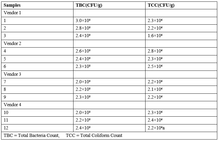
Table 1: Colony Forming Units of Bacterial Isolates (cfu/G)Keys
|
Isolate |
Shape |
Color |
Edge |
Elevation |
Surface |
Optical |
Suspected Organism |
|
A |
Cocci |
Cream |
Entire |
Raised |
Smooth |
Opaque |
Staphylococcus aureus |
|
B |
Rod |
White |
Entire |
Flat |
Rough |
Translucent |
Bacillus subtilis |
|
C |
Rod |
Blue |
Entire |
Flat |
Smooth |
Opaque |
Pseudomonas aeruginosa |
Table 2: Morphological Characteristics of Bacterial Isolates
|
Isolate |
Gram rxn |
Indole |
Catalase |
Methyl red |
Citrate |
Oxidase |
Coagulase |
Probable Organism |
|
A |
+ |
- |
+ |
+ |
+ |
- |
+ |
Staphylococcus aureus |
|
B |
+ |
- |
+ |
- |
+ |
+ |
- |
Bacillus subtilis |
|
C |
- |
- |
+ |
- |
- |
+ |
- |
Pseudomonas aeruginosa |
Table 3: Biochemical Tests of Probable Organisms
|
Vendor |
Number of Samples |
Isolates |
Frequency |
|
Vendor 1 Staphylococcus aureus |
03 |
Bacillus subtilis |
03 |
|
Staphylococcus aureus |
03 |
||
|
Vendor 2 |
03 |
Bacillus subtilis |
01 |
|
Pseudomonas aeruginosa |
02 |
||
|
Staphylococcus aureus |
02 |
||
|
Vendor 3 |
03 |
Bacillus subtilis |
03 |
|
Pseudomonas aeruginosa |
02 |
||
|
Vendor 4 |
03 |
Bacillus subtilis |
02 |
|
Staphylococcus aureus |
03 |
Table 4: Distribution of Microbial Isolates
Discussion
This study was conducted to investigate the bacteriological quality of Masa sold at Gwako Market. The result from this study showed that there is high bacterial count in the Masa samples. The total bacterial count (TBC) of bacterial isolates from four different vendors ranges from 2.0 x10⁸ to 3.0 x 10⁸ CFU/g and the total colifoms count (TTC) ranges from 1.5x10⁶ to 2.7x10⁶ indicate significant microbial contamination in Masa sold at Gwako Market which correlates with the findings of and on studies on ready-to-eat (RTE) foods, where high bacterial loads were attributed to poor hygiene practices during preparation and vending [15&16].
In this Study, three (3) pathogenic bacteria was isolated from Masa samples which were Bacillus subtilis, Pseudomonas specie and Staphylococcus aureus which is in agreement who in is study of microbial quality evaluation of Masa processed and sold within University of Maiduguri campus, Nigeria isolated different type of microorganism like Shigella spp, Salmonella spp, Pseudomonas spp, E coli and Staphylococcus [1]. The detection of Bacillus subtilis is of serious public health importance because of its ability to cause a wide range of infections especially food-borne intoxication, it is well known as a cause of food poisoning.
Bacillus cereus is a toxinproducing facultative anaerobic gram-positive bacterium. It commonly found in the environment and can contaminate Masa. When ingested, this toxin can cause gastrointestinal illness, which is the commonly known manifestation of the disease [17].The detection of Pseudomonas aeruginosa further highlights potential contamination from water sources used during Masa preparation, as noted in similar studies. According to the presence of P [18]. aeruginosa in RTE foods is often linked to the use of unclean water and inadequate storage conditions. In a study by the bacteria Pseudomonas aeruginosa were measured as the major cause of food spoilage and food-borne diseases [19].
This survey assesses the prevalence, antibiotic resistance properties, and virulence factors distribution in P. aeruginosa bacteria isolated from Masa products. reported S. aureus as a predominant contaminant in street foods, emphasizing its association with poor personal hygiene among food handlers [15]. According to, contamination with Staphylococcus aureus has been linked to carriage in nasal passages of food handlers or by infected workers [20]. S. aureus has been known to cause Staphylococcal food-borne disease (SFD), which is one of the most common food-borne diseases worldwide resulting from the contamination of food by preformed S. aureus enterotoxins [21].
Reported that the level and magnitude of sanitary practices/ hygienic practices by food vendors can have great influence on the bacterial load in vended foods [22]. Bacterial isolation and identification from food vendors is crucial for assuring food safety and protecting public health. This study's findings highlight the presence of harmful bacteria as well as the distributions and frequency of occurrence of bacteria isolated from food vendors, emphasizing the significance of implementing effective food safety measures.
Conclusion
This study was aimed at investigating the bacteriological quality of Masa sold at Gwako Market, presented in the isolation of three (3) bacterial isolates were identified in this study which are Bacillus subtilis, Staphylococcus aureus and Pseudomonas aeruginosa. The total bacterial load of Masa obtained from different vendors ranged from 2.0 x10⁸ to 3.0 x 10⁸ CFU/g and the total colifoms count (TTC) ranges from 1.5x10⁶ to 2.7x10⁶. The bacteria frequency of occurrence was determined from different vendors in descending order (from highest to lowest) Bacillus subtilis with percentage of 42.9%, Staphylococcus aureus with the percentage of 38.1% and Pseudomonas aeruginosa with percentage of 19.0%. This study suggests that Masa could pose a threat to health of consumer based on the types and numbers of pathogenic microorganisms isolated from the samples in this study and are therefore unfit for human consumption.
Recommendations
1. Vendors should undergo mandatory training on food safety and hygiene practices.
2. Regulatory bodies, such as public health agencies, should conduct periodic inspections of food vending sites.
3. Community education programs should be organized to raise awareness among consumers about the risks associated with consuming improperly handled foods.
4. Vendors should be educated on the importance of personal hygiene, including proper handwashing, wearing protective clothing (e.g., gloves, aprons, and hairnets), and avoiding direct hand contact with prepared foods.
References
- Badau, M., Shadrach, N., & Ogori, A. (2018). Microbial quality evaluation of masa processed and sold within University of Maiduguri campus. Journal of Bacteriology and Mycology Open Access, 6(3), 205-209.
- Da Silva, S. A., Cardoso, R. D. C. V., Góes, J. Â. W., Santos, J.N., Ramos, F. P., de Jesus, R. B., ... & da Silva, P. S. T. (2014). Street food on the coast of Salvador, Bahia, Brazil: A study from the socioeconomic and food safety perspectives. Food control, 40, 78-84.
- Adeleke, O. E., Abayomi, A. A. and Fola-Alabi, K. (2022). Microbial quality of street-vended foods in Nigeria: A review. Journal of Food Safety, 43(3): e12950.
- Olubunmi, O. A., Adetola, O. D. and Olanrewaju, T. O. (2023). Nutritional and microbiological evaluation of masa from different regions of Nigeria. International Journal of Food Science, 1-10.
- Igwe, E. C., Oyebode, Y. B., & Dandago, M. A. (2013). Effect of fermentation time and leavening agent on the quality of laboratory produced and market samples of masa (a local cereal based puff batter). African Journal of food, agriculture, nutrition and development, 13(5), 8415-8427.
- Nazni, P., & Jaganathan, A. (2014). Study on microbial analysis of street-vended food samples sold in Salem District. International Journal of Research in Biological Sciences, 4(3), 75-78.
- Bereda, T. W., Emerie, Y. M., Reta, M. A., & Asfaw, H. S. (2016). Microbiological safety of street vended foods in Jigjiga City, Eastern Ethiopia. Ethiopian journal of health sciences, 26(2), 163-172.
- da Silva, S. A., Cardoso, R. D. C. V., Góes, J. Â. W., Santos, J.N., Ramos, F. P., de Jesus, R. B., ... & da Silva, P. S. T. (2014). Street food on the coast of Salvador, Bahia, Brazil: A study from the socioeconomic and food safety perspectives. Food control, 40, 78-84.
- Hertanto, B. S., Nurmalasari, C. D. A., Nuhriawangsa, A.M. P., Cahyadi, M., & Kartikasari, L. R. (2018, January). The physical and microbiological quality of chicken meat in the different type of enterprise poultry slaughterhouse: a case study in Karanganyar District. In IOP conference series: Earth and environmental science (Vol. 102, p. 012051). IOP Publishing.
- World Health Organisation (2015). WHO’s first ever global estimates of foodborne diseases find children under 5 account for almost one third of deaths, viewed11 October 2024.
- World Health Organization (WHO). (2023). Food safety: Key facts. Retrieved from WHO website. Accessed 11-09-2024.
- Eze, O. M., Nwankwo, U. A. and Okafor, J. I. (2024). Bacteriological quality of traditional Nigerian foods: A focus on masa. African Journal of Food Science and Technology, 15(1): 55-62.
- Njoku, C. O., Afolabi, A. A. and Akintunde, J. O. (2023). Prevalence of foodborne pathogens in street-vended foods in Nigeria. Food Control, 145: 109402.
- Chukwuma, E. I. and Nwachukwu, N. I. (2022). The impact of poor hygiene on food safety: A case study of street food vendors in Nigeria. Nigerian Journal of Microbiology, 36(1): 200-210.
- Aboloma, R. I., Adebayo, O. and Tayo, A. (2020). Microbial safety of street-vended foods: A case study of selected markets in Nigeria. African Journal of Microbiology Research, 14(8): 205-212.
- Adewumi, A., Omole, A. R. and Alabi, J. O. (2022). Risk assessment of microbial contamination in street foods in southwest Nigeria. International Journal of Microbiology, 543-678.
- McDowell, R.H., Sands, E.M. and Friedman, H. (2023). Bacillus cereus. Treasure Island: StatPearls Publishing.
- Keshav, P., Das, S. and Gupta, R. (2021). Microbial hazards associated with water use in food preparation: A systematic review. Water Research Journal, 89(3): 567-575
- Rezaloo, M., Motalebi, A., Mashak, Z., & Anvar, A. (2022). Prevalence, antimicrobial resistance, and molecular description of Pseudomonas aeruginosa isolated from meat and meat products. Journal of Food Quality, 2022(1), 9899338.
- Beyene, G., Mamo, G., Kassa, T., Tasew, G., & Mereta, S. T. (2019). Nasal and hand carriage rate of Staphylococcus aureus among food handlers working in Jimma Town, Southwest Ethiopia. Ethiopian journal of health sciences, 29(5).
- Kadariya, J., Smith, T. C., & Thapaliya, D. (2014).Staphylococcus aureus and staphylococcal foodâ?borne disease: an ongoing challenge in public health. BioMed research international, 2014(1), 827965.
- Amissah,A., & Owusu, J. (2012).Assessing the microbiological quality of food sold around Koforidua Polytechnic Campus of Ghana. Annals Food Science and Technology, 13(1).