

Advances in Bioengineering and Biomedical Science Research(ABBSR)
ISSN: 2640-4133 | DOI: 10.33140/ABBSR

Impact Factor: 1.7

Review Article - (2018) Volume 1, Issue 1
Importance of Advanced Glycation End Products (AGE) in Human Disease and Diagnosis
Received Date: Apr 12, 2018 / Accepted Date: Apr 18, 2018 / Published Date: May 08, 2018
Copyright: ©Copyright: ©2018 Myat Thu Thu Win. This is an open-access article distributed under the terms of the Creative Commons Attribution License, which permits unrestricted use, distribution, and reproduction in any medium, provided the original author and source are credited.
Abstract
Advanced Glycation End (AGE) is a product of non-enzymatic reaction between reducing sugars of carbonyls and proteins. AGE accumulates with age. It may be derived from methylglyoxal (MG), glyoxal, and 3-deoxyglucosone (3DG) groups. AGEs are chemically heterogeneous group of compounds with only 25 AGE structures fully characterized. Among these Nε-carboxymethyl-lysine (CML) is the simplest and best characterized AGE. It is involved in the pathophysiological conditions like diabetes, cardiovascular diseases, atherosclerosis and aging disease like Alzheimer disease. So, it is important to measure the AGE level in human diseases. Skin autofluorescence (AF) is a noninvasive measurement of AGE accumulation. In this review, different methods of determination of AGE level in human will be discussed because of standard method of measurement of AGE have not been established yet. It is suggested that AGE may play an important in assessing diagnosis, prognosis and prevention of some high risk disease in near future.
Introduction
Advanced Glycation End (AGE) a heterogeneous derivative formed by irreversible dehydration, condensation and crosslinking of Amadori compounds which is a chemical linkage between the carbonyl groups and the amino group to form Schiff bases [1]. It was first described by Maillard that the formation of brown-colored substances resulted from nonenzymatic reaction between reducing sugar and proteins [2]. AGE formation is accelerated in diabetes due to hyperglycemia and 25 AGE structures are fully characterized. Nε-carboxymethyl-lysine (CML) is the simplest and best characterized AGE [3]. Formation and destabilization of coronary atherosclerotic plaques can occur reactive oxygen species generation in vascular wall cells by AGE [4,5]. Moreover, AGE is also associated with neurodegenerative diseases like Alzheimers’ and other non-communicable disease like cancers. In Alzheimer’s disease (AD), Aβ plaques in the AD patient brain contain high concentrations of advanced glycation end-products (AGEs) as well as transition metal ions [6].
It is important to assess the level of AGE accumulation in human diseases. Protein oxidation and glycation adducts can be measured quantitatively by liquid chromatography-tandem mass spectrometry (LC-MS/MS) to know level of exposure to potentially damaging protein modifications, protein inactivation in ageing and disease, metabolic control, protein turnover, renal function and other aspects of body function [7]. Noninvasive method for AGE measurement is skin autofluorescence (SAF) which is a marker for diabetic foot ulcer [8]. It is also a quick method to measure AGE to correlate the cardiovascular risk in dialysis patient. But, it is impossible to measure in patients with highly pigmented skin [9]. One of the highly reactive dicarbonyl compound methylglyoxal (MG) in human blood plasma was measured by high performance liquid chromatography (HPLC) with fluorescence detection [10]. In this review, AGE in human disease and diagnosis were discussed.
AGE
AGE is a brown-colored substance resulted from non-enzymatic reaction between reducing sugar and proteins which was first discovered by Millard. AGE formation and accumulation by increased reactive dicarbonyls production or reduced glyoxalase system detoxification or endogenous scavengers’ leads to a state of carbonyl stress [11]. Dietary AGE are absorbed from gastrointestinal tract (~10%), and approximately two-thirds remained in contact with tissues for >72 hr and others are rapidly excreted from the kidneys [12-14]. AGEs are broken down in the body by enzymatic degradation and receptors that binds, internalizes and degrades AGEs. Major degradative enzymes are glyoxalase I and II system [15]. AGE binds to AGE receptor called RAGE which composed of an extracellular region containing one V-type and two C-type immunoglobulin (Ig) domains that belongs to the Ig superfamily of cell surface molecules [16].

Figure 1: Formation of AGE
AGE and Diabetes
AGEs are risk factors which have great impact on metabolic health in diabetes [17]. Diabetes is a life-threatening disease and its complications such as cardiovascular disease, stroke and microvascular diseases. Extensive intracellular and extracellular formation of advanced glycation end products (AGE) is considered to be a causative factor in sustained hyperglycemia-induced vascular injuries in diabetes [3]. Researchers showed that glucose homeostasis improved in type 2 diabetes patients and in overweight healthy adults when dietary AGE intake reduced [18,19]. AGE may play an important role in the pathogenesis of diabetic vascular complications like diabetic nephropathy, diabetic retinopathy and diabetic neuropathy [3]. It was reported that an interaction between the renin-angiotensin system and the AGE-RAGE axis in podocytes. In diabetic nephropathy, intraglomerular angiotensin II levels are increased and there is an association between development of diabetic nephropathy, podocyte injury and inflammation [20]. The pharmacological blockage of RAGE by soluble RAGE (sRAGE) protected against glomerulosclerosis and other lesions of early diabetic nephropathy in db/db diabetic mice [21].
In nonproliferative stage (NPDR) and proliferative stage (PDR) diabetic retinopathy, there was association between CML accumulation and vascular endothelial growth factor (VEGF) expression [22]. VEGF plays an important growth factor in diabetic retinopathy and oxidative stress causes AGE formation and involved in retinal vascular dysfunction [23]. In one study showed that blood-retinal barrier breakdown and increased leukostasis in endothelial RAGE overexpressing mice which can be improved by sRAGE treatment [24].
Both autonomic and peripheral nerves are affected in diabetic neuropathy. Endothelial injury is the main target that may impair the blood flow which results in peripheral nerves hypoxia and oxidative stress [25]. Researchers explored that RAGE gene deletion protected animals from diabetic injuries though RAGE overexpression enhanced diabetic neuropathy. Furthermore, sRAGE could prevent loss of thermal pain perception in diabetic mice [26-29].
AGE and Neurodegenerative Disease
In aging, there an increase in the formation of advanced glycation end products (AGE). These glycation reactions induce the formation and aggregation of the β-sheet structure results in fibrillar structures that cause Alzheimer’s disease, Parkinson’s disease and other neuronal disease. Thus, modulation of the AGE-RAGE axis is now considered promising in the prevention of neurodegenerative diseases [30]. Dietary AGE decreased sirtuin 1 expression by increase production of β-amyloid and plaques via a disintegrin and metalloprotease domain–containing protein 10 in a mouse model of Alzheimer disease. It is necessary to explore the role of dietary AGEs or AGE precursors in neurodegeneration but metabolic factors like insulin resistance may involve in neurodegeneration that contributes to strengthening this relation [31,32].
AGE and Atherosclerosis
AGE stimulate reactive oxygen stimulation (ROS) generation in vascular wall cells. This will lead to the formation and destabilization of coronary atherosclerotic plaques by induction of redox-sensitive atherosclerosis related molecular expression such as Monocyte chemoattractant protein 1 (MCP-1), matrix metalloproteinase-9 (MMP-9) and plasminogen activator inhibitor1(PAI-1) [3]. In early stage of atherosclerosis, endothelial dysfunction occurred by AGE secondary to a reduced nitric oxide bioavailability mediated by free radicals which caused endothelium repair by endothelial progenitor cells [33]. Moreover, glycated low density lipoprotein (LDL) and high density lipoprotein (HDL) may also play an important role in atherogenesis. AGE inhibition can reduce the diabetes associated atheroma [34].
AGE and Cancer
AGEs have been observed not only in diseases such as atherosclerosis, rheumatoid arthritis, Alzheimer’s disease and diabetes but various forms of cancer [35]. People with diabetes are at a significantly higher risk of many forms of cancer according to epidemiologic evidence. Accumulation of AGEs is especially high in long-living proteins with low biological turnover. AGE binds with receptor of AGE (RAGE) and activation of AGE-RAGE axis produces hallmarks of cancer onset and tumor growth. This RAGE-mediated mechanism causes potential impact of extracellular matrix glycation on tumor progression [36]. Moreover, there is an up-regulation of oncogenic transmembranous receptor (RAGE) in various human cancers. Inhibition of AGE receptor with multiple siRNAs causes apoptosis stimulation, suppression of proliferation and inhibition of cervical squamous cancer cells migration [37]. It was suggested that primary breast cancer cells proliferation, tumorigenicity, invasion and migration were enhanced by AGEs via the extracellular signal-related protein kinases (ERK) and nuclear factor kappa-light-chain-enhancer of activated B cells (NF-kB) pathway [38]. In one study shown that AGEs increased prostate cancer cell proliferation directly but inhibition of macrophages for cytokine secretions [39].
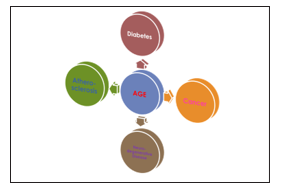
Figure 2: AGE involves in Diseases
Measurement of AGE
AGEs were originally measured according to their fluorescence and browning properties. Each AGE structure in pathological conditions can be demonstrated by high-performance liquid chromatography and gas chromatography mass spectrometry. In modernized techniques, AGE structures involved in diseases can be demonstrated by epitope-identified antibodies against AGEs [40].
Accumulation of AGE can also be measured noninvasively by skin autofluorescence (SAF) which is useful and convenient marker for diabetic vascular diseases in Caucasians [41]. SAF is useful to assess diabetic vascular complications, such as retinopathy, nephropathy in type 2 diabetes patients. In type 2 diabetes with carotid atherosclerosis patients, it may be a beneficial surrogate marker for evaluating carotid atherosclerosis [42]. In elderly people, accumulation of AGEs can be measured by skin autofluorescence (SAF) which may be a marker of metabolic memory [43].
Urinary AGEs can be measured by fluorescence intensity excitation at 370nm and emission at 440nm in a 96-well microplate using spectrophotometer in patients with metabolic syndrome. The results were adjusted according to the urinary creatinine levels [44]. Serum AGEs can be measured at excitation/ emission (370/435nm) to assess its relationship with diabetic chronic kidney disease (CKD) and cardiovascular disease (CVD) [45]. Nonfluorescence AGEs like carboxymethyllysine can be measured by using Enzyme linked immunosorbent assay (ELISA) kit [46].
Conclusion
In conclusion, AGEs are important role in the diabetic complications, atherosclerosis, aging disease and cancer. The accumulation of AGE can be measured by discussed methods and it is very useful for biologic marker to assess the risks and the relationships with diabetic chronic kidney disease (CKD) and cardiovascular disease (CVD). It is suggested that AGEs will be biomarker for inflammatory related disease and cancer for diagnosis, prognosis and prevention of high risk patients.References
- Yamamoto H, Watanabe T, Yamamoto Y, Yonekura H, Munesue S, et al. (2007) RAGE in diabetic nephropathy. Current molecular medicine 7: 752-757.
- Maillard LC (1912) Action des acides aminés sur les sucres; formation des mélanoïdines par voie methodique. Comptes R. Acad. Sci 154: 66-68.
- Win MT, Yamamoto Y, Munesue S, Saito H, Han D, et al. (2012) Regulation of RAGE for attenuating progression of diabetic vascular complications. Experimental Diabetes Research.
- Schmidt AM, Stern DM (2000) RAGE: a new target for the prevention and treatment of the vascular and inflammatory complications of diabetes. Trends in Endocrinology & Metabolism 11: 368-375.
- Goldin A, Beckman JA, Schmidt AM, Creager MA (2006) Advanced glycation end products: sparking the development of diabetic vascular injury. Circulation 114: 597-605.
- Fica-Contreras SM, Shuster SO, Durfee ND, Bowe GJ, Henning NJ, et al. (2017) Glycation of Lys-16 and Arg-5 in amyloid-β and the presence of Cu2+ play a major role in the oxidative stress mechanism of Alzheimer’s disease. JBIC Journal of Biological Inorganic Chemistry 22: 1211-1222.
- Thornalley PJ, Rabbani N (2014) Detection of oxidized and glycated proteins in clinical samples using mass spectrometry-a user’s perspective. Biochimica et Biophysica Acta (BBA)-General Subjects 1840: 818-829.
- Vouillarmet J, Maucort-Boulch D, Michon P, Thivolet C (2013) Advanced glycation end products assessed by skin autofluorescence: a new marker of diabetic foot ulceration. Diabetes technology & therapeutics 15: 601-605.
- Chaudhri S, Fan S, Davenport A (2013) Pitfalls in the measurement of skin autofluorescence to determine tissue advanced glycosylation content in haemodialysis patients. Nephrology 18: 671-675.
- Ogasawara Y, Tanaka R, Koike S, Horiuchi Y, Miyashita M, et al. (2016) Determination of methylglyoxal in human blood plasma using fluorescence high performance liquid chromatography after derivatization with 1, 2-diamino-4, 5-methylenedioxybenzene. Journal of Chromatography B 1029: 102-105.
- Thornalley PJ (1990) The glyoxalase system: new developments towards functional characterization of a metabolic pathway fundamental to biological life. Biochemical Journal 269: 1.
- Koschinsky T, He CJ, Mitsuhashi T, Bucala R, Liu C, et al. (1997) Orally absorbed reactive glycation products (glycotoxins): an environmental risk factor in diabetic nephropathy. Proceedings of the National Academy of Sciences 94: 6474-6479.
- Vlassara H, Cai W, Crandall J, Goldberg T, Oberstein R, et al. (2002) Inflammatory markers are induced by dietary glycotoxins: a pathway for accelerated atherosclerosis in diabetes. Proceedings of the National Academy of Sciences of the United States of America 99: 15596-15601.
- Cai W, Gao QD, Zhu L, Peppa M, He C, et al. (2002) Oxidative stress-inducing carbonyl compounds from common foods: novel mediators of cellular dysfunction. Molecular Medicine 8: 337.
- Cai W, He JC, Zhu L, Lu C, Vlassara H (2006) Advanced glycation end product (AGE) receptor 1 suppresses cell oxidant stress and activation signaling via EGF receptor. Proceedings of the National Academy of Sciences 103: 13801-13806.
- Neeper M, Schmidt AM, Brett J, Yan SD, Wang FE, et al. (1992) Cloning and expression of a cell surface receptor for advanced glycosylation end products of proteins. Journal of Biological Chemistry 267: 14998-15004.
- Borg DJ, Yap FY, Keshvari S, Simmons DG, Gallo LA, et al. (2018) Perinatal exposure to high dietary advanced glycation end products in transgenic NOD8. 3 mice leads to pancreatic beta cell dysfunction. Islets 10: 10-24.
- Uribarri J, Cai W, Ramdas M, Goodman S, Pyzik R, et al. (2011) Restriction of advanced glycation end products improves insulin resistance in human type 2 diabetes: potential role of AGER1 and SIRT1. Diabetes care 34: 1610-1616.
- de Courten B, de Courten MP, Soldatos G, Dougherty SL, Straznicky N, et al. (2016) Diet low in advanced glycation end products increases insulin sensitivity in healthy overweight individuals: a double-blind, randomized, crossover trial-3. The American journal of clinical nutrition 103: 1426-1433.
- Rüster C, Bondeva T, Franke S, Tanaka N, Yamamoto H, et al. (2009) Angiotensin II upregulates RAGE expression on podocytes: role of AT2 receptors. American journal of nephrology 29: 538-550.
- Wendt TM, Tanji N, Guo J, Kislinger TR, Qu W, et al. (2003) RAGE drives the development of glomerulosclerosis and implicates podocyte activation in the pathogenesis of diabetic nephropathy. The American journal of pathology 162: 1123-1137.
- Murata T, Nagai R, Ishibashi T, Inomata H, Ikeda K, et al. (1997) The relationship between accumulation of advanced glycation end products and expression of vascular endothelial growth factor in human diabetic retinas. Diabetologia 40: 764-769.
- Soulis T, Thallas V, Youssef S, Gilbert RE, McWilliam BG, et al. (1997) Advanced glycation end products and their receptors co-localise in rat organs susceptible to diabetic microvascular injury. Diabetologia 40: 619-628.
- Kaji Y, Usui T, Ishida S, Yamashiro K, Moore TC, et al. (2007) Inhibition of diabetic leukostasis and blood-retinal barrier breakdown with a soluble form of a receptor for advanced glycation end products. Investigative ophthalmology & visual science 48: 858-865.
- Cameron NE, Eaton SE, Cotter MA, Tesfaye S (2001) Vascular factors and metabolic interactions in the pathogenesis of diabetic neuropathy. Diabetologia 44: 1973-1988.
- Bierhaus A, Haslbeck KM, Humpert PM, Liliensiek B, Dehmer T, et al. (2004) Loss of pain perception in diabetes is dependenton a receptor of the immunoglobulin superfamily. The Journal of clinical investigation 114: 1741-1751.
- Toth C, Rong LL, Yang C, Martinez J, Song F, et al. (2008) Receptor for advanced glycation end products (RAGEs) and experimental diabetic neuropathy. Diabetes 57: 1002-1017.
- Yajima N, Yamamoto Y, Yamamoto H, Takeuchi M, Yaghihashi S (2004) Peripheral neuropathy in diabetic mice overexpressing receptor for advanced glycation end products (RAGE). InProceedings of the 8th International Symposium on the Maillard reaction 55.
- Vincent AM, Perrone L, Sullivan KA, Backus C, Sastry AM, et al. (2007) Receptor for advanced glycation end products activation injures primary sensory neurons via oxidative stress. Endocrinology 148: 548-558.
- Salahuddin P, Rabbani G, Khan RH (2014) The role of advanced glycation end products in various types of neurodegenerative disease: a therapeutic approach. Cellular & molecular biology letters 19: 407.
- Donmez G, Wang D, Cohen DE, Guarente L (2010) Advanced Molecular and Cellular Biology (2010 Autumn semester). Cell 142: 320.
- Talbot K, Wang HY, Kazi H, Han LY, Bakshi KP, et al. (2012) Demonstrated brain insulin resistance in Alzheimer’s disease patients is associated with IGF-1 resistance, IRS-1 dysregulation, and cognitive decline. The Journal of clinical investigation 122: 1316-1338.
- Raposeiras-Roubín S, Rodiño-Janeiro BK, Paradela-Dobarro B, Grigorian-Shamagian L, García-Acuña JM, et al. (2012) Predictive value of advanced glycation end products for the development of post-infarction heart failure: a preliminary report. Cardiovascular diabetology 11: 102.
- Forbes JM, Yee LT, Thallas V, Lassila M, Candido R, et al. (2004) Advanced glycation end product interventions reduce diabetes-accelerated atherosclerosis. Diabetes 53: 1813-1823.
- Wendel U, Persson N, Risinger C, Bengtsson E, Nodin B, et al. (2018) A novel monoclonal antibody targeting carboxymethyllysine, an advanced glycation end product in atherosclerosis and pancreatic cancer. PloS one 13: e0191872.
- Rojas A, Añazco C, González I, Paulina A (2018) Extracellular matrix glycation and RAGE activation. A missing piece in the puzzle of the association between diabetes and cancer. Carcinogenesis 24.
- Zhu X, Zhou L, Li R, Shen Q, Cheng H, et al. (2018) AGER promotes proliferation and migration in cervical cancer. Bioscience reports BSR 2017 1329.
- Lee KJ, Yoo JW, Kim YK, Choi JH, Ha TY, et al. (2018) Advanced glycation end products promote triple negative breast cancer cells via ERK and NF-κB pathway. Biochemical and biophysical research communications 495: 2195-2201.
- Chen Y, Filipov NM, Guo TL (2018) Dietary Glycation Products Regulate Immune Homeostasis: Early Glycation Products Promote Prostate Cancer Cell Proliferation through Modulating Macrophages. Molecular nutrition & food research 1: 62.
- Nagai R, Shirakawa JI, Ohno RI, Hatano K, Sugawa H, et al. (2016) Antibody-based detection of advanced glycation end-products: promises vs. limitations. Glycoconjugate journal 33: 545-552.
- Hangai M, Takebe N, Honma H, Sasaki A, Chida A, et al. (2016) Association of advanced glycation end products with coronary artery calcification in Japanese subjects with type 2 diabetes as assessed by skin autofluorescence. Journal of atherosclerosis and thrombosis 23: 1178-1187.
- Temma J, Matsuhisa M, Horie T, Kuroda A, Mori H, et al. (2015) Non-invasive measurement of skin autofluorescence as a beneficial surrogate marker for atherosclerosis in patients with Type 2 diabetes. The Journal of Medical Investigation 62: 126-129.
- Rajaobelina K, Cougnard-Gregoire A, Delcourt C, Gin H, Barberger-Gateau P, et al. (2015) Autofluorescence of skin advanced glycation end products: marker of metabolic memory in elderly population. Journals of Gerontology Series A: Biomedical Sciences and Medical Sciences 70: 841-846.
- Suehiro A, Uchida K, Nakanishi M, Wakabayashi I (2016) Measurement of urinary advanced glycation end-products (AGEs) using a fluorescence assay for metabolic syndrome-related screening tests. Diabetes & Metabolic Syndrome: Clinical Research & Reviews 10: 110-113.
- Ciobanu DM, Olar LE, Stefan R, Veresiu IA, Bala CG, et al. (2015) Fluorophores advanced glycation end products (AGEs)-to-NADH ratio is predictor for diabetic chronic kidney and cardiovascular disease. Journal of Diabetes and its Complications 29: 893-897.]
- Uribarri J, del Castillo MD, de la Maza MP, Filip R, Gugliucci A, et al. (2015) Dietary advanced glycation end products and their role in health and disease. Advances in nutrition 6: 461-473.