
International Journal of Forensic Research(IJFR)
ISSN: 2767-2972 | DOI: 10.33140/IJFR
Impact Factor: 1.9

Research Article - (2022) Volume 3, Issue 1
Identification of Species Origin of Wild Life Animals by using an Immunodiffusion Assay
Received Date: Nov 21, 2021 / Accepted Date: Nov 27, 2021 / Published Date: Jan 13, 2022
Copyright: ©Copyright: ©2022 Vilas S Harel, et al. This is an open-access article distributed under the terms of the Creative Commons Attribution License, which permits unrestricted use, distribution, and reproduction in any medium, provided the original author and source are credited.
Citation: Vilas S Harel, Sandip G Pawar, Bhausaheb P More and Sangita V Ghumatkar (2022) Identification of Species Origin of Wild Life Animals by using an Immunodiffusion Assay. In j Fore Res, 3(1), 89-92.
Abstract
Recently most of the forensic science laboratories do the species identification by forensic DNA techniques, however sometimes due to time and budget constraints it is practical for small laboratories to carry out species identification by serological methods. The immuno-electrophoretic method used in this study is rapid, specific and sensitive. It is very much useful for identification of species origin, and it can be considered a valid alternative method for the detection of animal species. Sometimes when DNA markers are not available electrophoresis can be invaluable tool for analysis of forensic samples.
Keywords
Species Identification, Immunodiffusion, Electrophoresis
Introduction
The illegal killing or poaching of wild animal species in India is becoming more and more serious concern, with animals being taken for fur and meat. Wild life species like tiger are threatened with extirpation or extinction. Forensic samples analysis of animal remains help to provide evidence for assailant [1, 2]. (Bertino and Bertino, 2015, Knecht, 2012 Use of immuno-electrophoretic methods is rapid specific and sensitive and is more useful for identification of species origin. Although DNA has widely replaced immunological methods of analysis in forensic cases, but in particular situations, immuno-electrophoretic methods may still be a valuable tool in criminology [3].
In forensic science laboratory in biology division received the cases under the “Animal Protection Act. “The tissue material or skin of wild life animal is received as cases for species identification. In the present case the skin portion of the family of tiger (Panthera Tigris) is received for identification of species, which belongs to the family as “Felidae”. The immunological discoveries of the last decade of the nineteenth century laid the groundwork for the exploitation of immunological species specificity in forensic work. Myers and Uhlenhuth independently noted that precipitating antibodies raised in rabbits would specifically distinguish the species of animals [4, 5].
A number of serological techniques are available for the species identification of tissue samples; all these rely on the specificity of the antiserum that is its ability to distinguish between the serum proteins of different species. A series of specific antisera are reacted with extracts of material under examination and the formation of a precipitate indicates the site of the antigen-antibody reaction. The immunological methods involving antigen- antibody precipitation reactions are applied. The electrophoresis and gel diffusion are the name of the techniques. In electrophoresis the exhibit extract is subjected to agar gel electrophoresis with known antisera of suspected species. The gel diffusion is another method in which the antigen and antibody are subjected to diffusion. At the cross over point the precipitin band is obtained if the animal is of related species [6, 7].
The test was performed called as Immunodiffusion. The antigen and antibody are allowed to diffuse until precipitation occurs. The test is performed in a liquid or semi liquid medium such as agarose gel, which stabilizes the diffusion process. In the agarose gel, Wells are created by punching holes in the gel layer at the desired locations; here we have created a pattern of four well surrounding the centre well. The antibody is loaded in the central well, and the questioned sample and the controls are loaded in the surrounding wells. The double diffusion between the antigen and antibody from the wells is allowed to occur during the incubation. A positive reaction is observed at the end of incubation if a precipitated ring / Square /line are observed (Kryndushkin et al., 2003) [8-11].
The Immunodiffusion techniques can be combined with electrophoresis to enhance the test results. Two arrays of opposing wells are created by punching the holes in the agarose gel. The antibody and samples are loaded in opposing wells arranged by pairs. Electrophoresis is used to drive antigen and antibody towards each other The wells contain antibody are at anode (+ end) end and antigens are at cathode end (- End) during gel electrophoresis the antigen and antibody migrates in the opposite directions and gives precipitate line between opposing wells [9, 10].
This technique is used for the species identification of stain and tissue extracts. The two reactants are placed into the wells punched in the gel close together along a line of electrophoretic movement, the tissue extract in cathodic well and the antiserum in the anodic well. The antigens in the tissue extracts are serum albumin and α and β globulins, where as the antibody in the antiserum are gamma-globulins. Under the conditions employed when the electrophoresis is performed, the movement of the gamma- globulins is towards cathode and all other proteins move towards the anode, when appropriate reactants meet in the area between the wells a precipitate is formed [12].
Materials
Sample collection and preparations:
1. Antigens for COE are from the forensic sample of questioned Skin Piece of the animal.
2. Elecrtophoresis buffer: The solution was prepared At pH 8.4, using the constituents of Tris Buffer: 4.5 gm, Glycine: 21.8 gm, Distilled water: 1000ml
3. Standard antisera of Human, cat, Dof, Fowl, Pig, Tiger, Sheep, and cow was used which was raised in New-Zealand White rabbits, three months old.
4. Coomassie Blue: Agarose gels are stained in a working solution of Coomassie blue, which is a blue colour stain in electrophoresis buffer.
Method
Cross Over Electrophoresis
Agarose gel preparation: Prepared solution of 1 % of agarose gel in a test tube, by heating it until it gets liquefied. The liquefied solution is cooled to 45 to 500 c and coated on the top surface of the glass slide; it was covered with moisture chamber and allowed to cool so as to solidify. The plate was hold by plastic holder and punched small wells of about 1 to 2 mm. The plugs of agar are removed by vaccum pipette.
Loading the sample: Prepared the tissue extract of the material by adding 50% of gel buffer solution. By using fine pipette, the three right hand holes on the agarose gel plate are filled with extract of the questioned material. The left-hand holes on the agarose gel plate are filled with appropriate antisera like anti human + other suspected animal species antisera (Figure 2).
Electrophoresis: The agarose gel plate was submerged in an electrophoresis tank to the depth of approx. 1mm which is already loaded with electrophoresis buffer, with the tissue extract being nearest to the cathode and antisera nearest to the anode. Closed the lid of the tank and attached the electrical leads. Switch on the power pack and was run for 20 min at 150 V.
Reading the plate: The plate was read with the help of the bench lamp. A fine precipitate line in the area between two holes of the pair indicates a positive reaction (Figure 1).
Staining: Soak the gel plate overnight in saline solution then is soaked in Deionized water, for 10 min. the process is repeated once the gel plate is dried for 20 min at oven. The gel is stained with Coomassie blue.
System for Cross over Electrophoresis:
|
Tank Buffer |
Tris Glycine D/W |
4.5 gm+21.8 gm+1000ml |
|
Gel Buffer |
Tris Glycine D/W |
4.5 gm+21.8 gm+1000ml |
|
Support medium |
1% Agar or agarose in Gel Buffer |
|
|
Temp and Conditions |
Room Temperature |
|
|
Voltage and Duration |
150V for 20 min |
|
|
Staining |
Coomassie Blue |
|
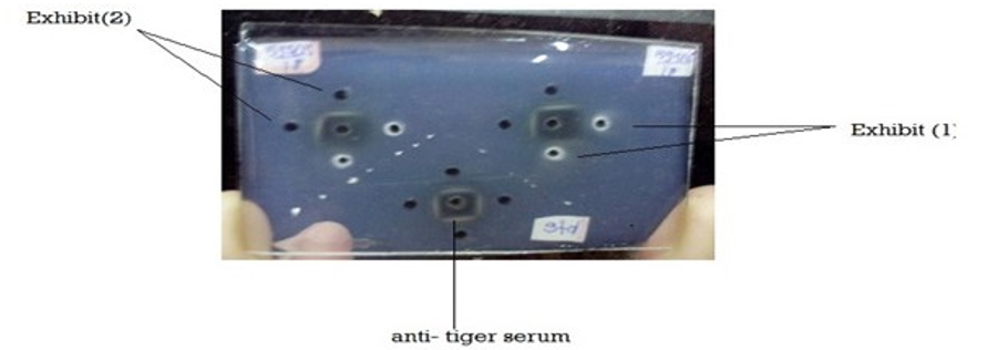
Figure1: Agarose Gel Electrophoresis plate

Figure1: Antigen-Antibody reaction
Results
The results obtained are as follows
|
Anti- Sera |
Results |
|
Anti- Human |
Negative |
|
Anti- cat |
Negative |
|
Anti- dog |
Negative |
|
Anti- tiger |
Positive |
|
Anti-pig |
Negative |
|
Anti- fowl |
Negative |
|
Anti- sheep |
Negative |
|
Anti-cow |
Negative |
Discussion
Species origin examination plays an important role in forensic investigation. In this method gel is used as the supporting media for the separation of biomolecules, under the influence of electric charge. The process of cross over electrophoresis is also called as counter Immuno-electrophoresis (CIE). It is a combination of Immunodiffusion and electrophoresis separates molecules according to the differences in their electrophoretic mobility. This technique is based on the precipitation reaction and is used for species identification in forensic laboratories. This process is used for the detection of species-specific muscle proteins in food products also for the detection of pork, beef, poultry, or in heat- processed meat products. As observed by Sherikar A.T. ET. Al [10-13].
Our results are similar to the available literature [5-10]. The present analysis revels that from the past decades electrophoresis can give us an accurate and effective approach for the examination of the species origin of an unknown biological sample. This method can be the best alternative for the examination of human and non- human samples. particularly it may be necessary to identify the animal species prior to further analysis. Electrophoretic analysis has been widely used in forensic science for analysis of trace evidence.
By using Cross-Over electrophoresis method the sample was confirmed to be of family Felidae. Also, it is proved that the inexpensive, simple, and sensitive method for the identification of species origin, where DNA markers are not available.
References
- Bertino AJ, Bertino P (2015) Forensic Science: Fundamentals & investigations (MindTap Course List) 2nd ed. Mason, OH: Cengage Learning.
- Knecht L (2012) The use of hair morphology in the identification of mammals. In: Huffman JE, Wallace JR, editors. Wildlife forensics: methods and applications. Chichester, UK: Wiley 2012: 129-143.
- Hugh Tuller, Rebecca Saunders (2012) The use of crossover immunoelectrophoresis to detect human blood protein in soil from an ambush scene in Kosovo. J Forensic Sci 57: 873-879.
- Walter Myers (1900) On Immunity Against Proteids. The Lancet 156: 98-100.
- Yenger N (1959) Applications of electrohoresis Technique in Forensci science. Current science 28: 316-319.
- Singh G, Walia S (2020) DNA Gel Electrophoresis. E-PG Pathshala. https://epgp.inflibnet.ac.in/Home/ ViewSubject?catid=16/
- Electrophoresis: Principle and types. (n.d.). BrainKart. https:// www.brainkart.com/article/Electrophoresis–Principle-and-Types_34123/
- Gambhir G, Gautam A. Electrophoresis. E-PGPathshala. https://epgp.inflibnet.ac.in/epgpdata/uploads/epgp_content/ S000016FS/P001352/M026975/ET/1516862256FSC_P4_M34_e-text.pdf.
- Parmar P (2016) Electrophoresis: Meaninh, Definition and classification (with diagram) Biotechnolgynotes.
- Sherikar AT, Khot JB, Jayarao BM, Pillai SR (1988) “Use of species-specific antisera to adrenal heat-stable antigens for the identification of raw and cooked meats by agar gel diffusion and counter immunoelectrophoretic techniques”. J Sci FD Agric 44: 63-73.
- Kryndushkin DS, Alexandrov IM, Ter-Avanesyan MD, Kushnirov VV (2003) Yeast [PSI+] prion aggregates are formed by small Sup35 polymers fragmented by Hsp10. Journal of Biological Chemistry 278: 49636.
- Lane D, Prentki P, Chandler M (1992) Use of gel retardation to analyse protein- nucleic acid interactions. Microbiol Rev 56: 509-528.
- Sharp PA, Sugden B, Sambrook J (1973) Detection of two restriction endonuclease activities in Haemophilus parainfluenzae using analytical agarose-ethidium bromide electrophoresis. Biochemistry 12: 3055-3063.