

Archives of Epidemiology & Public Health Research(AEPHR)
ISSN: 2833-4353 | DOI: 10.33140/AEPHR
Impact Factor: 1.98


Research Article - (2022) Volume 1, Issue 1
Histomorphological And Scanning Electron Microscopic Study of Explanatum (Digenea) In Native Cattle of Kerman Province, Iran
2Department of Basic Sciences, Faculty of Veterinary Medicine, Ardakan University, P.O. Box 184, Ardakan, Iran
3Research Center for Hydatid Disease in Iran, Kerman University of Medical Sciences, Kerman, Iran
4Department of Parasitology and Mycology, Faculty of Medicine, Mashhad University of Medical Sciences, Mashhad, Iran
5Department of Biodiversity, University of Limpopo, Sovenga, South Africa
Received Date: Aug 10, 2022 / Accepted Date: Aug 19, 2022 / Published Date: Aug 23, 2022
Copyright: ©Copyright: ©2022 Mingyuan Liu, et al. This is an open-access article distributed under the terms of the Creative Commons Attribution License, which permits unrestricted use, distribution, and reproduction in any medium, provided the original author and source are credited.
Citation: Mehdi Borhani Zarandi, Amin Ahmadi, Ashkan Faridi, Elham Moghaddas, and Mingyuan Liu et al. (2022). Histomorphological And Scanning Electron Microscopic Study of Explanatum Explanatum (Digenea) In Native Cattle of Kerman Province, Iran. Arch Epidemiol Pub Health Res, 1(1), 41- 45.
Abstract
Amphistomiasis is caused by different species of trematodes in the family paramphistomatidae and is one of the neglected parasitic diseases of livestock. Amphistomiasis has a wide geographic distribution, especially in subtropical and tropical regions. Although adult stage of most of the amphistomes are seen in the rumen and reticulum of ruminants, but sometimes can be seen in bile ducts too. A few parasites were collected from the bile ducts of a native cattle from the Kerman slaughterhouse, Kerman Province, southeastern Iran, and samples were studied by histomorphological as well as, Scanning Electron Microscopy. The worms were identified as Explanatum explanatum using identification keys and SEM. This trematode has been identified in livestock from only two other provinces of Iran but this is the first report of it in Kerman Province. Specific epidemiological studies with exact slaughter inspections are needed to recognize the extent of the parasite prevalence in native animals of Iran.
Keywords
Amphistomiasis, Explanatum, Iran, Kerman Province, Scanning Electron Microscopy, SEM
Introduction
Amphistomiasis is caused by different species of the family paramphistomatidae (Trematoda: Paramphistomidae). It is one of the neglected parasitic disease of domestic and wild rumi¬nants, which has a wide geographic distribution, especially in subtropical and tropical regions [1,2]. Generally, adult stage is nonpathogenic for host, but immature stages and their migration in anterior small intestinal mucosa can cause severe pathogen¬esis. Symptoms such as enteritis, haemorrhage, anorexia, poly-dipsia, unthriftiness, severe diarrhea, and even mortality [3,4]. In addition weight loss and reduction in animal production and subsequent economic loss have been observed due to amphisto¬miasis, but in general the importance of these parasites has been neglected [5]. Although adult stage of most of the amphistomes are seen in the rumen and reticulum of wild and domestic ru- minants, but in some cases such as Explanatum explanatum the adult stage exists in bile ducts [6]. This species has been reported in different parts of the world [6]. Although infection with Ex¬planatum has been reported from neighboring countries, i.e. Iraq and Afghanistan [7,8] but there is not much information about its epidemiology in Iran.
Different amphistomids have been reported in Iran, including Paramphistomum, Calicophoron, Gastrothylax, Cotylophoron, Carmyerius and Orthocoelium. E. explanatum has only been re¬ported in buffalo in southern part of Iran (Khuzestan Province) [9], cattle and sheep of central Iran (Shiraz) (Rajabloo et al. 2014) and cattle of Zabol, southeast Iran however limited data are available on this species from other parts of the country. The purpose of the present study was to investigate the presence of this parasite in Kerman province, southern Iran [10, 12-15].
In different studies Scanning Electron Microscope (SEM) has been used to study the surface topography of tegument of a number of species in the family paramphistomatidae [16–20]. However, there is not such study on the genus Explanatum so we decided to use a few of the collected samples for gross mi-croscopic, histomorphological evaluation as well as, and SEM.
Materials and Methods
The parasites were collected from the bile ducts of a native cattle from the Kerman slaughterhouse, Kerman Province, southeast-ern Iran (30.2907°N 57.0679°E). Recovered amphistomes were rinsed in saline, fixed and preserved in 70% ethanol. Later spec¬imens were transferred to the Research Center for Hydatid Dis¬ease (Kerman University of Medical Sciences, Kerman, Iran). Some of the worms were used for morphological study and the rest for SEM study. For morphology, specimens were stained with Carmine and mounted in Canada balsam. Measurements of morphological features were made with an optical microscope using a calibrated ocular micrometer (OLYMPUS CX31). For SEM study, samples were fixed in a 2. 5% glutaraldehyde in a 0.1 M sodium cacodylate buffer. Then, they were fixed in a 2.5% glutaraldehyde solution and 0.1 M phosphate buffer solution (pH 7.2) that was prepared from NaH2PO4 H2O and Na2HPO4 (anhydrous) salts for 24 h [21]. After washing with phosphate buffer solution (PBS 0.1 M), the trematodes were dehydrated in a graded ethanol series, dried in a critical-point dryer (SAN-DRI-780A), and coated with gold for 10 min in an ionizer (Ion Sputter JFC-1100, Jeol, Fine Coat). Coated worms were ob¬served under the SEM (TESCAN MIRA3 LM, Czech Republic) to check for the surface morphology at an accelerating voltage of 10 kV. For histomorphological study, the specimens were fixed in formalin %10, then paraffin blocks were made and were se-rially sectioned at 2 μm in thickness using microtome (AUTO-TECHNICON MONO, MOD. 2A &MICROTEC). These slides were stained by haematoxylin and eosin, and finally slides were examined and photographed by light microscope (OLYMPUS CX41).
Results
During veterinary inspection of cattle in Kerman municipal ab-attoir, 15 white ventrally curved helminths were found in the bile ducts of a native cattle. The helminths were attached to ep¬ithelia by mean of their acetabulum and there was some gross damage on attachment site. Gross microscopic studies showed characters like absence of an oral sucker, acetabulum enormous and subterminal and ventral 4-4.3 mm in external diameter in dorso-ventral direction, large body, pyriform to conical, tapering anteriorly, broad and rounded posteriorly, curved ventrally, 8.7-12 mm long; and net weight was 0.4- 0.6 gr (Fig 1), and thus based on these characters, the amphistomes were associated with Paramphistominae subfamily.
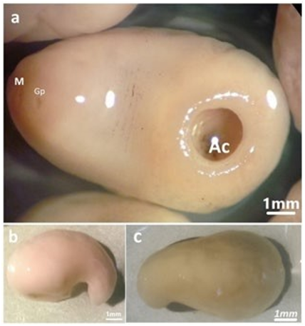
Figure 1: Gross view of E. explanatum under stereomicroscope. a) ventral view of whole-body shape, M: Mouth, Gp: Gonopore, ac: Acetabulum, b) lateral view of whole-body, c) dorsal view of whole-body
Further confirmation was provided by cross section of the sam¬ple as shown in [Figure 3]. In the section, observations were as well-developed pharynx, testis position, absence of esophageal bulb and sphincter, vitelline follicles in lateral sides, absence of genital sucker, ventral genital pore in midline, all confirming the identification as E. explanatum.
In SEM images [Figure 2], no body spines and armature were seen, but frequent and prominent tegumental papillae were no¬ticed that are characters of the genus Explanatum.

Figure 2: Scanning electron micrograph of E. explanatum. a) Ventral view of whole-body, M: Mouth, Gp: Gonopore, Ac: Acetabulum, b) M: Mouth, arrow shows the dome shaped papillae,c) Gp: Gonopore, arrow shows the dome shaped papillae, d & e) p: Dome shaped papillae, f) Star: tegument ridges and furrows.
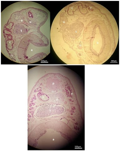
Figure 3: Cross sections of Explanatum. a: acetabulum, o: ovary, p: pharynx, u: uterus, t: testis, c: caeca, Gp: Genital pore, v: Vitelline follicles.
In search for the potential intermediate hosts (IH) for this para¬site, we found the shells of snails Family Planorbidae and fLym-naeidae in Kerman [Figure 4].
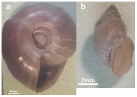
Figure 4: Potential intermediate hosts for E. explanatum in the region. a) Shell without operculum, discoid belongs to Family Planorbidae. b) Shell coiled shape, conical, dextral (aperture to right) belongs to Familyf: fLymnaeidae
Discussion
In the present study, we identified the amphistomes collected from native cattle in Kerman slaughterhouse, Iran. Further anal¬ysis proved that all these parasites were E. explanatum. Expla¬natum species have acetabulum, pharynx and ventral genitali-um, body has 7.86-14.30 mm long and acetabulum 3.50-4.82 mm in external diameter [22]. The measurements of our samples were consistent with the records in mentioned reference.
Family Paramphistomidae Fischoeder, 1901 has 19 genera and 70 species [23], one of these is Explanatum. This parasite is mostly found in the bile ducts and intrahepatic ducts and can cause granulomatous reactions together with fibrosis and thick¬ening of the bile ducts that is capable of imposing direct and indirect economic damages. Although it is a common parasite in buffaloes in India, Africa, Asia, and the Caribbeans [24], but lit¬tle information is available in terms of its epidemiology and ex¬tent of economic importance. The life cycle of this parasite is not much clear, specifically in Iran. Although we know freshwater snails of the family Lymnaeidae and Planorbidae are proposed as IH of the paramphistomatidae [25,26], but which exactly is involved in distribution of parasite in Iran is unclear. Although we accidentally found the snail shells of two mentioned fami¬lies during this study and this can introduce them as potential intermediate hosts, but proving this needs an extensive sam¬pling and in vitro tests to find the intermediate stages. Previous studies report the Planorbidae and Lymnaeidae snails in Ker¬man (GenBank accession numbers: KT267211.1, KT280457.1, KT365877.1, KT280430.1, KT280429.1), but unfortunately, there are no more epidemiological studies in this area.
Although E. explanatum was first reported in Iran in 1980, but they were collected from cattle imported from Afghanistan, Pa- kistan or India [9]. In the later stage this parasite was reported in indigenous buffaloes in southwest Iran [10], sheep and cattle with unknown origin in Shiraz slaughterhouse [14] and cattle with unknown origin in Zabol, southeast Iran [13]. As livestock is imported to Iran from different sources, if E. explanatum is really part of Iran fauna or the reports are with abroad (alien) or-igin needs further works, specially looking at intermediate hosts and checking the source of slaughtered animals more carefully. As this parasite has never been reported in Kerman Province and there is a possibility of introduction of this parasite through imported cattle, precise inspections of slaughtered animals and awareness of related authorities are required for clarifications. It should be noted that the unlawful transfer of livestock from Iran’s neighboring countries may have played a role in this oc-currence. However, a comprehensive study of native cattle and potential intermediate hosts may provide more insight into the occurrence of this parasite and potential life cycle of it. Such information may help the veterinarians for better control of this parasite.
Acknowledgement: We would like to thank the Research Cen-ter for Hydatid Disease in Iran for supporting the study.
Funding: None
Conflicts of interest: The authors of the present work declare no conflict of interest.
Ethical approval: Not required
References
-
- Arias, M., Lomba, C., Dacal, V., Vázquez, L., Pedreira, J., Francisco, I., ... & Paz-Silva, A. (2011). Prevalence of mixed trematode infections in an abattoir receiving cattle from northern Portugal and northâ?ÂÂwest Spain. Veterinary Record, 168(15), 408-408.
- Pfukenyi, D. M., Mukaratirwa, S., Willingham, A. L., & Monrad, J. (2005). Epidemiological studies of amphistome infections in cattle in the highveld and lowveld communal grazing areas of Zimbabwe. Onderstepoort Journal of Veterinary Research, 72(1), 67-86.
- Pfukenyi, D. M., & Mukaratirwa, S. (2018). Amphistome infections in domestic and wild ruminants in East and Southern Africa: A review. Onderstepoort Journal of Veterinary Research, 85(1), 1-13.
- Horak, I. G. (1971). Paramphistomiasis of domestic ruminants. Advances in parasitology, 9, 33-72.
- Lotfy, W. M., Brant, S. V., Ashmawy, K. I., Devkota, R., Mkoji, G. M., & Loker, E. S. (2010). A molecular approach for identification of paramphistomes from Africa and Asia. Veterinary Parasitology, 174(3-4), 234-240.
- Smith MC, Sherman DC. Goat medicine. Waverly Company, Philadelphia. John Wiley \& Sons; 2009.
- Kotrlá, B., Blazek, K., & Amin, A. (1976). Trematodes of domestic ruminants of Afghanistan and their role in pathology. Folia Parasitologica, 23(3), 217-220.
- Kadhim, J. K., Altaif, K. I., & Hawa, N. J. (1970). The occurrence of paramphistomes in ruminants in Iraq, with a description of Gigantocotyle explanatum in cattle and buffaloes. Bulletin of Endemic Diseases, 12(1/4).
- Otto, S., & Eslami, A. (1980). Review of amphistomes (Trematoda, paramphistomata) of Iranian domestic ruminants. Parasitologia Hungarica, 14, 61-65.
- Mazahery, Y., Razmyar, J., & Hoghooghi-Rad, N. (1994). Explanatum explanatum (Creplin, 1847) Fukui, 1929, in buffaloes in the Ahwaz area, southwest Iran. Veterinary Parasitology, 55(1-2), 149-153
- Coskun, S. Z., Eslami, A., Halajian, A., & Nikpey, A. (2012). Amphistome species in cattle in South coast of Caspian Sea. Iranian Journal of Parasitology, 7(1), 32
- Bagheri, H. A. (1962). Study on the species of paramphisto-mumes of cattle in slaughterhouse of Tehran. DVM Dissertation. Faculty of Veterinary Medicine, University of Tehran, 1-45
- Khedri, J., Radfar, M. H., Borji, H., & Mirzaei, M. (2015). Prevalence and intensity of Paramphistomum spp. in cattle from South-Eastern Iran. Iranian Journal of Parasitology, 10(2), 268
- Rajabloo, M., Namazi, F., Shayegh, H., & Alavi, A. M. (2014). Explanatum explanatum: an emerging liver fluke in cattle and sheep. Online Journal of Veterinary Research, 18(8), 675-679.
- Arfaa, F. (1962). A study of Paramphistomum microbothri-um in Khuzistan S.-W. Iran. Annales De Parasitologie Humaine Et Comparee, 37(4), 549-555.
- Anuracpreeda, P., Panyarachun, B., Ngamniyom, A., Tinikul, Y., Chotwiwatthanakun, C., Poljaroen, J., & Sobhon, P. (2012). Fischoederius cobboldi: a scanning electron microscopy investigation of surface morphology of adult rumen fluke. Experimental parasitology, 130(4), 400-407.
- Anuracpreeda, P., Phutong, S., Ngamniyom, A., Panyarachun, B., & Sobhon, P. (2015). Surface topography and ultra-structural architecture of the tegument of adult Carmyerius spa-tiosus Brandes, 1898. Acta tropica, 143, 18-28.
- Panyarachun, B., Sobhon, P., Tinikul, Y., Chotwiwatthanakun, C., Anupunpisit, V., & Anuracpreeda, P. (2010). Paramphistomum cervi: surface topography of the tegument of adult fluke. Experimental parasitology, 125(2), 95-99.
- Sanger, B., Swarnakar, G., & Roat, K. (2017). Ultrastruc-tural observations of rumen immature and mature Paramphistomum cervi (Trematoda: Digenea) in domestic buffalo of Udaipur district, Rajasthan. Int. J. Zool. Stud, 2, 63-69.
- Lenis, C., Galiano, A., Vélez, I., Vélez, I. D., Muskus, C., & Marcilla, A. (2018). Morphological and molecular characterization of Paragonimus caliensis Little, 1968 (Trematoda: Parago-nimidae) from Medellin and Pichinde, Colombia. Acta tropica, 183, 95-102.
- Bozzola, J. J., & Russell, L. D. (1999). Electron microscopy: principles and techniques for biologists. Jones & Bartlett Learning.
- Eduardo, S. L. (1984). The taxonomy of the family Paramphistomidae Fischoeder, 1901 with special reference to the morphology of species occurring in ruminants IV. Revision of the genusGigantocotyle Näsmark, 1937 and elevation of the subge-nusExplanatum Fukui, 1929 to full generic status. Systematic Parasitology, 6(1), 3-32.
- Jones, A. (2005). Superfamily Paramphistomoidea Fischoeder, 1901. In Keys to the Trematoda: Volume 2 (pp. 221-227). Wallingford UK: CABI Publishing.
- Haque, M., Mohan, C., & Ahmad, I. (2011). Natural trema-tode infection in liver of water buffalo (Bubalus bubalis): histo-pathological investigation. Journal of Parasitic Diseases, 35(1), 50-53.
- Santos, I. C. S., Laranja, R. J., Martins, J. R., & Cereser, V.H. (1986). Intermediate host of Paramphistomum (Fischoeder, 1901) Biomphalaria Tenagophila (Orbigny, 1935), Guaiba, Rio Grande do Sul, Brazil. Boletim do Instituto de Pesquisas Veteri-narias Desiderio Finamor (Brazil).
- Paiva, N. (1994). Epidemiología y control de Paramphistomum en Uruguay. Enfermedades Parasitarias de Importancia Económica en Bovinos. Hemisferio Sur, Montevideo Uruguay, 257-264.