
Journal of Electrical Electronics Engineering(JEEE)
ISSN: 2834-4928 | DOI: 10.33140/JEEE
Impact Factor: 1.2


Research Article - (2026) Volume 5, Issue 3
Features of Ultra-Weak Radiation of Hollow Resonators
Received Date: Mar 17, 2026 / Accepted Date: Apr 20, 2026 / Published Date: May 01, 2026
Copyright: ©2026 Andrey Batyanov. This is an open-access article distributed under the terms of the Creative Commons Attribution License, which permits unrestricted use, distribution, and reproduction in any medium, provided the original author and source are credited.
Citation: Batyanov, A. (2026). Features of Ultra-Weak Radiation of Hollow Resonators. J Electrical Electron Eng, 5(3), 01-09.
Abstract
This article discusses the hypothesis that the experimental setup used represents a physical system with a primary (other than thermal) coupling factor—coherent radiation with minimal energy—that is, a system with a correlation interaction of nonequilibrium, spatially separated electronically excited levels, where all the phenomena described, both in biological and model experiments, are determined by this principle. A series of experiments were conducted to confirm these propositions.
Introduction
Previous studies have demonstrated the specificity of hollow resonator radiation and its effect on photomultiplier tubes and other detectors, including biological objects [1-3]. Signal specificity is maintained even with maximum light isolation of the photomultiplier tube's photocathode, indicating that background radiation from the photomultiplier tube exhibits specificity for each sample under study. A noticeable response from the photomultiplier tube was also observed with minimal energy changes in the system. A series of experiments with photomultipliers were conducted with more complete cutoff of the UV, visible, and near-IR spectra—the emitter-resonator housings were made of various metals. Examples of the effect of ultra-weak radiation on organic glass from a quartz resonator, a mitochondrial suspension, and a yeast culture are provided, along with one medical observation.
Materials and Methods
The same basic experimental setup described in was used. The time of recording radiation on the photomultiplier (~90 min.) was divided into 5 equal segments (~15 min.): Phase-1 background radiation; Phase-2 resonator radiation without excitation; Phase-3 resonator radiation at the moment of photoexcitation of the light-insulated internal space of the resonator; Phase-4 resonator radiation without excitation (after excitation); Phase-5 background radiation. Main additions: radiation from metal hollow resonators (metal cups of different sizes) during photoexcitation of the light-insulated internal space was carried out using a photoelectric colorimeter (FEK-56) and a light guide, or a 6.3V incandescent lamp (see figure captions). The results obtained are shown in Figure ![]() 1+12 and the text.
1+12 and the text.
Results and Discussion
As in previous works, the response of the PMT without photocontact with metal emitter-resonators was observed, i.e. the specificity of the background radiation of the PMT for each studied sample. The specificity was preserved even with changes in the surface of the resonators, since chromium plating, bismuth-tin coating, chemical oxidation of the same resonators caused sharp differences in the response of the PMT (background radiation, phase-1 before and after coating Figure 1-2). With photoexcitation of the light-insulated inner surface of the resonators (wall thickness d-0.5 mm), the signal of the closed PMT also differed for individual samples (Figure 3-4). When using metal resonators with thinner walls (0.2 mm), the response of the PMT to changes in the ''color'' of the inner surface was more distinct (Figure 5).
A similar picture with wall colored inserts was observed for the plastic resonator. Considering the ultra-weak intensity of the resonator radiation, both in model experiments and with biological objects, insufficient to cause the described effects (noticeable response of the photomultiplier tube, visual changes in the surfaces coated with HgS, changes in the resistance of Ga AlN, effects on plants, mitochondria, yeast culture and the effect of clearing of photographic plates), it is possible to assume that these effects are caused by the correlation interaction of spatially separated nonequilibrium-excited electronic levels in the experimental system (molecules of the ADP-ATP complex and a complex of hybrid orbitals of the quartz or organic glass structure (incubation cells), as well as the photocathode of the photomultiplier tube for the model experiment; the structure of metals and semiconductors and the photocathode. Radiation appears when differences in nonequilibrium electronic states arise in the system, i.e., according to the thermal energy transfer scheme, energy transfer occurs only when there is a temperature difference. Thus, the excitation energy of the samples is necessary for the displacement (equilibrium) nonequilibrium excited system, and it does not matter (in the experimental system) how this is accomplished—by electrification, photoexcitation of the inserts (Figure 6, 7), or replacement of the resonator surface coating or the use of colored inserts (Figure 3, 4, 5). The electronically excited system of the photocathode responds to this shift in electronic equilibrium in the experimental system by the type of thermal radiation at a temperature difference. Energy exchange between waves and molecules ceases when the receiver and emitter structures contain the same number of identical, nonequilibrium electronically excited levels. Changes in electronic equilibrium in one of the system's elements, for example, photoexcitation of the internal light-insulated space of a hollow emitter-resonator, generate radiation in the system and changes in the PMT signals.
Apparently, the design of the resonator-emitter simulating the laser circuit (translucent walls: quartz, organic glass, plastic, thin metal, internal diaphragm, direction of the emitter to the recorder along the longitudinal axis) is a necessary condition for the generation and registration of hypothetical coherent radiation, both for bio and model experiments [1,2]. In this case, it can be assumed that the PMT responds not only to the intensity of the light flow, but also to changes in the external electronic structure of the system components - the exchange of energy and information in the non-optical range, i.e., the PMT, in this experiment can be considered as a non-selective receiver of electromagnetic radiation. The electrodes of the PMT can act as antennas. Radio frequency radiation can be generated. Radiation reception occurs not through the external photoelectric effect of the photomultiplier tube's photocathode, but apparently through interaction with the radiation from the photomultiplier tube's electrodes, which act as a dipole receiving radio frequency radiation. Radiation in this system occurs only when significant differences in the electronically excited states of the system's elements arise, similar to thermal radiation, which occurs only when there is a temperature difference.
The effect of very low radiation energies of an empty quartz resonator, a suspension of isolated mitochondria and a yeast culture on the molecular structure of organic glass is shown in Figure 8, 9, 10, 11.
An example of the long-range action of a plastic resonator (one observation).
• Radiation System: a plastic resonator with Pt (W-shaped foil, ~0.1 g) or pure Al for analysis, connected to -5V (unipolar electrification) from a constant power source during irradiation. The resonator is placed in a black, light-tight box (dimensions in the figure). Thus, the expected radiation propagates approximately 0.5 meters from the resonator, through a layer of air and the wall of the box (dry wood 12 mm thick, covered with several layers of light-absorbing enamel and a layer of photographic paper). The room is a dark room.
• Irradiated Object: basalioma of the temporal region of human skin Ulcus rodens (histologically confirmed).
• Irradiation Stages: 1.5 months, 1.5 hours per day, before surgical removal. Result: the edges of the ulcer cleared up, became less dense, and the ulcer itself became covered with an easily removable crust. Before surgery, the basal cell carcinoma took on the appearance of a ''treated basal cell carcinoma.''
After 1.5-2 months of surgery, elements of transitional forms of squamous cell carcinoma with keratinization (cancerous keratinous scales) appeared in the suture area. After 2-3 weeks of irradiation for 1.5 hours per day (a resonator with pure Al was used for analysis during the first week), the skin cleared significantly, and the keratinous scales became easily separated and fell off on their own. With repeated irradiation up to 5 times per year, stable improvement was observed. Follow-up is over 5 years. No additional treatment was performed after the surgery.
Since the given examples of the action of hollow resonators differ significantly from the classical theory, additional experiments are necessary for a more complete physical interpretation [4,5].
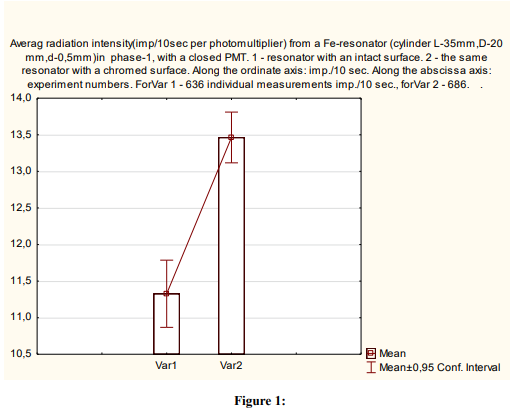
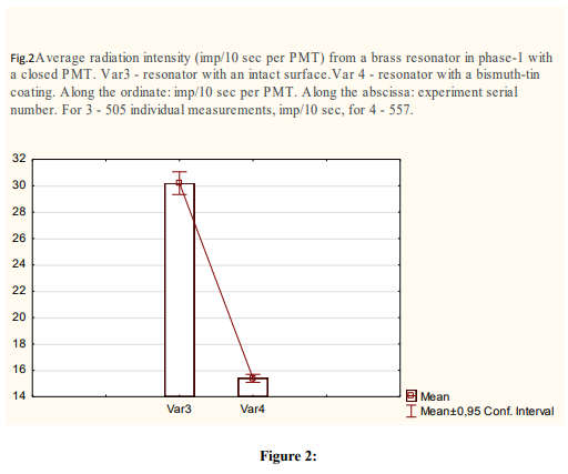
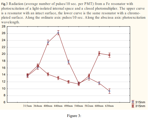
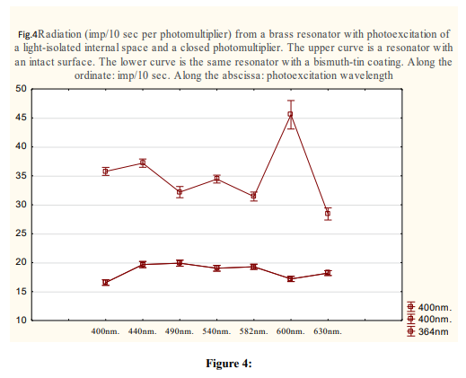
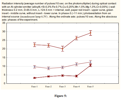
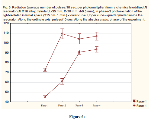
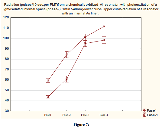
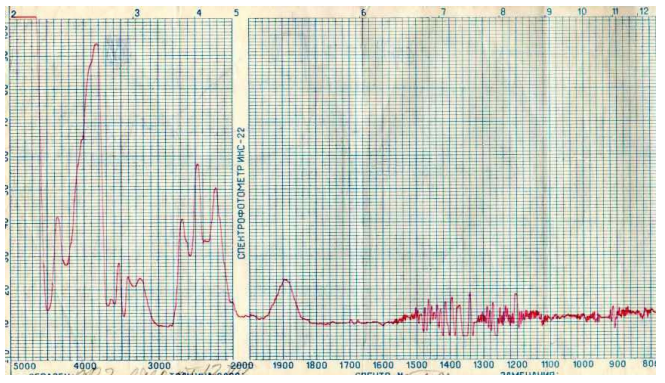
Figure 8: Changes in the IR absorption spectrum (5000-800 cm-1) of an organic glass plate after distant optical contact (10 min. through a 10 mm air layer) with a quartz cylinder cuvette. The quartz was pre-irradiated with reflected, scattered UV light from a mercury-quartz lamp. 10 min

Figure 9: Changes in the IR absorption spectrum (5000-2000 cm-1) of a plexiglass plate after distant optical contact with a suspension of isolated (shock) mitochondria in the substrate respiration phase (succinate). Exposure 5 min. 1-before contact, 2-after contact through a 5-6 mm air layer, 3-4 after contact through (5-6 mm air + 0.02 mm polypropylene)
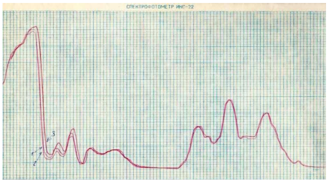
Figure 10: Changes in the IR absorption spectrum from 5000 cm-1 to 2000 cm-1 of a plexiglass plate after distant optical contact (5 mm air + 0.02 mm polypropylene) with a suspension of isolated (intact) mitochondria in the substrate respiration phase (succinate), exposure time 5 min. 1-2 before contact, 3 after contact

Figure 11: Changes in the IR spectrum (5000-800 cm-1) of absorption of an organic glass plate after distant optical contact (5 min. through a 10 mm air layer) with a 24-hour culture of Torula Utilis on an agar-agar block. 1 - before contact. Figure12^-need -cm.
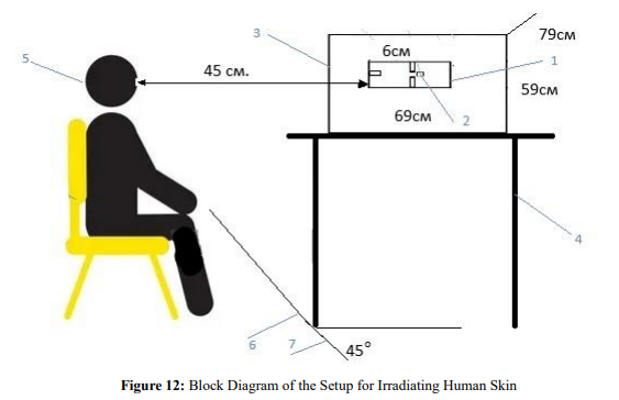
Figure 12: Block Diagram of the Setup for Irradiating Human Skin
1. Hollow Plastic Resonator
2. W-shaped Pt foil (~0.1g) or pure Al for analysis
3. Black box (dry wood 12mm thick coated with light-absorbing enamel + a layer of black paper on the inner wal ls)
4. Stand
5. Position the irradiated surface at ~45° to the side surface of the black box and the plastic resonator at a distance of ~450mm from the box surface
Declarations
Ethical Approval
Institutional Review Board Statement: The study was conducted according to the guidelines of the (DIRECTIVE 2010/63/EU OF THE EUROPEAN PARLIAMENT AND OF THE COUNCIL on the protection of animals used for scientific purposes of 22.09.2010.), and approved by the Institutional Ethics Committee of the Institute of General Pathology and Pathophysiology (final protocol # 1 of 01.02.2023).
Competing Interests
Informed Consent Statement
Informed consent was obtained from all subjects involved in the study.
Authors Contributions
Inadaptability
Funding
This research received no external funding.
References
- Batyanov, A., P. (2024). A Model Recording Ultra-Weak, Electromagnetic Radiation from Metals and Semiconductors. Journal Electrical Electron. Eng.3(5) 1-16
- Batyanov, A., P. (2024). Ultra-weak Electromagnetic Radiation of Mitochondria and Inorganic Systems. Sci Set J of Physics, 3(6), 01-09.
- Batyanov, A., P. (2025). The Effect of Ultra-Weak Radiation on Biological Objects. Journal Electrical Electron.Eng.4 (3) 01-07.
- Wichard, P. R. (1960). Electricity Theory. Seventeenth Revised and Enlarged Edition. Springer Publishing House. Berlin-Gottingen-Heidelberg.179-233.
- Wichard, P. R. (1963). Optics and Atomic Physics, Eleventh Revised and Expanded Edition. Springer Publishing, Berlin-Gottingen-Heidelberg.s.258-323.