

Advances in Bioengineering and Biomedical Science Research(ABBSR)
ISSN: 2640-4133 | DOI: 10.33140/ABBSR

Impact Factor: 1.7

Research Article - (2022) Volume 5, Issue 3
Effect of Low Level Laser Irradiation Preconditioning On Infarct Size and Ventricular Remodeling in the Rat Heart
2Department of Thoracic and Cardiovascular Surgery, Shandong Provincial, PKUcare Luzhong Hospital, Zibo, 255000, People’s Republic of, China
Received Date: Sep 05, 2022 / Accepted Date: Sep 10, 2022 / Published Date: Sep 20, 2022
Copyright: ©Copyright: ©2022 Xianen Fa, MD. This is an open-access article distributed under the terms of the Creative Commons Attribution License, which permits unrestricted use, distribution, and reproduction in any medium, provided the original author and source are credited.
Citation: itao Yang, Xian
Abstract
Objectives: To investigate the effects of Low level laser irradiation (LLLI) preconditioning on the infarct size and ventricular remodeling in the rat heart.
Background: LLLI can reduce the inflammatory reaction, attenuate the infarct size and formation of scar tissue, promote cardiomyocyte proliferation in the rat heart after myocardial infarction (MI). The effects of LLLI preconditioning on myocardium in the infarcted rat heart were rarely reported. Greater understanding of LLLI’s underlying photobiomodulative mechnisms would be helpful in translating photobiomodulation (PBM) therapy into the clinic.
Methods: 3 days before establishing the model of rat MI, the LLLI was applied for the heart through the intercostal muscles in the chest in the LLLI group. The models of rat MI were induced by ligation of the left anterior descending coronary artery (LAD). At 4 weeks post MI, the hearts were harvested for histological analysis to determine the infarct size , the left ventricular wall thickness and the percentage of collagen fibers in the infarcted area.
Results: In laser preconditioning group, the infarct size reduced significantly (15±9% to 35±10%, p<0.05), the left ventricular wall thickness increased (0.64±0.0 2mm to 0.31±0.0 3mm, p<0.05) and the percentage of collagen fibers in the infarcted area attenuated (35.67±2. 40% to 64.34±2. 20%) than that in the control group.
Conclusion: LLLI preconditioning could markedly attenuate infact size formation, increase ventricular wall thickness and attenuate the formation of collagen fibers. LLLI has benefits on improving ventricular remodeling after MI. This found may have an important beneficial effect on preventing ischemic heart disease.
Keywords
Low Level Laser Irradiation Preconditioning; Myocardial Infarction; Cardioprotection, Ventricular Remodeling
Introduction
Cardiovascular disease remains the leading cause of death in pa¬tients worldwide, especially the myocardial infarction (MI) [1]. After MI, the necrosis of myocardium will happen because of ischemia. However, adult cardiomyocytes that maintain a low capacity of self-renewal cannot be generated to compensate for the loss of necrotic tissue. Consequently, the deficiency in myo¬cardial tissue was be filled by a large amount of collagen pro¬duced by activated fibroblasts [2]. The stiffness of myocardial tissue increased and the cardiac contractile function decreased remarkedly. Great progress has been (MI) has been made in MI management, such as new pharmacological approaches, novel reperfusion strategies, and revascularization procedures repre¬sented by thrombolysis, percutaneous transluminal coronary angioplasty(PTCA) and coronary artery bypass grafting (CABG), to reduce myocardial infarct size, improve regional blood circulation and save ischemic myocardium rather than curing myo¬cardial infarction nosetiologically [3]. Unfortunately, cardiac remodeling following myocardial infarction is a complicated process associated with the loss of viable myocardium, all these measures cannot cut off the occurence of heart failure involved with myocardial cell necrosis and myocardial apoptosis [4]. In recent years, there has been a growing enthusiasm for rescuing critically injured cardiomyocytes with hopes of improving car¬diac function post-MI.
The investigation and application in medical practice of low level laser irradiation (LLLI) has been occurring for more than two decades since Dr. Goldman regarded as the father of lasers in medicine first applied the laser to medicine [5]. It has been shown that it is safe and can modulate various biological pro-cesses. LLLI, for example, has the potential to accelerate col-lateral circulation, improve microcirculation, facilitate wound healing, attenuate the inflammation reaction and promote the proliferation of osteoblasts, lymphocytes, articular cartilage cells and fibroblast cells [6-9]. Over the years, Oron and col-leagues found that LLLI can reduce formation of scar tissue and infarct size after myocardial infarction in rats and dogs [7, 8]. Jie Feng also demonstrated that LLLL could remarkably promote vascular endothelial growth factor (VEGF) and vascular endothelial cell proliferation, and Wenwen Zhang found the LLLI could improve cardiac function by ATP synthesis [10, 11]. Moreover, it has been demonstrated that LLLI could stimulate proliferation, growth factors secretion and myogenic differentia¬tion of bone marrow derived mesenchymal stem cells (BMSCs) and alter the viability of human umbilical vein endothelial cells (HUVECs) [12, 13]. Therefore, is there the same biomodulation if we apply the LLLI to precondition the myocardium? Unfor¬tunately, to our knowledge, there are few studies on the effects of LLLI preconditioning on myocardium. The purpose of this study was designed to investigate the effects on the infracted rat myocardial tissues by low level 635 nm diode laser irradiation preconditioning.
Methods And Materials
Animals
A total of 90 Female Spragur-Dawley rats (Laboratory Animal Center of Henan Province, Henan, China) weighing 250-350g, were included in this study. The rats were divided into sham group (30 rats), control group (30 rats) and LLLI precondition-ing group (30 rats) at random. The study was performed in ac-cordance with guidelines of the Measures for the Administration of Experimental Animals in Henan province, and the experiment protocol was approved by the local Ethics Committee for Ani¬mal Study in the Second Affiliated Hospital of Zhengzhou University.
Laser Irradiation Preconditioning
Three days before establishment of the rat myocardial infarction, the thoractomy was performed to expose the heart by invasion intercostal muscles between the 5th and 6th ribs (Fig 1). Briefly, the rats were anesthetized with 10% chloral hydrate solution (0.3 ml/100g body weight). After the tracheotomy, the tracheal incu¬bation was performed with an 18-gauge vein tube and mechan¬ical ventilation was used at 75 breaths/min, with a tidal volume of 3ml/100g (the HX-100E type of MS4000 ventilator of small animal, Chengdu Taimeng technology Ltd). The thoractomy was performed by invasion paralleled to the ribs of the intercostal muscles between the 5th and 6th ribs. The ribs were spread by two retractors to expose the heart. The pericardium around the heart apex was opened slightly and limitedly with two sharp for¬ceps. A diode (GaAs) laser, with 600 quartz optical fiber, contin¬uous wavelength of 635nm and an adjustable power output from 0 to 20mW, was used in this study (Model KDL-300, Beijing KeDian Microwave Electronics Co. Beijing, China). The output power was set at 6mW continuously with uninterrupted wave mode. The optical fiber tip, in all experiments, was placed 15mm above the surface of heart to allow the laser beam diameter of 10mm. The power density on the myocardium was 7.64mW/ cm2. Thus, the laser beam could spread over most of lateral wall of left ventricle. The irradiation lasted for 125 seconds con¬stantly. Apparently, the energy density to the myocardium was 0.96J/cm2. The rats in the sham and control group underwent the same process above described as laser irradiated rats, but the laser power was not switched on. The rats with MI for the laser irradiation and no irradiation were chosen at random. After la¬ser irradiation, the chest was closed when the infarct heart beats regularly. The rat was subjected to extubation after sponstaneous respiratory restoration and recovered in a special warm chamber with 30 degrees Celsius until fully awake. Benzylpenicillin with 10×103 U was injected into the thigh muscles at 1 day before the operation and for 3 days post operation continuously. Food and water were supplied ad libitum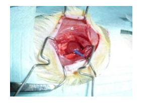
Figure 1: The exposure of heart by invasion intercostal muscles between the 5th and 6th ribs
Establishment of Myocardial Infarction Model
Myocardial infarction was induced by ligation of the left anterior descending (LAD) coronary artery [14]. The thoractomy was performed again as described previously to expose the heart by invasion intercostal muscles between the 4th and 5th ribs to re¬duce the risk of infection. After thoractomy, the heart was then exteriorized by exerting some pressure on the two sides of the chest. The ligation of the LAD was performed at the site 2.0-3.0mm below the anterior- inferior edge of the left atrium with 6-0 polypropylene thread, along a line connecting the middle of the left auricular appendage and the pulmonary trunk with the apex of the heart. Before the LAD occluded, 0.05ml 2% lido-caine carbonate injection was dropped in the surface of the heart to avoid arrhythmia. The chest was closed and the rats were tak-en the same procedure as post reconditioning of LLLI.
Histological Analysis and Massons Staining
The hearts were excised at 4 weeks after MI, immediately soaked in the cold saline to remove blood by the heart beating and rinsed clean the residual blood in the chamber. Then the hearts were fixed in the 10% neutral buffered formalin for at least 48 hours, dehydrated in different concentration of the alcohol and embed¬ded in paraffin. 8-micrometer thick sections were prepared in a plane parallel to the atrioventricular groove and in the middle of the infarcted area. The serial sections were stained with haema-toxylin-eosin (Hâ?»E) to observe the morphological changes and determine the infarct size and the left ventricular anterior wall thickness (Fig 1) and the Masson’s trichrome to delineate fibrous tissue from viable myocardial tissue and determine the percent¬age of fibrous in the left ventricular anterior wall (Fig.2). The in¬farct size was described as the percentage of infarct wall length from the circumferential length of the left ventricular wall. The left ventricular wall thickness was calculated by measuring the endocardial and epicardial length of the infarcted area. The percentage of fibrous in the left ventricular anterior wall was determined in different visual field of each histological section. Tow observers who were blind to the experiment analyzed the histological slices.
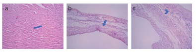
Figure 2: Representative light micrographs of Hâ?»E-stained cross-section of intact heart (a), non-LLLI preconditioned (b) and LLLI preconditioned (c) hearts 4 weeks after myocardial infarction. Note that the ventricular wall thickness in b and c decreased significantly compared with normal one (p<0.05). The normal myocardium (regular arrowhead in a) disappear in infarcted myocardium contained mainly collagenous materials (dovetail arrowhead in e) and capillaries (v shaped arrowhead in c). (H&E staining, original magnification 100×).
Statistical Analysis
The data were collected and reported as means ± SE. The SPSS 25.0 software was used for statistical analysis. The group data comparison was done by one-way analysis of variances (ANO-VA) and multiple comparisons with the least significant differ-ence (LSD) test. A value of P<0.05 was considered statistically significant.
Results
Status of Establishing Myocardial Infarction Model
The mortality of the rat model was 70.6%. Unrecovered breathing, acute left ventricular failure, incision infection and fatal ventricular arrhythmia were the main causes of the death. The 3 days after LAD ligation was the high-risk period. One third of the MI model suffered left ventricular aneurysm (Fig 3). There was no significant difference in mortality between control and LLLI rats after laser irradiation. 2 rat died of fatal ventricular arrhythmia.
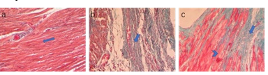
Figure 3: Typical light micro-photographs of intact normal rat heart (a) and infarcted rat heart with (b) or without laser irradia¬tion preconditioning (c). Note the normal myocardium (stained in red) in a marked with arrows. Note typical collagen tissues stained green (dovetail arrowhead in a, b and c) and viable myo¬cardium ( v shaped arrowhead in c) in the infarcted heart with laser irradiation. (Masson’s trichrome staining, original magnification 200×) .
Assessment of the Infarct Size and Left Ventricular Wall Thickness
Infarct size decreased significantly in the LLLI preconditioning group than that of the control group (15±9% to 35±10% , re-spectively, p<0.05). The left ventricular wall thickness in LLLI preconditioning rats is 0.64±0.02mm while the control rats with 0.31±0.03mm . And the left ventricular wall thickness of the shame rats was 1.32±0.04mm. The differences were all signif-icant. (p<0.05).
Evaluation of Collagen Fibers in the Left Ventricular Wall
The collagen fibers were colored green while myocardium and red cells were dyed red. The percentage of collagen fibers in the left ventricular wall in the sham rats was 4.81±0.40%. The per-centage of the collagen fibers (35.67±2. 40%) in the laser irra-diation preconditioning group decreased statistically compared to the control rats without LLLI preconditioning (64.34±2.20%) (Fig 4).
Discussion
Myocardial infarction (MI), an important cause of global death, is an irreversible heart muscle damage resulting from prolonged myocardium ischemia. A series of events after interruption of blood flow to cardiac tissue, such as inflammation, leads the cardiomyocytes to dead. Then the repair fibrosis will replace the non-functional muscles, the ventricle’s wall thickness will decrease and the cardiac pump function deteriorate, heart fail-ure (HF) will happen. The loss, impairment of contractility and hypertrophy of cardiomyocytes promote a compensatory rear-rangement in the myocardium when reaching three morpholog-ical and molecularly distinct regions: infarcted region, border region of the MI, and remote region to the MI [14].
The process of necrotic myocardium replaced by fibrosis was complicated, involving acute inflammatory response, modifica-tion of the extracellular matrix, and metabolic alteration. The newly formed extracellular matrix, especially rich in collagen, deposited in the infarcted tissue and in the transition zone to the viable tissue, culminating in the formation of the infarct scar afterward [15]. With remnant tissue overload, chronic inflamma-tory process, and involvement of neurohumoral factors present post-MI, cardiac remodeling begins.
These changes that occur in cardiac tissues may be clinically expressed by changes in the structure, shape, and function of cardiomyocytes (hypertrophy and depression of contraction and relaxation), extracellular matrix (interstitial and perivascular fi¬brosis), and blood vessels (reduction of capillarity, hypertrophy of smooth muscle, vascular dysfunction) [16]. Based on these pathophysiological changes, different therapeutic strategies, such as administration of mesenchymal stem cells (MSCs) into the infarcted area, recombinant adenovirus-mediated transfer of genes encoding antioxidants to the myocardium, introducing the growth factors (mainly of the vascular endothelial growth factors [VEGF] family), were applied with the objective of reversing or preventing damage in the different regions of the infarcted heart [17-19]. Unfortunately, these techniques may be less efficient. Photobiomodulation therapy (PBMT) is considered a novel and promising approach in injured tissues and has minimal deleteri-ous effects on healthy, uninjured cells. It may induce biochemi¬cal, bioelectrical, and bioenergetics modifications, promoting an increase in metabolism, proliferation and cell maturation and a decrease of inflammatory mediators [20]. So LLLI also could strengthen the capacity of the myocardial ischemia tolerance. And our study proved this. The main findings of the present study are that LLLI precondition remarkedly reduce the infarct size and the left ventricular wall thickness and attenuate the for¬mation of collagen fibers.
Low level laser irradiation, that is physical treatment with non-invasive and no significant harm, comes into work via so-called “light biological stimulation effect” rather than heating effect. In the heart, it was demonstrated the LLLI have anti-inflammatory effects, could stimulate proliferation and differentiation of bone marrow-derived mesenchymal stem cells (BMSCs) and attenu¬ate the hostile pathological microenvironment of damaged myo¬cardium and have cardioprotective effect in MI animal models [21, 22]. Unfortunately, the exact mechanism of the biological effects has not been fully understood. Someone think the un¬derlying mechanism of cardioprotective effect of LLLI includes increasing the mitochondrial respiration and ATP synthesis, stimulating angiogenesis, reducing the inflammatory responses, decreasing the number of injured cardiomyocytes, and reducing scarring [7-9, 23]. In our study, we found the LLLI precondition¬ing also had cardioprotective effect of stimulating angiogenesis, reducing scarring.
The biostimulatory effect of LLL seems to be influenced by sev-eral parameters including the wave length of the laser, its power density, energy density, and frequency. In our study, the energy density of laser irradiation, 0.96J/cm2, was used in this study to gain maximum benefit and avoid heating injury. Yaakobi T and co-workers in their previous studies demonstrated that the ener¬gy density of approximately 1 J/cm2 was the “optional” dose to get beneficial effects for the ischemic heart diseases [24].
The process of cardiac remodeling characterized by variations in geometry, volume, mass, and constitution of the heart is compli-cated. The inflammation due to ischemia and cell death, and the role of cytokines after MI are well-established remarks of car¬diac remodeling [25, 26]. Our present study demonstrated that LLLI precondition could reduce the infarct size and attenuate the formation of collagen fibers. These are in accordance with the effect of LLLI. Uri Oron and his team found that LLLI with proper energy density could reduce infarct size by 65% and at¬tenuate formation of scar tissue in rats and dogs with myocardial infarction [7, 8]. In addition, LLLI could stimulate proliferation, increase growth factors and facilitate myogenic differentiation of bone marrow mesenchymal stem cells [12]. Therefore, LLLI could not only modulate cardiac remodeling, but LLLI recondi¬tioning also could improve ventricular remodeling. This finding provides a new approach to improve the capacity of myocardial ischemia tolerance and has an important beneficial effect on pre¬venting ischemic heart disease.
At last, although a myriad of studies have reported the possible mechanisms involving in LLLI, the exact mechanisms of this biological effect are still not clearly understood. So further stud-ies should be conducted to clarity the molecular mechanisms of LLLI and LLLI preconditioning. A large number of preclinical and clinical studied are also required before its clinical applica¬tion for diseases.
Conclusion
The present study demonstrates that the LLLI preconditioning has a cardioprotective effect on the infarcted myocardium, could improve the ventricular remodeling. Future work need to inves¬tigate the application of LLLI in myocardial infarction.Acknowledgement
This study is supported by the important research and invention special foundation of Henan province, China (221111310700). The authors thank Mrs Xiuhua Ren for assistance in histological preparation and morphometric analysis and Mr Shanfeng Zhang for assistance for technical assistance.
Abbreviations
LLLI, low level laser irradiation; MI, myocardial infarction, LAD, left anterior descending coronary artery; PBMT, photo-biomodulation therapy; HF, heart failure; MSCs, mesenchymal stem cells; VEGH, vascular endothelial growth factor
The authors have no competing financial interests
The authors have no competing financial interests.References
- Roth, G. A., Forouzanfar, M. H., Moran, A. E., Barber, R.,Nguyen, G., Feigin, V. L., ... & Murray, C. J. (2015). Demographic and epidemiologic drivers of global cardiovascular mortality. New England Journal of Medicine, 372(14), 1333-1341.
- Fishbein, M. C., Maclean, D., & Maroko, P. R. (1978). Experimental myocardial infarction in the rat: qualitative and quantitative changes during pathologic evolution. The American journal of pathology, 90(1), 57.
- Buja, L. M. (1998). Modulation of the myocardial response to ischemia. Laboratory investigation, 78(11), 1345-1373.
- Francis, J., Weiss, R. M., Wei, S. G., Johnson, A. K., & Felder, R. B. (2001). Progression of heart failure after myocardial infarction in the rat. American Journal of Physiol-ogy-Regulatory, Integrative and Comparative Physiology, 281(5), R1734-R1745.
- Gavish, L., Perez, L., & Gertz, S. D. (2006). Low-level laser irradiation modulates matrix metalloproteinase activity and gene expression in porcine aortic smooth muscle cells. Lasers in Surgery and Medicine: The Official Journal of the American Society for Laser Medicine and Surgery, 38(8), 779-786.
- Wu, Y. H., Wang, J., Gong, D. X., Gu, H. Y., Hu, S. S., &Zhang, H. (2012). Effects of low-level laser irradiation on mesenchymal stem cell proliferation: a microarray analysis. Lasers in medical science, 27(2), 509-519.
- Ad, N., & Oron, U. (2001). Impact of low level laser irradiation on infarct size in the rat following myocardial infarc-tion. International journal of cardiology, 80(2-3), 109-116.
- Oron, U., Yaakobi, T., Oron, A., Hayam, G., Gepstein, L., Rubin, O., ... & Haim, S. B. (2001). Attenuation of infarct size in rats and dogs after myocardial infarction by low-energy laser irradiation. Lasers in Surgery and Medicine: The Official Journal of the American Society for Laser Medicine and Surgery, 28(3), 204-211.
- Peplow, P. V., Chung, T. Y., & Baxter, G. D. (2010). Laser photobiomodulation of proliferation of cells in culture: a review of human and animal studies. Photomedicine and Laser Surgery, 28(S1), S-3.
- Feng, J., Zhang, Y., & Xing, D. (2012). Low-power laser irradiation (LPLI) promotes VEGF expression and vascular endothelial cell proliferation through the activation of ERK/ Sp1 pathway. Cellular signalling, 24(6), 1116-1125.
- Zhang, W., Gao, X., Wang, X., Li, D., Zhao, Y., Zhang, T., ... & Yang, B. (2021). Light Emitting Diodes Photobiomodulation Improves Cardiac Function by Promoting ATP Synthesis in Mice With Heart Failure. Frontiers in Cardiovascular Medicine, 8.
- Hou, J. F., Zhang, H., Yuan, X., Li, J., Wei, Y. J., & Hu,S. S. (2008). In vitro effects of low-level laser irradiation for bone marrow mesenchymal stem cells: Proliferation, growth factors secretion and myogenic differentiation. Lasers in Surgery and Medicine: The Official Journal of the American Society for Laser Medicine and Surgery, 40(10), 726-733.
- Terena, S. M. L., Mesquita-Ferrari, R. A., de Siqueira Araú-jo, A. M., Fernandes, K. P. S., & Fernandes, M. H. (2021). Photobiomodulation alters the viability of HUVECs cells. Lasers in Medical Science, 36(1), 83-90.
- Jackson, B. M., Parish, L. M., Gorman III, J. H., Enomoto, Y., Sakamoto, H., Plappert, T., ... & Gorman, R. C. (2005). Borderzone geometry after acute myocardial infarction: a three-dimensional contrast enhanced echocardiographic study. The Annals of thoracic surgery, 80(6), 2250-2255.
- Sun, Y., Zhang, J. Q., Zhang, J., & Lamparter, S. (2000). Cardiac remodeling by fibrous tissue after infarction in rats. Journal of Laboratory and Clinical Medicine, 135(4), 316-323.
- Feliciano, R. D. S., Atum, A. L. B., Ruiz, É. G. D. S., Serra,A. J., Antônio, E. L., Manchini, M. T., ... & Silva Junior,J. A. (2021). Photobiomodulation therapy on myocardial infarction in rats: transcriptional and posttranscriptional implications to cardiac remodeling. Lasers in Surgery and Medicine, 53(9), 1247-1257.
- Woo, Y. J., Zhang, J. C., Vijayasarathy, C., Zwacka, R. M., Englehardt, J. F., Gardner, T. J., & Sweeney, H. L. (1998). Recombinant adenovirus-mediated cardiac gene transfer ofsuperoxide dismutase and catalase attenuates postischemic contractile dysfunction. Circulation, 98, II-255.
- Safavi, S. M., Kazemi, B., Esmaeili, M., Fallah, A., Modar-resi, A., & Mir, M. (2008). Effects of low-level He–Ne laser irradiation on the gene expression of IL-1β, TNF-α, IFN-γ, TGF-β, bFGF, and PDGF in rat’s gingiva. Lasers in medical science, 23(3), 331-335.
- Donahue, J. K. (2004). Gene therapy for cardiac arrhythmias. Annals of the New York Academy of Sciences, 1015(1), 332-337.
- Tuby, H., Maltz, L., & Oron, U. (2006). Modulations of VEGF and iNOS in the rat heart by low level laser therapy are associated with cardioprotection and enhanced angiogenesis. Lasers in Surgery and Medicine: The Official Journal of the American Society for Laser Medicine and Surgery, 38(7), 682-688.
- Guerra, F. D. R., Vieira, C. P., Marques, P. P., Oliveira, L. P., & Pimentel, E. R. (2017). Low level laser therapy accelerates the extracellular matrix reorganization of inflamed tendon. Tissue and Cell, 49(4), 483-488.
- Yang, J., Huang, Z., Zhou, Y., Sai, S., Zhu, F., Lv, R., & Fa,X. E. (2013). Effect of low-level laser irradiation on oxygen free radicals and ventricular remodeling in the infarcted rat heart. Photomedicine and Laser Surgery, 31(9), 447-452.
- Manchini, M. T., Serra, A. J., Feliciano, R. D. S., Santana,E. T., Antonio, E. L., de Tarso Camillo de Carvalho, P., ... & Silva Jr, J. A. (2014). Amelioration of cardiac function and activation of anti-inflammatory vasoactive peptides expression in the rat myocardium by low level laser therapy. PLoS One, 9(7), e101270.
- Yaakobi, T., Shoshany, Y., Levkovitz, S., Rubin, O., Ben Haim, S. A., & Oron, U. (2001). Long-term effect of low energy laser irradiation on infarction and reperfusion injury in the rat heart. Journal of applied physiology, 90(6), 2411-2419.
- Nah, D. Y., & Rhee, M. Y. (2009). The inflammatory response and cardiac repair after myocardial infarction. Korean circulation journal, 39(10), 393-398.
- Burchfield, J. S., Xie, M., & Hill, J. A. (2013). Pathological ventricular remodeling: mechanisms: part 1 of 2. Circulation, 128(4), 388-400.