
World Journal of Radiology and Imaging(WJRI)
ISSN: 2835-2440 | DOI: 10.33140/WJRI


Case Report - (2024) Volume 3, Issue 1
Ectopic Vaginal Ureterocele of Left Renal Duplex Collecting System Presenting as Vaginal Gartner’s Cyst
2Diagnostic Radiology Department, Republican Clinical Hospital after Mirgasymov, Azerbaijan
3Diagnostic Radiology Department, West Hospital, Azerbaijan
4Diagnostic Radiology Department, Universal Hospital, Azerbaijan
Received Date: Sep 20, 2024 / Accepted Date: Oct 14, 2024 / Published Date: Oct 24, 2024
Copyright: ©©2024 Vusala Rasif Hasanova et al. This is an open-access article distributed under the terms of the Creative Commons Attribution License, which permits unrestricted use, distribution, and reproduction in any medium, provided the original author and source are credited.
Citation: Bayramov, R. B., Hasanova, V. R., Hidayatov, A. K., Ismayilzada, K. I. (2024). Ectopic Vaginal Ureterocele of Left Renal Duplex Collecting System Presenting as Vaginal Gartner
Abstract
Gartner’s duct cysts are vaginal cyst that can develop along parts of the mesonephric duct (Wolffian duct), when the duct has failed to regress. The remnant of the mesonephric duct is known as Gartner duct. Gartner’s duct cyst associated some renal anomalies, such as renal agenesis, ipsilaterally renal dysplasia, cross fused renal ectopia. The ectopic ureter opening into the Gartner’s duct cyst is a very rare congenital malformation. In this case we describe a 41 years old woman with complete duplex collecting system on the left kidney and ectopic ureter which draining the lower part of the left kidney opens into Gartner’s duct cyst. The patient recently underwent a partial nephrectomy due to infection and grade IV hydronephrosis in lower part of the left kidney. During the control ultrasound examination after partial nephrectomy, cyst and tubular structure was found in the left side of pelvis.
Keywords
Gartner’s Duct Cyst, Wolffian Duct, Duplex Collecting System, Ectopic Ureter
Abbreviations
MRI-Magnetic Resonance Imaging
Introduction
Remaining Gartner duct are identified in approximately 25 % of all adult individuals with vaginas, however, only 1% will develop Gartner’s duct cyst [1]. During embryological development , the mesonephric (Wolffian) ducts develop, from their predetermined structures, and later regress. Remnants often remain, however, until they develop a secretory mechanism, cause dilation of surrounding cells, and thus yield a Gartner’s duct cyst, most often during and after late adolescence [2]. Gartner’s duct cysts are typically associated with congenital malformations of the urinary tract, such as ipsilateral renal agenesis, renal dysplasia, or atypical development of the kidney, urethral diverticulum, or the formation of pockets along the urethra, ectopic ureter, a ureter hat does not connect to the bladder and drain different site. We report a case in 41 years old women a cyst and tubular structure were detected in the pelvis who underwent partial nephrectomy. Before surgery in the left kidney there was a duplex collecting system with a fourth-degree hydronephrosis and infection in the lower part of the left kidney. The upper part of the left kidney was normal. The ureter which collects upper pole of the left kidney connects to the urinary bladder and was not dilated. The ureter which collecting the lower part of the left kidney merges the Gartner’s duct cyst. Right kidney and ureter are normal. Gartner’s duct cyst did not open into the bladder.
Case Report
A 41 years old women applied to the clinic for a follow-up examination after left sided partial nephrectomy.In the ultrasound examination, a cyst and tubular structure was found on the left side of the pelvis. MRI of the pelvis was performed on the patient.
On non-enhanced MRI was detected a cyst with regular contours at the left anterolateral wall of the proximal 1/3 part of the vagina(Figure 1, Figure 2, Figure 3
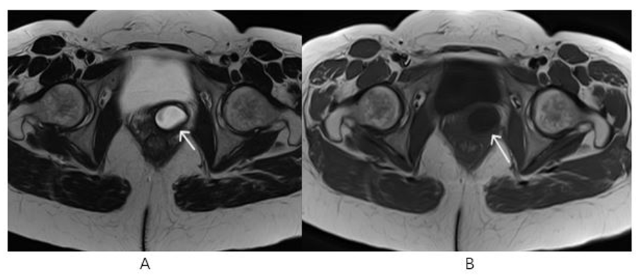
Figure 1, 2: Pelvic MR Non-Enhanced Axial T2 (A) and Axial T1 (B) Images ( White Arrows): Cystic Structure with Regular Contours at the Left Anterolateral Wall of the Proximal 1/3 Part of the Vagina.
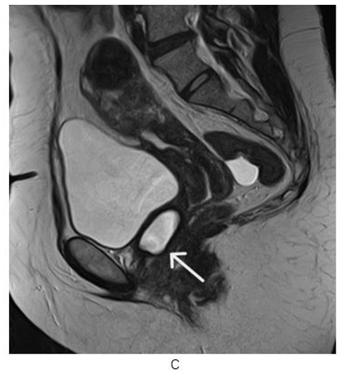
Figure 3: Pelvic MR Non-Enhanced Sagital T2 (C) image : Cystic Structure with Regular Contours at the Left Anterolateral Wall of the Proximal 1/3 Part of the Vagina ( White Arrow)
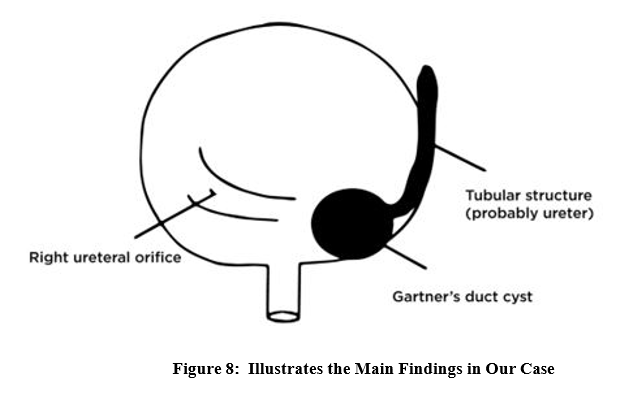
A tubular structure is observed, in left side pelvis extending and opening Gartner’s duct cyst, and ending blindly (Figure 4, Figure 5)
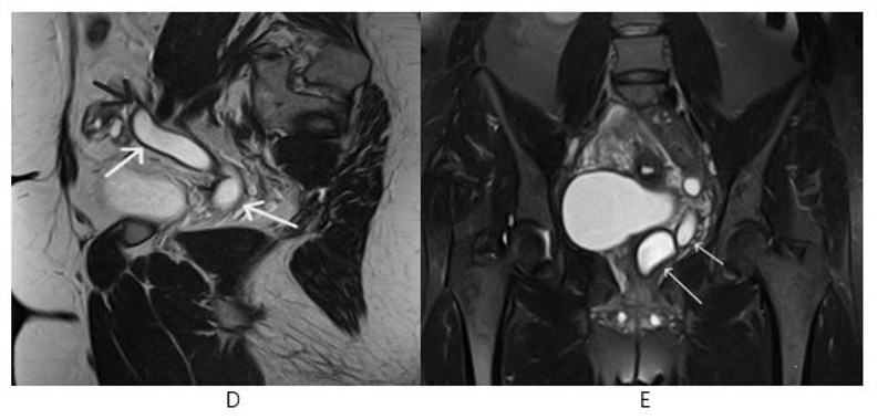
Figure 4, 5: Pelvic MR Non-enhanced Sagital T2 (D) Image: Tubular structure ( White Arrows) Ending blindly (Black Arrow) and Coronal STIR (E) Image: Gartner’s Duct Cyst (Long Arrow) and Tubular Structure (Short Arrow)
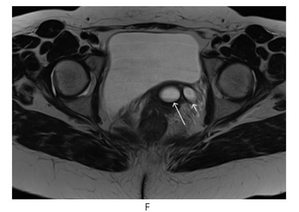
Figure 6: Pelvic MR Non-Enhanced Axial T2 (F) Tubular Structure ( White Short Arrow) and Gartner’s duct cyst (White Long Arrow)
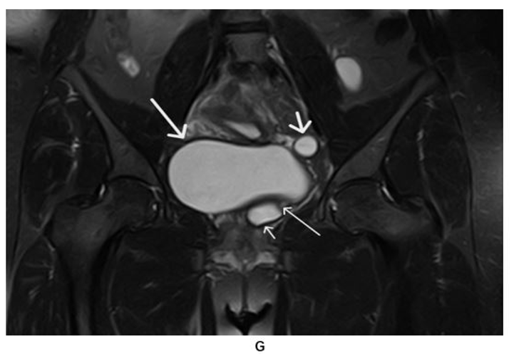
Figure 7: Pelvic MR Non-Enhanced Coronal STIR (G) Image: Gartner’s Duct Cyst (Short Thin Arrow), Urinary Bladder (Long Thick Arrow), Tubular structure (Short Thick Arrow), Tubular Structure Opening into Gartner’s Duct Cyst (Long thin Arrow)
Discussion
Embryologically, the urogenital system is derived from intermediate mesoderm and the primitive urogenital sinus which is a part of cloaca. The kidney is formed from two sources, excretory tubules derived from metanephros and collecting part is formed by ramification of the ureteric bud which arises from the mesonephric duct. Ureter arises from the ureteric bud. The distal part of the ureteric bud eventually incorporates into the bladder to from the trigone. Premature branching of the ureteric bud results in an incomplete duplex with ureters that meet before the bladder, or a bifid renal pelvis. If more than one bud develops and migrates to the metanephros a duplex kidney with two separate ureters form. When the buds are close to each other, the ureteric orifices are in the bladder in the normal position. When the buds are widely separated, the orifices may be ectopic [3-5]. In the female, a similar high origin of the ureter off the mesonephric duct should logically result in a persistence of the caudal portion of the wolffian duct (Gartner’s duct or cyst) with the ectopic ureter draining into it [6,7].
Gartner’s duct cysts arise from remnants of the mesonephric ducts (Wolffian ducts) that don’t regress successfully during development of the reproductive and urinary systems. The mesonephric duct refers to an embryonic structure that exists in all embryos but is maintained only by the presence of increased testosterone in order to from the testicles, epididymis and prostate in embryos with XY chromosomes. Occasionally, parts of the duct may remain in embryos with XX chromosomes, which may later present Gartner ‘s duct cysts. [1]
This malformation is often discovered due to the frequent occurrence of urinary incontinence, urinary tract infection, vaginal discharge, etc., and these findings show that communication between the cyst and bladder or vagina exist in many cases [8]. On the other hand, in a case such as this one, in which there is no communication with the cyst, the patient shows no symptoms and the malformation is only discovered incidentally.
Conclusion
Ureter inserting ectopically into Gartner’s duct cyst as in our case is rare and not much literature is available regarding it. This can lead to hydronephrosis and kidney infection. Therefore, it should be carefully interpreted.
Conflict of interest
The authors state that there is no conflict interest.
References
- Nikol Natalia Armata, Gartner duct Cyst “What Is It? Symptoms, and More”
- Letizia, M. J., & Kelly, J. V. (2011). Case report: Gartner's duct cyst. Emergency Medicine News, 33(5), 35.
- Narula, H., Mittal, S., Vohra, A., & Jindal, G. (2016). Ectopic vaginal ureterocele presenting as vaginal gartner’s cyst. Journal of OBGYN, 3(1), 61-65.
- Ureteroceles on duplex ureters. In: Stephens F, Smith E, Hutson J. 2002 editors. Congenital anomalies of the kidney, urinary and genital tracts. London: Martin Dunitz. 243-262
- Glassberg, K. I., Braren, V., Duckett, J. W., Jacobs, E. C.,King, L. R., Lebowitz, R. L., ... & Stephens, F. D. (1984). Suggested terminology for duplex systems, ectopic ureters and ureteroceles. The Journal of urology, 132(6), 1153-1154.
- Kate, D., & Shinde, R. (2015). Duplex kidney–an anatomical and clinical insight. IOSR J Dent Med Sci, 14(4), 14-17.
- Lee, M. J., Yoder, I. C., Papanicolaou, N., & Tung, G. A. (1991). Large Gartner duct cyst associated with a solitary crossed ectopic kidney: imaging features. Journal of computer assisted tomography, 15(1), 149-151.
- Koroku, M., Sakaiy, S., Yanase, M., & Shimamura, S. (1995). A case of Gartner's duct cyst with a right aplastic kidney. International journal of urology, 2(3), 211-213.