

Journal of Agriculture and Horticulture Research(JAHR)
ISSN: 2643-671X | DOI: 10.33140/JAHR
Impact Factor: 1.12

Research Article - (2026) Volume 9, Issue 3
Differential Redox Regulation and Antioxidant Dynamics in Tomato Fruits Under Mercury Stress
 , Sanders Junglee 2 , Huguette Sallanon 2 , Laurent Urban 1 and Felicie Lauri 1,2
 2QualiSud UMR95, CIRAD, Montpellier Université, InstitutAgro, F-34000 Montpellier, France
Received Date: May 28, 2026 / Accepted Date: Jun 22, 2026 / Published Date: Jul 02, 2026
Copyright: ©2026 Ramzi Murshed, et al. This is an open-access article distributed under the terms of the Creative Commons Attribution License, which permits unrestricted use, distribution, and reproduction in any medium, provided the original author and source are credited.
Citation: Murshed, S., Junglee, S., Sallanon, H., Urban, L., Lauri, F. (2026). Differential Redox Regulation and Antioxidant Dynamics in Tomato Fruits Under Mercury Stress. J Agri Horti Res, 9(3), 01-08.
Abstract
The present study investigated the impact of mercury exposure on plant water status, oxidative stress markers, and antioxidant defense systems in leaves and fruits of tomato (Solanum lycopersicum L. cv. Micro-Tom). Plants were exposed to increasing concentrations of HgCl2 for 24 and 48 h. Mercury treatment led to a significant reduction in predawn leaf water potential, whereas other water-related parameters in both leaves and fruits remained largely unaffected. Oxidative stress was predominantly observed in leaves, as indicated by elevated hydrogen peroxide (H2O2) and malondialdehyde (MDA) levels, while fruit tissues showed no significant accumulation of these oxidative markers. In contrast, fruits exhibited a marked activation of antioxidant defenses, including increased activities of superoxide dismutase (SOD) and catalase (CAT), along with concentration- dependent modulation of ascorbate–glutathione cycle enzymes (APX, MDHAR, DHAR, and GR) and their corresponding transcript levels. Alterations in the ascorbate pool, reflected by changes in reduced ascorbate (AsA) and dehydroascorbate (DHA), further indicated a dynamic regulation of cellular redox status in response to mercury exposure. Collectively, these findings demonstrate that mercury induces tissue-specific oxidative responses and rapidly activates antioxidant mechanisms in fruits, thereby contributing to the maintenance of redox homeostasis and protection against oxidative damage.
Keywords
Oxidative Stress, Antioxidant Enzymes, Ascorbate–Glutathione Cycle, Mercury Toxicity, Solanum Lycopersicum
Introduction
Heavy metal contamination of soils has emerged as a critical environmental constraint with far-reaching implications for agricultural sustainability and food safety, primarily due to the potential transfer of toxic elements into trophic systems. Among these contaminants, mercury (Hg) is particularly problematic owing to its persistence, high mobility, and strong bioaccumulative behavior in both terrestrial and aquatic compartments. Agricultural practices may inadvertently introduce mercury into soils through amendments such as sewage sludge, mineral fertilizers, pesticides, and organic residues, while anthropogenic inputs, including industrial effluents and domestic discharges, further intensify Hg loading in aquatic environments [1,2]. At the cellular and molecular levels, mercury exerts pronounced phytotoxic and genotoxic effects [3]. Its reactivity with biomolecules enables mercuric ions to interact with nucleic acids, leading to the formation of stable covalent adducts with DNA, and to the induction of chromosomal instability, including sister chromatid exchanges [4]. In addition, mercury disrupts key physiological processes in plants, impairing photosynthetic performance, water relations, and pigment biosynthesis, while simultaneously promoting membrane lipid peroxidation and metabolic dysfunction [5].
Exposure to elevated concentrations of heavy metals is commonly associated with the onset of oxidative stress, a condition arising from an imbalance between reactive oxygen species (ROS) generation and detoxification [6]. Under stress conditions, perturbations in chloroplast photochemistry enhance the leakage of excess excitation energy toward oxygen, resulting in the formation of ROS such as superoxide radicals, singlet oxygen, hydrogen peroxide, and hydroxyl radicals [7]. These highly reactive species can severely compromise cellular integrity by inducing lipid peroxidation, protein oxidation, enzyme inhibition, pigment degradation, and genomic instability [8]. To maintain redox homeostasis, plants have evolved a complex and highly coordinated antioxidant network. Metal toxicity becomes critical when intracellular concentrations exceed the sequestration and detoxification capacities of plant tissues [9]. The primary defense system consists of enzymatic components, where superoxide dismutase (SOD) catalyzes the dismutation of superoxide radicals into hydrogen peroxide, which is subsequently detoxified by catalase (CAT) or ascorbate peroxidase (APX) [10-12].
A central hub of redox regulation is the ascorbate–glutathione cycle, which operates through a tightly regulated sequence of redox reactions. In thiss pathway, APX mediates the oxidation of ascorbate (AsA) to monodehydroascorbate (MDHA), which is either enzymatically reduced back to AsA via monodehydroascorbate reductase (MDHAR) or spontaneously converted into dehydroascorbate (DHA). DHA is subsequently reduced to AsA by dehydroascorbate reductase (DHAR) using reduced glutathione (GSH) as an electron donor, while glutathione reductase (GR) regenerates GSH from its oxidized form (GSSG) in an NADPH-dependent manner [13,14]. This cycle plays a pivotal role in sustaining cellular redox buffering capacity and preventing ROS overaccumulation. Complementary to enzymatic defenses, low-molecular-weight antioxidants, including ascorbate, glutathione, tocopherols, and carotenoids, contribute to the fine-tuning of redox balance and oxidative stress mitigation [15]. Despite extensive recognition of heavy metal toxicity, the mechanistic basis of mercury-induced phytotoxicity remains incompletely resolved, particularly with respect to its interaction with oxidative metabolism. The scarcity of integrative data hampers a precise evaluation of the contribution of oxidative stress to mercury toxicity in plants. Therefore, the present study aims to elucidate whether oxidative stress constitutes a central component of mercury-induced damage in tomato plants, and to characterize the dynamic responses of enzymatic and non-enzymatic antioxidant systems under Hg exposure [16].
Materials and Methods
Plant Material and Experimental Design
Tomato plants (Solanum lycopersicum L., cv. Micro-Tom) were cultivated in 4 L plastic pots containing a peat–vermiculite mixture (1:1, v/v) under controlled growth chamber conditions [17]. Environmental parameters were maintained at 60 ± 5% relative humidity, with a day/night temperature regime of 25/20 °C and a 16/8 h photoperiod under a light intensity of 300 µmol m-2 s-1 provided by fluorescent lamps [18]. Mercury treatments were initiated during the fruit development stage by supplementing the growth substrate with HgCl2 at concentrations of 2, 5, 10, and 20 ppm. Untreated plants were used as controls. Fruits at the mature green stage (35–40 days post-anthesis) were collected after 24 and 48 h of exposure. Immediately after harvest, samples were snap-frozen in liquid nitrogen, finely ground, and stored at −80 °C until further analyses.
Determination of Water Status Parameters
Predawn leaf water potential (LΨw) was measured using a pressure chamber following the approach of Scholander et al. (1965). Fruit water potential (FΨw) was determined using a dew point microvoltmeter (HR-33T) equipped with C-52 chambers (Wescor Inc., USA), while fruit osmotic potential (FΨO) was assessed using a vapor pressure osmometer (VAPRO, Wescor) [18]. To evaluate water flux, plants were excised approximately 1 cm above the substrate surface. Fruit fresh weight (FW) was recorded immediately after sampling, whereas dry weight (DW) was obtained after oven-drying at 70 °C for 72 h. Fruit water content (FWC) was calculated according to the equation:FWC = (FW − DW) / FW × 100 [19].
Quantification of Hydrogen Peroxide
Hydrogen peroxide (H2O2) levels were quantified according to a microplate-adapted protocol. Approximately 0.25 g of frozen fruit tissue was homogenized in 1 mL of cold 0.1% (w/v) trichloroacetic acid (TCA) and centrifuged at 12,000 × g for 15 min at 4 °C. Aliquots of the supernatant (100 µL) were mixed with potassium phosphate buffer (10 mM, pH 7.0) and potassium iodide (KI), and the reaction mixture was incubated prior to absorbance measurement at 390 nm using a microplate reader. Hydrogen peroxide concentration was calculated based on a standard calibration curve generated using known H2O2 concentrations [20].
Lipid Peroxidation Assay
Lipid peroxidation was estimated by quantifying malondialdehyde (MDA) using the thiobarbituric acid (TBA) method. Frozen tissue (0.25 g) was homogenized in 0.1% (w/v) TCA and centrifuged as described above. An aliquot of the supernatant was reacted with TBA solution (0.5% in 20% TCA) and incubated at high temperature for 30 min [21-25]. The reaction was terminated by rapid cooling on ice. Absorbance was measured at 532 nm, and non-specific turbidity was corrected by subtracting readings at 600 nm. MDA concentration was calculated using an extinction coefficient of 155 m-1 cm-1 [26].
Determination of Ascorbate Pool Components
Total ascorbate (AsA + DHA) and reduced ascorbate (AsA) contents were determined using a modified microplate protocol. Frozen samples (0.5 g) were extracted in cold 6% (w/v) TCA and centrifuged at 16,000 × g for 15 min at 4 °C [27]. For total ascorbate quantification, extracts were treated with dithiothreitol (DTT) to reduce dehydroascorbate (DHA), followed by neutralization with N-ethylmaleimide (NEM). Subsequently, a colorimetric reaction mixture containing orthophosphoric acid, bipyridyl, and ferric chloride was added. After incubation at 42 °C, absorbance was recorded at 525 nm [28-30]. AsA content was measured using the same procedure without DTT and NEM. DHA concentration was calculated as the difference between total ascorbate and reduced ascorbate. Standard curves were established using L-ascorbic acid.
Gene Expression Analysis
Total RNA was extracted from frozen fruit tissues using Tri Reagent according to the manufacturer’s protocol. RNA quantity and purity were assessed spectrophotometrically (A260/A280), and integrity was verified by agarose gel electrophoresis [31]. First-strand cDNA synthesis was performed from 4 µg of total RNA using an oligo(dT) primer system following thermal denaturation and reverse transcription steps. Quantitative real-time PCR (qRT-PCR) was conducted using SYBR Green chemistry on a Realplex system (Eppendorf). Gene expression levels were normalized against the reference gene SlActin, and relative transcript abundance was calculated accordingly. Gene-specific primers targeting antioxidant-related genes were designed based on sequences retrieved from the GenBank database using DNAMAN software [32]. PCR products were purified, sequenced, and deposited in GenBank under the corresponding accession numbers.
Antioxidant Enzyme Activities
Protein Extraction
Proteins were extracted from frozen fruit tissues homogenized in MES/KOH buffer (50 mM, pH 6.0) containing KCl, CaCl2, and ascorbate. After centrifugation at 16,000 × g for 15 min at 4 °C, the supernatants were collected for enzymatic assays [33]. Protein concentration was determined using the Bradford method.
Enzyme Assays
Enzyme activities were measured spectrophotometrically in microplate-based kinetic assays at 25 °C. Activities of APX, DHAR, MDHAR, and GR were determined according to established protocols [34]. Superoxide dismutase (SOD) activity was evaluated based on its ability to inhibit the photochemical reduction of nitro blue tetrazolium (NBT) under light exposure, with absorbance measured at 560 nm. Catalase (CAT) activity was determined by monitoring the decomposition rate of hydrogen peroxide at 240 nm. Enzyme activities were calculated using appropriate extinction coefficients and expressed relative to protein content [35].
Statistical Analysis
All experiments were conducted in triplicate [36]. Data are presented as mean values ± standard error (SE). Statistical differences among treatments were evaluated by analysis of variance (ANOVA) using MINITAB software, with significance set at p < 0.05.
|
PCR fragment |
Encoded protein |
Primer sequence (5’ → 3’) |
Size (bp) |
Accession number |
|
SlAPXt |
Thylakoid-bound ascorbate peroxidase |
Sense : TTCACCCAATGACTTCCCT Antisense: TATCATTTAGTCCCATTCTGT |
699 |
FJ532352 |
|
SlAPXcyto |
Cytosolic ascorbate peroxidase |
Sense : GTTGAAGGTCGCTTGCCG Antisense: CCAAGGTATGGGCACCAG |
118 |
FJ532353 |
|
SlDHAR1 |
Dehydroascorbate reductase |
Sense : TGCCTCTGTGGGCTCGAA Antisense: ACCACCCTGCGATGACGT |
335 |
FJ532354 |
|
SlDHAR2 |
Dehydroascorbate reductase |
Sense : ACAACTCCTAACAAGCTCGG Antisense: TCCAAGCGACAAATCAGCA |
430 |
FJ532355 |
|
SlMDHAR |
Monodehydroascorbate reductase |
Sense : GGAGAAGTTTCGTTGCTGCT Antisense: GAGCAGCTTTCCTGAATTGT |
371 |
FJ544908 |
|
SlGRcp |
Chloroplastic glutathione reductase |
Sense : TGGAGGCGTCGGTGGCAC Antisense: TATAGCATACTCGCTTCCAG |
362 |
FJ544906 |
|
SlGRcyto |
Cytosolic glutathione reductase |
Sense : GCAAAGAATTATGGATGGGA Antisense: CACAGCACGCTTTGGTAA |
333 |
FJ544907 |
Table 1: Sets of Primers used to Amplify Gene-Specific Regions, Corresponding Size and Accession Number of the Amplified Product
Results
Plant Water Status
Mercury exposure resulted in a pronounced reduction in predawn leaf water potential (LΨw), with the magnitude of decline being both concentration- and time-dependent (Table 2). In contrast, no significant alterations were observed in fruit water potential (FΨw), fruit osmotic potential (FΨO), or water content across fruits, leaves, and peduncles (FWC, LWC, PWC) (Table 2) [37]. Water transport dynamics were, however, affected by mercury, as evidenced by a progressive decline in water flux up to 10 ppm HgCl2, followed by a complete cessation at 20 ppm, indicating a threshold beyond which hydraulic conductivity is severely impaired [38].
|
24 h |
48 h |
|||||||||
|
|
C |
2 ppm |
5 ppm |
10 ppm |
20 ppm |
C |
2 ppm |
5 ppm |
10 ppm |
20 ppm |
|
LΨw |
- 0.34 ± 0.02a |
- 0.48 ± 0.02b |
- 0.63 ± 0.03c |
- 1.15 ± 0.07d |
- 1.37 ± 0.06e |
- 0.35 ± 0.02a |
- 0.45 ± 0.02b |
- 0.65 ± 0.03c |
- 1.35 ± 0.03 e |
- 1.60 ± 0.04f |
|
FΨw |
- 0.45 ± 0.04a |
- 0.38 ± 0.09a |
- 0.48 ± 0.10a |
- 0.41 ± 0.02a |
- 0.42 ± 0.09a |
- 0.44 ± 0.03a |
- 0.42 ± 0.05a |
- 0.45 ± 0.03 a |
- 0.43 ± 0.08 a |
- 0.46 ± 0.05a |
|
FΨO |
- 0.72 ± 0.01a |
- 0.68 ± 0.03a |
- 0.77 ± 0.03a |
- 0.83 ± 0.01a |
- 0.82 ± 0.02a |
- 0.73 ± 0.02a |
- 0.76 ± 0.01a |
- 0.78 ± 0.02a |
- 0.78 ± 0.03 a |
- 0.75 ± 0.01a |
|
FWC |
94 ± 0.5 a |
94 ± 0.4 a |
94 ± 0.5 a |
95 ± 0.4 a |
94 ± 0.2 a |
94 ± 0.3 a |
95 ± 0.4 a |
94 ± 0.4a |
94 ± 0.6 a |
95 ± 0.3a |
|
LWC |
91 ± 0.4 a |
91 ± 1.8 a |
92 ± 0.6 a |
91 ± 0.3 a |
90 ± 0.3 a |
91 ± 0.4 a |
90 ± 0.2 a |
90 ± 0.4a |
90 ± 0.8 a |
89 ± 1.5a |
|
PWC |
86 ± 5.0 a |
89 ± 4.5 a |
86 ± 0.8 a |
82 ± 5.6 a |
84 ± 0.4 a |
86 ± 4.2 a |
83 ± 2.4 a |
83 ± 0.2 a |
83 ± 0.6 a |
82 ± 0.8a |
Table 2: Predawn Leaf Water Potential (LΨw; MPa ), Fruit Water Potential (FΨw; MPa ), Fruit Osmotic Potential (FΨO; MPa) and Water Content of Fruits (FWC; %), Leaves (LWC; %) and Fruit Peduncles (PWC; %) of Control Plants (C) and Plants Treated with 2, 5, 10, and 20 ppm of HgCl2 for 24 and 48 hours. Values are the mean (± S.E.) of Five Replicates, and Different Letters within lines Indicate Significant Differences (P<0.05).
Oxidative Markers: H2O2 and Lipid Peroxidation
Mercury treatments triggered a marked accumulation of H2O2 in leaves, particularly at moderate to high concentrations during early exposure (24 h), while prolonged exposure (48 h) led to a decline at the highest concentration tested (Table 3). Lipid peroxidation, assessed via MDA levels, showed a clear induction under mercury stress, especially after prolonged exposure (Table 3) [39]. Notably, these oxidative responses were restricted to leaf tissues, as fruit tissues did not exhibit significant changes in either H2O2 or MDA levels (Table 3), suggesting tissue-specific sensitivity to mercury-induced oxidative stress [40].
|
Leaves |
Fruits |
|
||||||
|
|
24h 48h |
24h 48h |
||||||
|
|
H2O2 |
MDA |
H2O2 |
MDA |
H2O2 |
MDA |
H2O2 |
MDA |
|
C |
849.36 ± 17.64 a |
34.04 ± 0.19a |
850.31 ± 16.48a |
34.25 ± 0.27a |
56.77 ± 3.24a |
34.74 ± 5.24a |
58.82 ± 3.29a |
33.59 ± 4.37 a |
|
2 ppm |
804.19 ± 34.19 a |
33.19 ± 0.95a |
856.76 ± 37.30a |
37.14 ± 0.71b |
51.47 ± 4.41a |
34.37 ± 6.83a |
55.29 ± 49.71a |
33.60 ± 5.97 a |
|
5 ppm |
1081.70 ± 107.62 b |
33.36 ± 0.53a |
1063.76 ± 29.65c |
38.92 ± 0.66b |
54.41 ± 7.06a |
35.13 ± 6.62a |
55.30 ± 6.47a |
35.08 ± 5.41a |
|
10 ppm |
1013.49 ± 87.27 b |
32.16 ± 1.42a |
1212.60 ± 22.26d |
43.51 ± 0.68c |
57.35 ± 2.47a |
37.04 ± 3.21a |
59.71 ± 5.88a |
39.39 ± 3.03a |
|
20 ppm |
1211.88 ± 93.64b |
48.67 ± 0.65b |
476.93 ± 21.36e |
44.26 ± 1.57c |
58.53 ± 2.35a |
35.09 ± 5.66a |
51.18 ± 4.94a |
34.65 ± 6.90a |
Table 4: Total ascorbate, Ascorbate (AsA) and dehydroascorbate (DHA) concentrations (nmol. g-1 FW) and ascorbate redox state (AsA/Total) in leaves and fruit of control plants (C) and plants treated with 2, 5, 10, and 20 ppm of HgCl2 for 24 and 48 hours. Values are the mean (± S.E.) of five replicates, and different letters within columns indicate significant differences (P<0.05).
|
|
24h |
48h |
|
|
|
|
|
|
|
|
|
|
|
|
|
C |
SlDHAR1 |
SlDH AR2 |
SlMDH AR |
SlAPX cyto |
SlAP Xt |
SlGR cyto |
SlGR cp |
SlDHA R1 |
SlDHA R2 |
SlMDH AR |
SlAPXc yto |
SlAP Xt |
SlGR cyto |
SlGR cp |
|
2 ppm |
1.00 ± 0.2 a |
1.00 ± 0.2 a |
1.00 ± 0.2 a |
1.00 ± 0.2 a |
1.00 ± 0.2 a |
1.00 ± 0.2 a |
1.00 ± 0.2 a |
1.00 ± 0.2 a |
1.00 ± 0.2 a |
1.00 ± 0.2 a |
1.00 ± 0.2a |
1.00 ± 0.2 a |
1.00 ± 0.2 a |
1.00 ± 0.2 a |
|
|
|
|
|
|
|
|
|
|
|
|
|
|
|
|
|
5 ppm |
1.71± 0.2 b |
1.61± 0.2 b |
4.10± 0.5 b |
2.05± 0.2 b |
1.41± 0.05 b |
2.63± 0.3 b |
0.31± 0.02 b |
1.93± 0.2 b |
2.43± 0.3 b |
2.59± 0.3 b |
1.06± 0.1 a |
5.55± 0.6 b |
4.75± 0.5 b |
2.41± 0.3 b |
|
|
|
|
|
|
|
|
|
|
|
|
|
|
|
|
|
10 ppm |
3.17± 0.4 c |
2.73± 0.3 c |
4.75± 0.4 b |
4.47± 0.5 c |
1.79± 0.2 c |
6.07± 0.7 c |
0.46± 0.03 c |
5.47± 0.7 c |
3.84± 0.5 c |
2.57± 0.3 b |
1.08± 0.2 a |
4.26± 0.5 c |
4.62± 0.6 b |
2.02± 0.2 b |
|
20 ppm |
1.90± 0.2 b |
4.72± 0.5 d |
5.65± 0.6 b |
1.21± 0.1 a |
2.37± 0.3 d |
4.30± 0.3 d |
0.44± 0.02 c |
0.54± 0.04 d |
0.20± 0.02 d |
1.79± 0.2 b |
0.28± 0.02 b |
2.66± 0.3 d |
0.90± 0.1 a |
0.12± 0.02 c |
|
|
0.22± 0.03 d |
0.68± 0.06 e |
0.50± 0.06 c |
0.37± 0.04 d |
3.39± 0.4 e |
1.33± 0.1 a |
0.00± 0.0 d |
0.00± 0.0 e |
0.00± 0.0 e |
0.23± 0.02 c |
0.00± 0.0 c |
0.58± 0.04 e |
0.19± 0.02 c |
0.00± 0.0 d |
Table 5: Changes in the levels of mRNA encoding antioxidant enzymes in fruits of control plants (C) and plants treated with 2, 5, 10, and 20 ppm of HgCl2 for 24 and 48 hours. Data were expressed as relative values with respect to the value found in the control fruits. Each value is the mean of nine replicates, and different letters within columns indicate significant differences (P<0.05).
Expression of Antioxidant-Related Genes
The transcriptional profiles of genes associated with the ascorbate–glutathione cycle revealed complex and concentration-dependent regulation patterns (Table 5). While moderate mercury levels generally stimulated the expression of genes such as SlAPXt, SlDHAR1, SlDHAR2, and SlMDHAR, higher concentrations and longer exposure periods led to transcriptional repression. Distinct isoform-specific responses were also observed [42]. For instance, cytosolic and chloroplastic glutathione reductase genes (SlGRcyto and SlGRcp) displayed divergent expression patterns (Table 5), reflecting differential compartmental regulation of redox homeostasis.
Antioxidant Enzyme Activities
Mercury exposure induced a substantial activation of antioxidant enzymes in fruits. SOD activity increased consistently under all treatments, with a peak response at intermediate concentrations (Figure 1). CAT and APX activities were also strongly stimulated, indicating enhanced detoxification capacity for hydrogen peroxide (Figure 1). Enzymes involved in ascorbate recycling (MDHAR, DHAR, and GR) showed significant activation, although their responses varied with mercury concentration (Figure 1). Notably, maximal induction was often observed at moderate concentrations, whereas higher concentrations resulted in partial inhibition, suggesting enzyme sensitivity to excessive mercury levels.
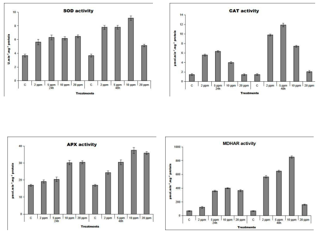
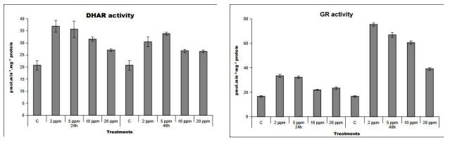
Figure 1: Activity of superoxide dismutase (SOD), catalase (CAT), ascorbate peroxidase (APX) monodehydroascobate reductase (MDHAR), dehydroascobate reductase (DHAR) and glutathione reductase (GR) in fruits of control plants (C) and plants treated by 2, 5, 10, and 20 ppm of HgCl2 for 24 and 48 hours
Discussion
The decline in leaf water potential observed under mercury exposure (Table 2) likely reflects impaired membrane transport processes, particularly those mediated by aquaporins, which are known targets of mercury inhibition. Despite this disruption at the leaf level, fruit water relations remained relatively stable (Table 2), indicating a degree of hydraulic buffering or compartmentalization. The accumulation of reactive oxygen species in leaves, coupled with increased lipid peroxidation (Table 3), confirms that mercury induces oxidative stress primarily in vegetative tissues. The absence of similar changes in fruits (Table 3) suggests the existence of more efficient antioxidant systems in reproductive tissues, a hypothesis supported by the enhanced enzymatic activities observed (Figure 1). The upregulation of SOD, CAT, and APX activities (Figure 1) indicates an increased flux through ROS detoxification pathways. However, the decline in enzyme efficiency at higher mercury concentrations suggests potential inhibitory effects of mercury on protein structure and function. The ascorbate–glutathione cycle appears to play a central role, as reflected by changes in enzyme activities (Figure 1), gene expression patterns (Table 5), and ascorbate redox state (Table 4). Nevertheless, discrepancies between transcript levels and enzyme activities point toward additional regulatory mechanisms beyond transcriptional control. In fruits, the reduction in ascorbate redox state under prolonged exposure (Table 4) reflects a shift toward oxidative conditions despite the activation of enzymatic defenses, indicating potential limitations in sustained stress tolerance.
Conclusions
Mercury exposure induces oxidative stress in tomato plants, as evidenced by increased ROS accumulation and lipid peroxidation in leaves (Table 3), alongside significant modulation of antioxidant systems (Figure 1, Table 4). Although fruit tissues initially maintain redox stability, likely through efficient antioxidant responses, prolonged exposure results in measurable disturbances in redox balance (Table 4). These findings emphasize the importance of antioxidant mechanisms in mercury tolerance and highlight differential responses between plant organs.
Declarations
Funding
The authors gratefully acknowledge the financial support provided by the Scholar Rescue Fund (SRF) and the PAUSE Program (Programme d’aide à l’accueil en urgence des scientifiques et des artistes en exil), which made the publication of this article possible.
Author Contributions
Ramzi MURSHED : Conceptualization, Investigation, Methodology, Writing – original draft. Sanders JUNGLEE : Methodology. Huguette SALLANON : Supervision, Validation, Writing – review and editing. Laurent URBAN : Validation, Writing – review and editing. Félicie LAURI : Methodology, Project administration, Writing – review and editing.
Data Availability Statement
Data available upon reasonable request.
References
- Patra, M., & Sharma, A. (2000). Mercury toxicity in plants.The botanical review, 66(3), 379-422.
- Huang, D., Niu, C., Zeng, G., Wang, X., & Lv, X. (2015).A highly sensitive protocol for the determination of Hg2+ in environmental water using time-gated mode. Talanta, 132, 606-612.
- Israr, M., Sahi, S.V., Jain, J. (2011). Accumulation and effects of mercury on plants: a review. Environmental Chemistry Letters. 9, 161–176.
- Sharma, A., Talukder, G. (1989). Metals as clastogens-some aspects of study. in: Sharma, A.K., Sharma, A. (Eds.), Advances in Cell and Chromosome Research. IBH Publ, New Delhi, Oxford, pp. 197–213.
- Cho, U.H., Park, J.O. (2000). Mercury-induced oxidative stress in tomato seedlings. Plant Science. 156, 1–9.
- Yamamoto, Y., Hachia, A., Matsumoto, H. (1997). Oxidative damage to membranes by a combination of aluminium and iron in suspension-cultured tobacco cells. Plant Cell Physiology. 38, 1333-1339.
- Mittler, R., 2002. Oxidative stress, antioxidants and stress tolerance. Trends Plant Science. 7, 405- 410.
- Halliwell, B., 2006. Reactive species and antioxidants. Redox biology is a fundamental theme of aerobic life. Plant Physiology. 141, 312-322.
- Sinha, S., Gupta, M., Chandra, P. (1996). Bioaccumulation and biochemical effects of mercury in the plant Bacopa monnieriL. Environmental Toxicology. 11, 105–112.
- Fridovich, I. (1986). Superoxide dismutases. Advances in Enzymology and Related Areas of Molecular Biology. 58, 61-97.
- Willekens, H., Inze, D., Van Montagu, M., Van Camp, W. (1995). Catalases in plants Molecular Breeding. 1, 207-228.
- Bowler, C., Van Montagu, M., Inze, D. (1992). Superoxide dismutase and stress tolerance. Annual Review of Plant Physiology and Plant Molecular Biology. 43, 83-116.
- Foyer, C.H., Lelandais, M., Kunert, K.J. (1994). Photooxidative stress in plants. Physiol Plant. 92, 696–717.
- Noctor, G., Foyer, C.H. (1998). Ascorbate and Glutathione: keeping active oxygen under control. Annual Reviews Plant Physiology, Plant Molecular Biology. 49, 249-279.
- Foyer, C.H., Noctor, G. (2005). Redox homeostasis and antioxidant signalling: a metabolic interface between stress perception and physiological responses. Plant Cell. 17, 1866-1875.
- Aebi, H. (1984). Isolation, purification, characterization, and assay of antioxygenic enzymes: catalase in vitro. Methods in Enzymology. 105, 121-126.
- Allen, R.D. (1995). Dissection of oxidative stress tolerance using transgenic plants. Plant Physiology. 107, 1049–1054.
- Bradford, M.M. (1976). A rapid and sensitive method for the quantitation of micrograms quantities of protein utilizing the principle of protein–dye binding. Analytical Biochemistry. 72, 248–254.
- Cargnelutti, D., Tabaldi, L.A., Spanevello, R.M., Jucoski, G.O., Battisti, V., Redin, M., Linares, C.E.B., Dressler, V.L., Flores, E.M.M., Nicoloso, F.T., Morsch, V.M., SchetingerM.R.C. (2006). Mercury toxicity induces oxidative stress in growing cucumber seedlings. Chemosphere. 65, 999-1006.
- Chaoui, A., Mazhouri, S., Ghorbal, M.H., Ferjani, E.E. (1997). Cadmium and zinc induction of lipid peroxidation and effects on antioxidant enzyme activities in bean (Phaseolus vulgaris L.). Plant Science. 127, 139–147.
- Dhindsa, R.S., Plumb-Dhindsa, P., Thorpe, T.A. (1981). Leaf senescent: correlated with increased levels of membrane permeability and lipid peroxidation, and decreased levels of superoxide dismutase and catalase. Journal of Experimental Botany. 32, 93–101.
- Elstner, E.F., Wagner, G.A., Schultz, W. (1988). Activated oxygen in green plants in relation to stress situations. Plant Physiology and Biochemistry. 7, 159–187.
- Foyer, C.H., Halliwell, B. (1976). The presence of glutathione and glutathione reductase in chloroplasts: a proposed role in ascorbic acid metabolism. Planta. 133, 21–25.
- Foyer, C.H., Trebst, A., Noctor, G. (2005). Protective and signalling functions of ascorbate, glutathione and tocopherol in chloroplasts. in: Demmig-Adams, B., Adams, W.W. (Eds.), Advances in Photosynthesis and Respiration: Photoprotection, Photoinhibition, Gene Regulation, and Environment. Dordrecht, The Netherlands, pp. 241–268.
- Gomez, L.D., Noctor, G., Knight, M., Foyer, C.H. (2004). Regulation of calcium signaling and gene expression by glutathione. Journal of Experimental Botany. 55, 1851–1859.
- Ishikawa, T., Dowdle, J., Smirnoff, N. (2006). Progress in manipulating ascorbic acid biosynthesis and accumulation in plants. Physiologia Plantarum. 126, 343–355.
- Janero, D.R. (1990). Malondialdehyde and thiobabituric acid-reactivity as diagnostic indices of lipid peroxidation and peroxidative tissue injury. Free Radical Biology and Medicine. 9, 515–540.
- Jimenez, A., Creissen, G., Kular, B., Firmin, J., Robinson, S., Verhoeyen, M., Mullineaux, P. (2001). Changes in oxidative processes and components of the antioxidant system during tomato fruit ripening. Planta. 214, 751-758.
- Kampfenkel, K., Van Montagu, M., Inze, D. (1995). Extraction and determination of ascorbate and dehydroascorbate from plant tissue. Analytical Biochemistry. 225, 165-167.
- Mittova, V., Guy, M., Tal, M., Volokita, M. (2002). Response of the cultivated tomato and its wild salt-tolerant relative Lycopersicon pennellii to salt-dependent oxidative stress: Increased activities of antioxidant enzymes in root plastids. Free Radical Research. 36, 195-202.
- Mondal, K., Sharmma, N.S., Malhotra, S.P., Dhawan, K., Singh, R. (2004). Antioxidant systems in ripening tomato fruits. Biologia plantarum. 48, 49-53.
- Murshed, R., Lopez-Lauri, F., Keller, C., Monnet, F., Sallanon,H. (2008). Acclimation to Drought Stress Enhances Oxidative Stress Tolerance in Solanum lycopersicum L. Fruits. Plant Stress. 2, 145-151.
- Murshed, R., Lopez-Lauri, F., Sallanon, H. (2008). Microplate quantification of enzymes of the plant ascorbate–glutathione cycle. Analytical Biochemistry. 383, 320–322.
- Potters, G., De Gara, L., Asard, H., Horemans, N. (2002). Ascorbate and glutathione: guardians of the cell cycle, partners in crime. Plant Physiology and Biochemistry. 40, 537-548.
- Prasad, K., Saradhi, P.P., Sharmila, P. (1999). Concerted action of antioxidant enzymes and curtailed growth under zinc toxicity in Brassica juncea. Environmental and Experimental Botany. 42, 1–10.
- Scholander, P.F., Hammel, H.T., Bradstreet, E.D., Hemmingsen, E.A. (1965). Sap pressure in vascular plants. Science. 148, 339-346.
- Shaw, B.P. (1995). Effects of mercury and cadmium on the activities of antioxidative enzymes in the seedlings of Phaseolus aureus. Plant Biology. 37, 587–596.
- Sonnefeld, C., Van der Wees, A. (1984). Voedingsoplossing voor de teelt van tomaten in steenwol. Proefstation voor de Tuinbouw onder glas-Consulentshap voor de Tuinbouw te Naaldwijk, Informatiereeks, Naaldwijk, Netherlands, pp. 63.
- Veljovic-Jovanovic, S.D., Pignocchi, C., Noctor, G., Foyer, C.H. (2001). Low ascor bic acid in the vtc-1 mutant of Arabidopsis is associated with decreased growth and intracellular redistribution of the antioxidant system. Plant Physiology. 127, 426- 435.
- Zhang, W.H., Tyerman, S.D. (1999). Inhibition of water channels by HgCl2 in intact wheat root cells. Plant Physiology. 120, 849- 858.
- Zhou, Z.S., Guo, K., Abdou Elbaz, A., Yang, Z.M. (2009).Salicylic acid alleviates mercury toxicity by preventing oxidative stress in root of Medicago sativa. Environmental and Experimental Botany. 65, 27-34.
- Zhou, Z.S., Wang, S.J., Yang, Z.M. (2008). Biological detection and analysis of mercury toxicity to alfalfa (Medicago sativa) plants. Chemosphere. 70, 1500-1509.