
International Journal of Women's Health Care(IJWHC)
ISSN: 2573-9506 | DOI: 10.33140/IJWHC
Impact Factor: 1.011


Research Article - (2017) Volume 2, Issue 2
Diagnostic Accuracy of Ultrasound Scanning In In-Utero Prediction of Fetal Gender
2Life Institute For Endoscopy Division of Life specialist Hospital, Limited, Nnewi, Nigeria
3Effective Care Research Unit, Department of Obstetrics and Gynaecology, Nnamdi Azikiwe University, Nnewi Campus, PMB 5001, Nnewi, Nigeria
Received Date: Jul 17, 2017 / Accepted Date: Jul 24, 2017 / Published Date: Aug 02, 2017
Copyright: ©George Uchenna Eleje, et al. This is an open-access article distributed under the terms of the Creative Commons Attribution License, which permits unrestricted use, distribution, and reproduction in any medium, provided the original author and source are credited.
Abstract
Background: Until recently, the ultrasound was the only non-invasive scientific way to learn the gender of the unborn baby. In developed countries, ultrasound practice has far been in existence for decades. However, in developing world, the advent of ultrasound was in the last three decades in majority of the centres. Thus gender assessment using ultrasound is an expertise that was acquired in the developing world in less than three decades in generality of cases.
Objectives: This was to determine the accuracy of ultrasound in predicting the sex of baby in-utero.
Methods: This study was conducted in Nnamdi Azikiwe University Teaching Hospital, and Life Specialist Hospital, both in Nnewi, south-east Nigeria. The accuracy of the ultrasound was related to the gestational age at which the ultrasound was done, body mass index (BMI), the presentation of the fetus and the experience of the Sonographer. The ultrasound scans were done by a Registrar in department of Obstetrics, a consultant in the same department and a consultant radiologist. Analysis was done using SPSS Package version [19].
Results: Three hundred and fifty one cases met the inclusion criteria and were finally used for analysis. This study revealed that the overall accuracy was 96%. In all, 14 cases were misdiagnosed out of 351 cases. Further analysis showed that 100% accuracy was obtained by the scans done by the consultants but 88.0% by the ones done by the registrar. Apart from experience, the BMI and presentation of the fetuses were contributory to the high accuracy. In patients with body mass index (BMI) <25 kg/m2 , the accuracy was 98.1% and 72.7% for cephalic and breech presenting fetuses respectively (p>0.05). Similarly, in patients with BMI of ≥25 but <30 kg/m2 , the accuracy was 93.3% and 66.7% for cephalic and breech presenting fetuses respectively. This too was not statistically significant (p>0.05). There was also no statistical significant difference in the accuracy of fetal sex determination between the two groups of women with respect to their BMI (p>0.05). None of the fetuses were in transverse lie and none had malformations of the external genitalia on delivery.
Conclusion: Ultrasound remains a very important tool in the prediction of the sex of the fetus especially in developing country setting where there is high penchant for male babies. A good number of factors contribute to this accuracy.
Introduction
Recently there have been so many advances in ultrasound for prenatal diagnosis [1, 2]. When ultrasound was newly introduced into obstetric practice in our region, the sonographer was not encumbered by a lot of parameters to be assessed [3, 4]. However, recently, our pregnant women have joined the rising trend worldwide in requesting to know the sex of their fetus and other parameters before they are born [1,2].
As a result, the majority request for ultrasound simply to know the sex of their fetuses [5,7]. Most of these women will prefer to spend additional money to repeat the ultrasound if the first fails to reveal the sex of their fetuses. Cases of women who had repeated ultrasound up to four times before the sex of the fetus was revealed to them had been recorded [5,6].
A number of factors may be responsible for this trend. However, in our environment the penchant for a male offspring had resulted in increasing number of abandonment of newly delivered mother and the female baby in the hospital if she had repeatedly given birth to a female baby without any male baby. Some of these men do not stop at abandoning their wives in the hospital but go further to divorce them. Some may also consider the option of termination of the pregnancy if the sex of the baby is not their expectation [8-15].
In spite of these developments, it is imperative that sonographers are conversant with appropriate fetal landmarks/ markers for accurate determination of fetal gender prenatally. Unlike first trimester sex determination, based mainly on the visualization of the angle of the genital tubercle (the sagittal sign), second trimester fetal sex determination is based on actual visualization of the penis and scrotum in males, and the labia majora and minora represented by 2 or 4 parallel lines, respectively, in females [3]. In addition to visualizing the external genitalia, others have reported that the accuracy of fetal sex determination can be enhanced by measuring the anogenital distance and the ischiogenital angle [3,8].
In difficult cases, the other sonographic findings that can be useful in fetal sex determination include testicular descent, penis size and visualization of maturation in males [3-8]. Surprisingly most obstetricians are often unwilling to divulge the gender of the baby prenatally using ultrasound. This is because of cases of threat to separation of marriage as a result of repeated female delivery even while the mother is still pregnant. However, those who insist on knowing the gender of their baby prenatally are adequately counselled, especially on the fact that the definite diagnosis of the fetal gender is made at birth [1-3].
In Nnewi, Nigeria, the accuracy of ultrasound in determining the sex of an unborn baby has largely been uninvestigated. Against this backdrop, this study is aimed at determining the accuracy of ultrasound in predicting the sex of the unborn baby.
Materials and Methods
Study site
This study was conducted in Nnamdi Azikiwe University Teaching Hospital (NAUTH), Nnewi and Life Specialist Hospital, Ltd, Nnewi. NAUTH is a tertiary hospital that serves as a referral center for many cases from Anambra State and environs such as Enugu, Abia, Delta, Imo, Ebonyi and Rivers States. NAUTH also has well equipped radiological and ultrasound facilities which are at disposal of the antenatal women who access them. Life Specialist Hospital is a specialist hospital that to that is run by Obstetrician-Gynaecologist, Paediatrician and Radiologist. It also has well equipped radiological and ultrasound facilities which are at the disposal of the antenatal women who access them.
Study area
This study was conducted in the Nnewi North Local Government Area (NNLGA) one of the 21 local government areas in Anambra State. Nnewi is a semi-urban town and the headquarters of Nnewi North Local Government Area of Anambra State, South-east Nigeria. It is the 2nd largest city in Anambra State with an estimated population of 391,227 with women of reproductive age making 22% of its population (2007 Census) and area of 2,789 km2 giving a population density of about 140/km2. It is a fast growing town often referred to as the industrial and commercial hub in south-East Nigeria. The town has the largest motor and motor¬cycle spare parts market in West Africa region. The occupation of the people is mainly trading and the population is predominantly Igbo’s [7]. Nnewi also has a handful of professionals as staff in the numerous financial and health-care institutions. The people are predominantly Christians with a few traditionalists.
Study population
The study population consisted of women attending antenatal clinic (ANC) in NnamdiAzikiwe University Teaching Hospital, Nnewi and Life Specialist Hospital, Nnewi between January 2009 and January 2011.
Study design
The study was a descriptive cross- sectional study.
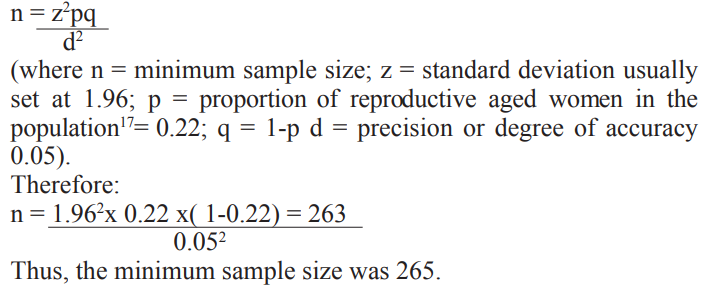
The sample size for the study group was derived using the formula by Araoye [16]:
Sampling technique
Simple random sampling was used to select cases that were used in this study. All ultrasound reports of pregnant women within the period of study were retrieved totalling 550. The patients’ case files were numbered from the earliest date to the latest date of scanning. Simple random sampling was subsequently used to select 400 cases which was 1.5 times the minimum sample size. This was to ensure that after incomplete and lost to follow up were subtracted, the sample would still well be above the minimum required sample size (attrition).
Data collection
The case files of the patients were retrieved from the medical record department. The sex of baby, fetal presentation, gestational age, expected date of delivery and cadre of scanner were retrieved from the ultrasound reports in the case files of the pregnant women. The other data extracted include the booking weight, height and the phone numbers of the patients. The body mass index (BMI) of the patients was also determined. The predicted sex of the babies by the scan reports were compared with the sex of the baby after delivery. The two-dimensional trans-abdominal ultrasound of 3.5MHz probe by Siemens© was used in this study. Women who did not deliver in the Teaching Hospital or Life Specialist Hospital were traced by calling their phone numbers to determine the sex of their child. Those who could not be reached via phone were considered missing and excluded from the study. All cases of multiple pregnancy and patients whose BMI was ≥30kg/m2 were also excluded from the study.
Data analysis
The data obtained in the study were analyzed using the statistical package SPSS version 19. Chi-square and cross-tabulations were used to compare proportions between variables. Statistical significance was set at p-values <0.05.
Results
The total number of pregnant women enrolled for this study was 400. Of this number, 49 were lost to follow-up. This resulted in a final sample size of 351.
The mean age of the patients was 30.1± 4.9. The modal and median parity was one. Almost all the patients were married. This is shown in (Table I).
Table I: SOCIO-DEMOGRAPHIC CHARACTERISTICS OF THE STUDY POPULATION
|
CHARACTERISTICS |
FREQUENCY (%) [N=351] |
|
AGE |
|
|
16-20 |
7(2.0) |
|
21-25 |
49(14.0) |
|
26-30 |
132(37.6) |
|
31-35 |
118(33.6) |
|
36-40 |
41(11.7) |
|
41-45 |
4(1.1) |
|
PARITY |
|
|
0 |
101(28.8) |
|
1 |
119(33.9) |
|
2 |
56(15.9) |
|
3 |
27(7.7) |
|
4 |
16(4.6) |
|
≥5 |
32(9.1) |
|
GESTATIONAL AGE AT SCANNING |
|
|
14-26 |
76(21.7) |
|
27-40 |
275(78.3) |
|
LEVEL OF EDUCATION |
|
|
No formal education |
7(2.0) |
|
Primary Education |
14(4.0) |
|
Secondary Education |
228(65.0) |
|
Tertiary Education |
102(29.0) |
|
MARITAL STATUS |
|
|
Single |
7(2.0) |
|
Married |
344(98.0) |
No transverse lie was observed amongst the patients and none of the babies had malformations of the external genitalia on delivery. The relationship between maternal BMI, fetal presentation, fetal gender at ultrasound and at birth is shown in Table II.
TABLE II: THE RELATIONSHIP BETWEEN MATERNAL BMI, FETAL PRESENTATION, FETAL GENDER AT ULTRASOUND AND AT BIRTH
|
BMI (Kg/m2) |
FREQUENCY (%) |
FETAL PRESENTATION AT U/S (FREQ) |
GENDER AT U/S (FREQ) |
GENDER AT BIRTH (FREQ) (%) |
ACCURACY |
P-Value |
|
< 25 |
273(77.8) |
CEPHALIC (262) |
M (149) |
*M (154) |
|
|
|
|
|
|
F (113) |
F (108) |
98.1 |
0.7235 |
|
|
|
BREECH (11) |
M (6) |
*M (9) |
|
|
|
≥25 to <30 |
|
|
F (5) |
F (2) |
72.7 |
0.3599 |
|
|
78 ( 22.2) |
CEPHALIC (75) |
M (43) |
*M (48) |
|
|
|
|
|
|
F(32) |
F (27) |
93.3 |
0.5036 |
|
|
|
BREECH (3) |
M(2) |
*M (3) |
|
|
|
|
|
|
F(1) |
F (0) |
66.0 |
1.0000 |
|
TOTAL= 351 |
|
|
|
|
|
|
Key: *There was no misdiagnosis of male gender at ultrasound.
BMI=Body mass index
%=Percentage
U/S= Ultrasound
FREQ=Frequency
M=Male
F=Female
In patients with Body mass index (BMI) of <25 kg/m2, the accuracy of fetal gender determination at ultrasound was 98.1% and 72.7% for cephalic and breech presenting foetuses respectively (p>0.05). Similarly, in patients with BMI of ≥25 but <30 kg/m2, the accuracy of fetal gender determination at ultrasound was 93.3% and 66.7% for cephalic and breech presenting fetuses respectively. This too was not statistically significant (p>0.05). This is shown in table III. There was also no statistical significant difference in the accuracy of fetal sex determination between the two groups of women with respect to their BMI (p>0.05).
As shown in table III, the accuracy was higher for female gender. There was no recorded misdiagnosis of male fetal gender at ultrasound. The overall accuracy of fetal sex determination was 96.0%.
The relationship between the rank of the scanner and the accuracy of determination of fetal gender is shown in table III. The accuracy was 100 % in both consultant radiologist and consultant obstetrician while it was 88.0% in the junior resident. This difference was not statistically significant (p>0.05).
Table III: The Relationship Between the Rank of the Scanner and the Accuracy of determination of Fetal Gender.
|
SCANNER |
GENDER AT U/S (FREQ) |
GENDER AT BIRTH (FREQ) |
ACCURACY (%) |
P- Value |
|
Consultant |
|
|
|
|
|
Radiologist |
M (67) |
M (67) |
|
- |
|
|
F (50) |
F (50) |
100.0 |
- |
|
Consultant |
|
|
|
|
|
Obstetrician |
M (67) |
M (67) |
|
|
|
|
F (50) |
F (50) |
100.0 |
0.8875 |
|
Junior |
|
|
|
|
|
Resident |
M (66) |
*M (80) |
|
0. 4948 |
|
|
F(51) |
F (37) |
88.0 |
0.34470 |
Key: *There was no misdiagnosis of male gender at ultrasound.
%=Percentage
U/S= Ultrasound
FREQ=Frequency
M=Male
F=Female
Discussion
There are increasing awareness of our women about the sensitivity of ultrasound in determination of the sex of the unborn fetus. As a result, an appreciable number of our pregnant women present for ultrasound in pregnancy solely to determine the sex of their baby or following ultrasound for other obstetric indications. The gender was determined by visually inspecting the ultrasound image for the penis or labial folds in the second and third trimesters of pregnancy.
In this study, the accuracy of ultrasound in determining the sex of the fetus was 96.0%. This was similar to the study done by Michailidis et al in 1st trimester of pregnancy in which the accuracy was 85.3% [18]. The lower accuracy of the study in comparison to ours might be largely due to the earlier gestation in which the study was undertaken. Additionally, this study was done in 2nd and 3rd trimesters. It has been reported that ultrasound done in 2nd and 3rd trimesters are more accurate in predicting the sex of the baby than those done in first trimesters [7, 8].
Apart from the trimester of gestation, the presentations of the fetus contribute towards the accuracy of prediction of the sex by ultrasound. Out of the 351 cases, 14 were in breech presentation and the ultrasound predicted the sex correctly in 71.4% of the cases while in cephalic presented fetus, there was 99.4% accuracy in predicting the sex.
Also, in a study done by Adeyinka et al in the 1st trimester, 90.3% and 83.2% of the female and male fetuses respectively were correctly predicted. In some studies, accuracy was higher for male gender, whereas in others, it was higher for females [19-25]. Mielke et al however reported no difference in identification rate between the two sexes [26]. In this study, accuracy was higher for female gender. This could be due to undescended testis and inability to visualize the median raphe and in these conditions the scrotum might be interpreted as labia. There has been reported comparable similarity between the scrotum and labia majora in fetuses in the late first and second trimesters, and unless the penis is clearly demonstrable one tends to interpret that the fetus is female [21-23]. It is our belief that if further studies of this type are conducted, increased determination to demonstrate a penis will result in a lower error rate. Alternatively, one could restrict any gender determination to only male fetuses. In order to reduce the error rate, it is imperative to consider displaying the appropriate genitalia such as the phallus or scrotum while the remainder is considered indeterminate.
In this study, none of the fetuses scanned had malformation of the external genitalia. However in fetuses without congenital malformations of the external genitalia, diagnosis of fetal sex based on the sonographic findings have been documented to be accurate in 90% to 100% of cases [3, 4]. Since fetuses with malformations of the external genitalia often represent a diagnostic challenge, clues to the presence of congenital malformations of the external genitalia include: non-visualization of the fetal bladder, curvature of the phallus; scrotal-phallus mal-position, undescended testis in the third trimester, and absence of labial or scrotal structures [3, 4].
As revealed in this study, the accuracy of determination of fetal gender is less when the presentation of the fetus was breech and BMI of ≥25- <30 kg/m2 (overweight). A possible explanation to this could be that the dimension of the lower segment of the uterus is smaller than the upper segment, the breech usually fix tightly preventing the baby to expose the perineum for visualization. Additionally, women who are overweight are more likely to have higher abdominal fat than their average-weight counterparts, thereby affecting the resolution of the ultrasound.
Finally, the experience of the sonographer was shown by this study to contribute to the accuracy of this prediction. The junior resident failed to predict the sex correctly in 14 out the 117 cases, giving the accuracy of 88.0%. The consultant Obstetrician and consultant Radiologist however, recorded 100% accuracy in predicting the gender of the fetuses. All the fourteen fetuses missed were predicted as females while their actual sexes were male.
In conclusion, ultrasound is a very important tool in the prediction of the sex of the fetus especially in developing country settings where there is high penchant for male babies. A good number of factors contribute to this accuracy.
References
- Chibueze EC, Parsons AJQ, Lopes KDS, Yo T, Swa T, et al. (2017) Diagnostic Accuracy of Ultrasound Scanning for Prenatal Microcephaly in the context of Zika Virus Infection: A Systematic Review and Meta-analysis.Sci Rep 7: 2310.
- LeibovitzZ, Daniel-Spiegel E, Malinger G, Haratz K, Tamarkin M, et al. (2016) Prediction of microcephaly at birth using three reference ranges for fetal head circumference: can we improve prenatal diagnosis? Ultrasound Obstet Gynecol 47:586-592.
- Odeh M, Grinin V, Kais M, Ophir E, Bornstein J. (2009) Sonographic Fetal Sex Determination. Obstet Gynaecol Surv 64: 50-57.
- Chelli D, Methni A, Dimassi K, Boudaya F, Sfar E, et al. (2009). Fetal Sex Assignment by First Trimester Ultrasound: A Tunisian Experience. Prenat Diagn 29: 1145-1148.
- Efrat Z, Akinfenwa OO, Nicolaides KH. (1999) First-trimester determination of fetal gender by ultrasound. Ultrasound Obstet Gynecol 13: 305-307.
- Whitlow BJ, Lazanakis MS, Economides DL. (1999) the sonographic identification of fetal gender from 11 to 14 weeks of gestation. Ultrasound Obstet Gynecol 13: 301-304.
- Bronshtein M, Rottem S, Yoffe N, Blumenfeld Z, Brandes JM. (1990) Early determination of fetal sex using transvaginal sonography: technique and pitfalls. J Clin Ultrasound 18: 302-306.
- Natsuyama E. (1984) Sonographic Determination of Fetal Sex from TwelveWeeks of gestation. Am J Obstet Gynaecol 149: 748-757.
- Emerson DS, Felker RE, Brown DL. (1989) the sagittal sign. An early second trimester sonographic indicator of fetal gender. J Ultrasound Med 8: 293-297.
- Stocker J, Evens L. (1977) Fetal sex determination by ultrasound. Obstet Gynecol 50: 462-466.
- Braithwaite JM, Armstrong MA, Economides DL. (1996) Assessment of fetal anatomy at 12 to 13 weeks of gestation by transabdominal and transvaginal sonography. Br J Obstet Gynaecol 103: 82-85.
- Michailidis GD, Papageorgiou P, Economides DL. (2002) Assessment of Fetal Anatomy in the First Trimester using Two- and Three-Dimensional Ultrasound. Br J Radio 75: 215-219.
- Lev-Toaff AS, Ozhan S, Pretorius D, Bega G, Kurtz AB, et al. (2000) Three-dimensional Multiplanar Ultrasound for Fetal Gender Assignment: Value of the Mid-sagittal Plane. Ultrasound Obstet Gynaecol 16: 345-350.
- Pedreira DA, Yamasaki A, Czeresnia CE. (2001) Fetal phallus ‘erection’ interfering with the sonographic determination of fetal gender in the first trimester. Ultrasound Obstet Gynecol 18: 402-404.
- Harrington K, Armstrong V, Freeman J, Aquilina J, CampbellS. (1996) Fetal Sexing by ultrasound in the second trimester: maternal preference and professional ability. Ultrasound Obstet Gynecol 8: 318-321.
- Araoye MO. (2003) Subject Selection and Sample Size Determination. In: Araoye MO (ed). Research Methodology with Statistics for Health and Social Sciences. Nathadex Publishers 115-120.
- Adinma JI, Adinma ED. (2011) Impact of Reproductive Health on Socio-economic Development: A Case Study of Nigeria. Afr J Reprod Health 15: 7-12.
- Michailidis GD, Papaqeorgiou P, Morris RW, Economides DL. (2003) “The use of three- dimensional ultrasound for fetal gender determination in the first trimester”. Br J Radiology 76: 448-51.
- AdeyinkaAO, OgunloyeAM, Idris S. (2005) “Ultrasonographic assessment of fetal gender”. Afr J Med Sci 34: 345-348.
- Hsiao CH, Wang HC, Hsieh CF, Hsu JJ. (2008)“fetal gender screening by ultrasound at 11 to 13 (+6) weeks”. Acta Obstetrics and Gynaecology Scand 87: 8-13.
- Shalev E, Weiner E, Zukerman H. (1981) Ultrasound Determination of Fetal Sex. Am J Obstet Gynaecol 141: 582-583.
- Dunne GM, Cunat JS. (1983) Sonographic Determination of Fetal Gender before 25 Weeks Gestation. AJR Am J Roentgenol 140: 741-743.
- Reece EA, Winn HN, Wan M, Burdine C, Green J, et al. (1987). Can Ultrasonography Replace Amniocentesis in Fetal Gender Determination during the Early Second Trimester? Am J Obstet Gynaecol 156: 579-581.
- Watson WJ. (1990) Early-second Trimester Fetal Sex Determination with Ultrasound. J Reprod Med, 35: 247-249.
- Efrat Z, Perri T, Ramati E, Tugendreich D, Meizner I.(2006) “Fetal gender assignment by first trimester ultrasound. Ultrasound in Obstetrics and Gynaecology. 27: 619-621.
- Mielke G, Kiesel L, Backsch C, Erz W, Gonser M. (1998) Fetal Sex Determination by High Resolution Ultrasound in Early Pregnancy. Eur J Ultrasound 7: 109-114.