
Journal of Gastroenterology & Digestive Systems(JGDS)
ISSN: 2640-7477 | DOI: 10.33140/JGDS
Impact Factor: 1.13


Case Report - (2026) Volume 10, Issue 1
Delayed Bile Leak Presenting One Week after Laparoscopic Cholecystectomy due to Biliary Ascariasis: A Case Report with Review of Literature
2Clinical Registrar, Department of Surgery, Jawaharlal Nehru Medical College, India
3Resident Doctor, Department of Surgery, Jawaharlal Nehru Medical College, India
Received Date: Apr 24, 2026 / Accepted Date: May 14, 2026 / Published Date: May 20, 2026
Copyright: ©2026 Hazique Jameel, et al. This is an open-access article distributed under the terms of the Creative Commons Attribution License, which permits unrestricted use, distribution, and reproduction in any medium, provided the original author and source are credited.
Citation: Ali, W. M., Yadav, S. S., Jameel, H. (2026). Delayed Bile Leak Presenting One Week after Laparoscopic Cholecystectomy due to Biliary Ascariasis: A Case Report with Review of Literature. J Gastro & Digestive Systems, 10(1), 01-03.
Abstract
Background: Bile leak is a recognized but uncommon complication of laparoscopic cholecystectomy, with an incidence ranging between 0.3% and 2%. Most cases occur within a few days post-operatively; however, delayed presentation beyond one week is rare and can be diagnostically challenging.
Case Presentation: We present a case of a 28-year-old female who developed upper abdominal pain and low-grade fever one week following laparoscopic cholecystectomy. Ultrasound revealed a localized subhepatic fluid collection. Patient underwent USG-guided pigtail insertion in the subhepatic space and peri-splenic space. MRCP revealed mild beaking of postoperative fluid towards the cystic duct remnant region, likely a bile leak originating from the cystic duct stump. The patient underwent ERCP for stenting, and incidentally, a worm was seen in the CBD. The patient was managed successfully with ERCP-guided CBD stenting and worm removal from the CBD.
Conclusion: Delayed bile leak, though uncommon, should be suspected in any patient presenting with abdominal pain and distension of the abdomen. Early imaging and a minimally invasive management strategy can prevent serious morbidity.
Keywords
Bile Leak, Delayed Complication, Laparoscopic Cholecystectomy, ERCP, Cystic Duct Stump, Bilioma
Introduction
Laparoscopic Cholecystectomy (LC) has become the gold standard for treating symptomatic gallstone disease due to its low morbidity and short recovery time. However, bile duct injuries and leaks remain among the most serious complications, with reported incidences of 0.3–2% [1,2]. Bile leaks may originate from the cystic duct stump, accessory biliary ducts (Ducts of Luschka), or inadvertent injury to the extrahepatic bile ducts [3]. While most leaks occur within the first few postoperative days, delayed bile leaks presenting beyond a week are rare and may be mistaken for abscesses or postoperative seromas [4]. Recognition and management are crucial to prevent peritonitis, abscess formation, and sepsis. This report highlights a case of delayed bile leak presenting one week after laparoscopic cholecystectomy and discusses the available literature on its management.
Case Presentation
We present a case of a 28-year-old female who developed upper abdominal pain and low-grade fever one week following laparoscopic cholecystectomy. Ultrasound revealed a localized subhepatic fluid collection. Patient underwent USG-guided pigtail insertion in the subhepatic space and peri-splenic space. MRCP revealed mild beaking of postoperative fluid towards the cystic duct remnant region, likely a bile leak originating from the cystic duct stump (Figure 1). The patient underwent ERCP for stenting, and incidentally, a worm was seen in the CBD (Figure 2). The patient was managed successfully with ERCP-guided CBD stenting and worm removal from the CBD.

Figure 1: Cystic Duct Stump Leak (MRCP)
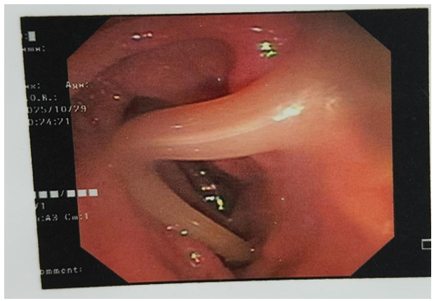
Figure 2: CBD with Worm
Clinical Findings
The patient was febrile on admission and had mild right hypochondrial tenderness. The laparoscopic port sites were healthy.
• Investigations
![]() Hemoglobin: 8.6g/dL
Hemoglobin: 8.6g/dL
![]() Total Leucocyte Count: 19000/mm³ (Mild Leukocytosis)
Total Leucocyte Count: 19000/mm³ (Mild Leukocytosis)
![]() Serum Bilirubin: 2.1 mg/dL
Serum Bilirubin: 2.1 mg/dL
![]() Alkaline Phosphatase: Mildly elevated
Alkaline Phosphatase: Mildly elevated
![]() Ultrasound Abdomen: Revealed a 100 ml subhepatic collection.
Ultrasound Abdomen: Revealed a 100 ml subhepatic collection.
![]() MRCP: localized collection with communication from the cystic duct stump, suggesting a Type A (Strasberg) bile leak.
MRCP: localized collection with communication from the cystic duct stump, suggesting a Type A (Strasberg) bile leak.
The patient underwent ERCP, which demonstrated contrast extravasation from the cystic duct stump and a worm in the CBD. A 10 Fr biliary stent was placed after removing the worm, and percutaneous pigtail drainage was inserted to evacuate the bilioma. The patient's condition improved gradually. The drain output reduced to less than 10 mL/day over 5 days and was removed on postoperative day 6 after intervention. The stent was removed endoscopically after six weeks. The patient remained asymptomatic during three months of follow-up.
Discussion
Bile leak remains a challenging postoperative complication after LC. The most common sites include the cystic duct stump (75%), the ducts of Luschka (19%), and the common bile duct (6%) [3,5]. In most cases, bile leaks manifest within the first 3–5 days after surgery. Delayed bile leaks (>7 days postoperatively) are rare and often result from gradual clip slippage, cystic duct necrosis, or infection-related ductal erosion [6].
Diagnosis
Patients typically present with right upper quadrant pain, fever, or persistent bilious drainage.
Ultrasound can identify fluid collections, but MRCP offers better delineation of the biliary anatomy and site of leakage. Hepatobiliary Scintigraphy (HIDA Scan) is highly sensitive in detecting active bile leaks, particularly when the leak is intermittent or of small volume [7].
Management
Management depends on the severity and location of the leak:
• Minor leaks (Strasberg type A): Managed with ERCP and sphincterotomy ± biliary stenting [3].
• Major duct injuries (Strasberg types D or E): Require surgical repair such as Roux-en-Y hepaticojejunostomy [8].
• Associated Biloma: Managed by percutaneous catheter drainage.
Early endoscopic intervention reduces intraductal pressure and facilitates closure of the leak [9]. The combination of ERCP with stent placement and percutaneous drainage is now considered the gold standard for most minor bile leaks.
Review of Literature
Several studies have reported successful management of post-cholecystectomy bile leaks with minimally invasive approaches.
• Sandha et al., proposed an ERCP-based classification and reported endoscopic therapy success in 93% of patients [3].
• Schmidt et al., reviewed 79 patients with major bile duct injuries and found that early recognition significantly improved long-term outcomes [8].
• Sharma et al., emphasized the role of HIDA scan in detecting bile leaks when MRCP findings were equivocal [7].
• Bhandari et al., reported that delayed leaks (>7 days) were more likely related to clip displacement or ductal necrosis and had favorable outcomes with ERCP stenting alone [10]. Overall, literature supports a stepwise, minimally invasive approach beginning with imaging, ERCP, and drainage, reserving surgical intervention for refractory or major duct injuries.
Conclusion
Delayed bile leak following laparoscopic cholecystectomy is an uncommon but important postoperative complication. It should be suspected in patients presenting with right upper quadrant pain, fever, or bilious discharge beyond the first postoperative week. Early diagnosis through MRCP or HIDA scan, and prompt management with ERCP-guided stenting and drainage, results in excellent outcomes. A multidisciplinary approach involving surgeons, interventional radiologists, and gastroenterologists ensures optimal recovery.
References
- Stewart, L., & Way, L. W. (1995). Bile duct injuries during laparoscopic cholecystectomy: factors that influence the results of treatment. Archives of Surgery, 130(10), 1123-1128.
- Kapoor, V. K. (2007). Bile duct injury repair: when? what? who?. Journal of hepato-biliary-pancreatic surgery, 14(5), 476-479.
- Sandha, G. S., Bourke, M. J., Haber, G. B., & Kortan, P. P. (2004). Endoscopic therapy for bile leak based on a new classification: results in 207 patients. Gastrointestinal endoscopy, 60(4), 567-574.
- Strasberg, S. M., Hertl, M., & Soper, N. J. (1995). An analysis of the problem of biliary injury during laparoscopic cholecystectomy. Journal of the American College of Surgeons, 180(1), 101-125.
- Deziel, D. J., Millikan, K. W., Economou, S. G., Doolas,A., Ko,S. T., & Airan, M. C. (1993). Complications of laparoscopic cholecystectomy: a national survey of 4,292 hospitals and an analysis of 77,604 cases. The American journal of surgery, 165(1), 9-14.
- Kaman, L., Behera, A., Singh, R., & Katariya, R. N. (2004). Management of major bile duct injuries after laparoscopic cholecystectomy. Surgical Endoscopy and Other Interventional Techniques, 18(8), 1196-1199.
- Sharma, D., Jain, S., Bansal, N., et al. (2019). Role of HIDA scan in the evaluation of postoperative bile leaks: an underutilized tool. Indian J Surg, 81(2), 135-139.
- Schmidt, S. C., Langrehr, J. M., Hintze, R. E., & Neuhaus, P. (2005). Long-term results and risk factors influencing outcome of major bile duct injuries following cholecystectomy. Journal of British Surgery, 92(1), 76-82.
- Verma, D., Kapadia, A., Eisen, G. M., et al. (2006). Efficacy of endoscopic management of bile leaks after cholecystectomy. Gastrointest Endosc, 63(7), 895-899.
- Bhandari, L., Kaur, R., Singh, G., et al. (2018). Delayed bile leaks following laparoscopic cholecystectomy: incidence, presentation and management. Int Surg J, 5(7), 2329-2333.