

International Journal of Endocrinology Research and Reviews(IJERR)
ISSN: 2993-656X | DOI: 10.33140/IJERR

Research Article - (2022) Volume 2, Issue 1
COVID-19 is a contributing risk factor for development of DKA in typeI DM
2Department of Clinical pathology, Faculty of Medicine, Minia University, El Minya, Egypt
Received Date: Mar 02, 2022 / Accepted Date: Jun 12, 2022 / Published Date: Mar 22, 2022
Copyright: ©Copyright: ©2022 Noura Mohammed ElBakry. This is an open-access article distributed under the terms of the Creative Commons Attribution License, which permits unrestricted use, distribution, and reproduction in any medium, provided the original author and source are credited
Citation: Noura M. El Bakry, Ayat M.Mohamed ,Yosra S. Fadl (2022) COVID-19 is a contributing risk factor for development of DKA in typeI DM, International Journal of Endocrinology Research and Reviews 2(1): 01-07.
Abstract
Background: Viral infections trigger type 1 diabetes (T1D) in susceptible children, and may increase incidence of T1D and/or diabetic ketoacidosis (DKA) during the COVID-19 pandemic. We aimed to describe the frequency of diabetic ketoacidosis in children and to determine if COVID contribute to the risk of diabetic ketoacidosis.
Objective: To study the incidence of DKA due to new-onset T1D during the COVID-19 pandemic, and whether SARS-CoV-2 infection is a triggering factor for the development of DKA in type I DM in Minia university hospital, Egypt.
Methods: This retrospective cohort study of children admitted to PICU and inpatient wards due to new-onset T1D or due to DKA and diagnosed as have positive covid -19 in Mina University Hospital from 1 April to 31 October 2021. We compared the incidence, number and characteristics of children with newly diagnosed T1D and DKA during the pandemic periods.
Results: An increase in the number of children with newly diagnosed type 1 diabetes (T1D) has been reported during the COVID-19 pandemic and more children with new-onset T1D now present with severe diabetic ketoacidosis (DKA).
Conclusion: COVID contribute to the risk of diabetic ketoacidosis and new onset T1D among children
Keywords
COVID-19, T1D.DKA
List Of Abbreviations
|
DKA |
Diabetic Ketoacidosis |
|
COVID |
Corona Virus Induced Disease |
|
RBG |
Random Blood Glucose |
|
SARS-Cov-2 |
Severe Acute Respiratory Syndrome Due to Corona Virus 2 |
|
Hcovs |
Human Corona Viruses |
|
FBG |
Fasting Blood Glucose |
|
ROC |
Receiver-Operating Curve |
|
OGTT |
Oral Glucose Tolerance Test |
|
T1D |
Type One Diabetes |
|
PICU |
Pediatric Intensive Care Unit |
|
AUC |
Area Under the Curve |
Background
Diabetes mellitus is a group of metabolic diseases characterized by chronic hyperglycemia resulting from defects in insulin se-cretion, insulin action, or both [1]. Diabetes in children usually insulin dependent due to severe insulin deficiency result mostly from immune destruction of Beta cells in pancreas usually af¬ter environmental triggers in genetic susceptible children [2] Re-garding the pathogenesis of T1D, Individuals have an absolute deficiency of insulin secretion and are prone to DKA and most cases are primarily due to T-cell mediated pancreatic islet β-cell destruction, which occurs at a variable rate, and becomes clinically symptomatic when approximately 90% of pancreatic beta cells are destroyed [3].
The environmental triggers (chemical and/or viral) which initiate pancreatic beta cell destruction remain largely unknown, but the process usually begins months to years before the manifestation of clinical symptoms [4].
Enterovirus infection has been associated with development of diabetes associated auto-antibodies in some populations, [5] and enteroviruses have been detected in the islets of individuals with diabetes [6].
In most western countries, T1D accounts for over 90% of child-hood and adolescent diabetes [7], although less than half of indi-viduals with T1D are diagnosed before the age of 15 years [8].
Gender differences in incidence are found in some, but not all, populations with more common in males [9].
A seasonal variation in the presentation of new cases is well de-scribed, with the peak being in the winter months [10], Despite familial aggregation, which accounts for approximately 10% of cases of type 1 DM (T1D) [9]. Insulin dependent diabetes or TID usually presents with charac-teristic symptoms such as polyuria, polydipsia, blurring of vision, and weight loss, in association with glycosuria and ketonuria [2].
In most severe form, ketoacidosis [DKA] or rarely a non-ketotic hyperosmolar state may develop and lead to stupor, coma and in absence of effective treatment, death [3].
The diagnosis of DKA is usually confirmed quickly by measure-ment of a marked elevation of the blood glucose level. In this sit-uation if ketones are present in blood or urine, treatment is urgent [4].
In the absence of symptoms or presence of mild symptoms of di¬abetes, hyperglycemia detected incidentally or under conditions of acute infective, traumatic, circulatory or other stress may be transitory and should not in itself be regarded as diagnostic of di¬abetes [5].
The diagnosis of diabetes should not be based on a single plasma glucose concentration. Diagnosis may require continued observa¬tion with fasting and/or 2 hour post-prandial blood glucose levels and/or an oral glucose tolerance test (OGTT), and usually TID di¬agnosed by found auto antibodies positive [6].
The severe acute respiratory syndrome due to Corona vi¬rus 2 (SARS-Cov-2), responsible for the Corona virus disease COVID-19, was first identified in China in 2019 [10].
“Fever of Unknown Origin with pneumonia” had been contin-uously increasing in Wuhan. The causative agent of this unex-plained infected pneumonia was identified as severe acute respira¬tory syndrome corona virus 2 (SARS-CoV-2) which was not only has a strong human-to-human transmission but also caused severe pneumonia to death [11-15].
Corona viruses are positive-strand ribonucleic, large viruses. Only two genera of the viruses can infect humans: A &B types and they are called human corona viruses (HCoVs) [11].
Common symptoms at the onset of COVID-19 pneumonia for these children included a fever, cough and shortness of breath, whereas less common symptoms were myalgia, malaise, sore throat and diarrhoea [16-18].
The impact of SARS-CoV2 on the incidence of new-onset T1D and DKA is unclear [11]. Diabetes has been recognized as a major risk factor for outcomes and mortality in children with COVID-19 [11].
COVID-19 may damage the pancreas or through autoimmune pro¬cess and initiate the manifestation of diabetes [12].
Diabetes is associated with an increased risk of infections, in-cluding viral ones due to defects in the innate immunity affecting phagocytosis, neutrophil chemotaxis, and cell-mediated immunity [13].
Diabetes is associated with endothelial dysfunction and increased platelet aggregation and activation leading to the hypercoagulable state [13] and this is associated with the low-grade chronic inflam¬mation might promote the cytokine storm, a severe complication of COVID-19 which characterized by excessive production of in¬flammatory cytokines [14].
Furthermore, COVID-19 spike protein gains entry to its target cells by using the angiotensin-converting enzyme 2 (ACE2) as the receptor, which has protective effects primarily regarding in¬flammation [15], COVID-19 infection reduces ACE2 expression inducing cellular damage, inflammation, and respiratory failure and can lead to a direct effect on β-cell function, which not only explains that diabetes might be a risk factor for a severe form of COVID-19 disease but also explains that infection could induce new-onset diabetes [16-18].
Besides, most patients present multiple stresses with COVID-19 including respiratory failure and sepsis requiring intravenous in-fusion of insulin and fluid balance to control the glycemia levels [19]. In addition, potassium balance must be carefully considered in the context of insulin treatment, as hypokalemia is a common feature of COVID-19 and may be exacerbated after the introduc¬tion of insulin [19].
In diabetic childerns with COVID, laboratory tests indicated that lymphopenia is also likely to occur. Additionally, increased con-centrations of D Dimer and serum ferrtin might be one of the clini-cal manifestations. However, none of these symptoms was present in every patient [20-24].
Radiological, chest CT might have a high diagnostic value because of its typical images of virus infection, high accuracy with a low false negative rate, and time efficiency [24].
Methods
Sixety one children were enrolled in this study, The enrolled pa-tients were 2-17years old fulfilled the following inclusion criteria: Children admitted to pediatric intensive care unit (PICU) with hy-perglycemia, Acidosis or Ketosis eiher known diabetic or newly diagnosed diabetic and have respiratory distress with low O2 sat-uration. Type 1 diabetes was defined according to autoantibody positivity and by OGT RBG (Random Blood Glucose > 200 mg/ dL, FBG (Fasting Blood Glucose >126 mg/dL, and 2 hour post-prandial blood glucose >200 mg/dL [1].
DKA was defined according to International Society for Pediat-ric and Adolescent Diabetes criteria (blood glucose >11 mmol/L [ 200 mg/dL], venous pH <7.3 or bi-carbonate <15 mmol/L, ke-tonemia and ketonuria) [10].
We evaluate the number of patients diagnosed with T1D, how many had DKA and, how many were diagnosed with COVID-19, and how many patients need PICU admission
Regarding T1D patients were asked about compliance, recuurent admission by DKA and insulin therapy and if there abnormal re¬spiratory manifestations like cough repiratory distress cyanosis or symptoms of infectons like fever anorexia headache malaise O2 saturation using pulse oximetery & Chest CT were done to all children. Among children with T1D, SARS-CoV-2 was detected by PCR
Children admitted to PICU due to other diseases were excluded from our study.
The study conducted according to the principles of Helsinki and agreed by the faculty of medicine, Minia university, Ethical com¬mittee (No: 116-5-2020). Informed written consents from the pa-tient’s caregiver were obtained.
Sample collection: 10 ml of venous blood samples were taken for RBG, white blood cells count with differential, HbA1C, and ABG (Arterial Blood Gases) using fully automated chemical auto-ana-lyzer Dimension-ES, USA, also PCR for Corona virus was done
Statistical analysis: The numerical data were presented as means – standard deviations while non numerical data were presented as percentage. Two tailed t-tests were used to analyze differences between the control and patients groups. P-values less than 0.05 were considered statistically significant. The magnitude of correla¬tions was determined by Pearson’s correlation coefficient. All the data were analyzed by statistical package Prism 3.0 (Graph Pad software, San Diego, CA, USA). Figures were done by Microsoft Office Excel 2007.
Results
Total number of patients admitted to endocrine unit were 61 by hyperglycemia, thirty eight patients had DKA (Group I) and other twenty three hadn’t DKA (Group II) (Table 1)
There were no difference between both groups regarding age, sex (p-value <0.190, <0.241respectively) (Table 1)
Table 1: Demographic Data of Patients
|
Variables |
|
With DKA Group I |
No DKA Group II |
P value |
|
N=38 |
N=23 |
|||
|
Age |
Median |
6.1 |
7.8 |
0.190 |
|
IQR |
(1.3-10.2) |
(3-11.2) |
||
|
Sex |
Male |
24(63.2%) |
11(47.8%) |
0.241 |
|
Female |
14(36.8%) |
12(52.2%) |
There were significant difference between both groups regarding ABG profile; HCO3 CO2 PO2 (P-value <0.001*,<0.015*,<0.001* respectively) (Table 2)
There were significant difference between both groups regarding O2 saturation &Chest CT finding (p-value<0.001*, <0.025* respectively) (Table 2)
There were significant difference between both groups regarding severe hyperglycemia and lymphocytes count (p-value<0.010*,<0.035* respectively) (Table 2)
There was slight difference between both groups regarding ph value (p-value= 0.492) (Table 2)
There were no difference between both groups regarding HBA1c %(p-value=0.496) (Table 2)
|
|
|
With DKA Group I |
No DKA Group II |
P value |
|
N=38 |
N=23 |
|||
|
pH |
Range |
(6.5-11.2) |
(7.3-7.5) |
0.492 |
|
Mean ± SD |
7.2±1 |
7.4±0 |
|
|
|
HCO3 |
Range |
(3-23.3) |
(16-29.2) |
<0.001* |
|
Mean ± SD |
9.8±5.6 |
20.3±3.5 |
|
|
|
CO2 |
Range |
(20-67) |
(34.2-46.7) |
0.015* |
|
Mean ± SD |
47.7±13.2 |
41.9±3.7 |
|
|
|
PO2 |
Range |
(45-128) |
(20.6-70) |
<0.001* |
|
Mean ± SD |
93.6±28.1 |
48±13.2 |
|
|
|
RBS |
Range |
280 |
209 |
0.010* |
|
Mean ± SD |
(216-501) |
(190-240) |
|
|
|
HBA1c % |
Range |
(8.6-15.6) |
(8.9-15.6) |
0.496 |
|
Mean ± SD |
11.4±2.5 |
11±2 |
|
|
|
Lymphocytes |
Median |
1300 |
2500 |
0.035* |
|
IQR |
(1000-2500) |
(1200-4500) |
|
|
|
Saturation |
Range |
(49-98.9) |
(49-87) |
<0.001* |
|
Mean ± SD |
85±16.3 |
64.4±9.3 |
|
|
|
Chest CT |
-Ve |
26(68.4%) |
9(39.1%) |
0.025* |
|
+Ve |
12(31.6%) |
14(60.9%) |
|
On admission, COVID diagnosed patients were seventy percent were presented by DKA, twenty two % were presented by hyperglycemia only without DKA while only 8% were within normal blood sugar ( Figure I )

Figure 1: COVID Patients
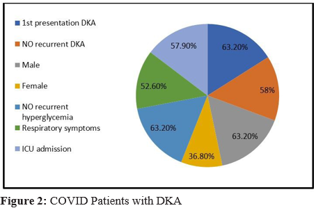
Patients who were diagnosed to have COVID and presented by DKA were 63.20% male&36.80% female 63.20% of them were presented by new onset diabetes. However, 57.90% need ICU ad¬mission and 63.20% of them entre in recurrent hypherglycemia after hospital discharge in three months duration (Figure II)
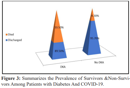
Prevalence of non-survivors among patients with DKA and COVID-19was 10.50% of patients (Figure III)
Discussion
Data of COVID-19 in children are limited, and clinical manifesta¬tions are nonspecific and might delay the diagnosis, which might lead to severe complications. In this clinical case, we describe new-onset diabetes with consciousness impairment as an atypical revealing way of COVID-19 [18].
Diabetes might facilitate infection by COVID-19 due to increased viral entry into cell and impaired immune response. COVID-19 could have effect on the path-physiology of diabetes [20].
Total number of patients admitted to endocrine unit were 61 by hyperglycemia, thirty-eight patients had DKA (Group I) and oth¬er twenty-three hadn’t DKA (Group II), There were no difference between both groups regarding age, sex (p-value <0.190, <0.241 respectively)
There was significant difference between both groups regard¬ing ABG profile; HCO3 CO2 PO2 (P-value <0.001*, <0.015*, <0.001* respectively) On admission, COVID diagnosed patients were seventy percent were presented by DKA, twenty two % were presented by hyperglycemia only without DKA while only 8% were within normal blood sugar.
Among children, SARS-cov-2 was detected by PCR, Patients who were diagnosed to have COVID and presented by DKA were 63.20% male&36.80% female 63.20% of them were presented by new onset diabetes and this is suggesting that SARS-cov-2 infec¬tion was the primary trigger for more severe presentation of T1D and for the increase in children diagnosed with T1D.
This study found of a significant increase in the number (57.90%) of children requiring PICU care for severe ketoacidosis and 63.20% of them entre in recurrent hypherglycemia after hospital discharge in three months duration also found a significant increase in DKA at diabetes diagnosis in children during the COVID-19 pandemic in Minia university hospital and the increase we observed in the total number of children with new-onset T1D is surprising, and Underlying causes may be multifactorial and reflect reduced med¬ical services, fear of approaching the health care system, and more complex psychosocial factor and this was in agree with Clemens K., et.al, 2020 study Who explain that there are several precedents for a viral cause of ketosis-prone diabetes, including other corona viruses that bind to ACE2 receptors.
Our study show greater incidences of hyperglycemia and acute-on-set diabetes have been reported among patients with SARS corona virus 1 pneumonia than among those with non-SARS pneumonia, in agree with Clemens K., etal.,2020 study Who reported high prevalence of diabetes is seen in patients with COVID-19 and the presence of diabetes is a determinant of severity and mortality
Blood glucose control is important not only for patients who are infected with COVID-19, but also for those without the disease [21].
Guo. Et al., (2020) reported that hyperglycaemia persist for 3 years after recovery from SARS indicating a transient damage to beta cells [22].
During the observation period, eight % patients were diagnosed with COVID-19, all of whom were asymptomatic or had only mild symptoms and all were positive by PCR with normal blood glu-cose on admission
Prevalence of survivors and non-survivors among patients with diabetes and COVID-19 during the observation period, only three patients were diagnosed with COVID-19 died from severe respi-ratory manifestations and there chest imaging showed CORAD 3 due to severe viral load
Unsworth R., etal., (2020) found the prevalence of non-survivors was also higher in diabetic subjects with COVID-19
Our findings are in line with recent Italian, German, UK and Aus-tralian studies reporting an increased incidence of DKA in children with new-onset T1D during the COVID-19 pandemic [25].
In contrast, a German study reported no increase in the incidence of T1D [26].
As T1D may be triggered by viral infections in susceptible chil-dren, the potential association of SARS-cov-2 with either increas-ing incidence of T1D or more severe disease presentation needs to be addressed [27].
As we noticed that the virus plays a direct role in the increased incidence or more severe presentation of T1D in children.
Delays in the diagnostic process of T1D are likely to explain the increase in the number of children with DKA, as many of the chil-dren admitted to the PICU had been symptomatic for longer than the patients in previous months and more complex associations, influencing the threshold of families to seek medical attention and the accessibility of health services, seem to have been involved. Our results show in a setting with high COVID-19 incidence. With a 70 % seropositivity of COVID-19 in children with acute infec¬tions in the PICU presented by DKA, and only 22% with hypher¬glycemia
Our study suggest that patients of COVID-19 with diabetes are more often associated with severe or critical disease, also Elbar-bary NS. Et al., (2020) in a study of 138 patients reported that 72% patients of COVID-19 with co morbidities including diabetes required admission in ICU, compared to 37% of patients without co morbidities [28].
Conclusions
COVID-19 can result in asymptomatic to severe illness in childern . Fortunately, children without underlying chronic illness appeared to have more severe DKA.
COVID-19 pandemic might have altered diabetes presentation and DKA severity and requires strategies to educate and reassure parents about timely emergency department attendance for non–COVID-19 symptoms.
Limitation of the study:
The smaller sample size which due to refusal of many patients to share in this study.
Conflict of interest: None.
Financial Disclosure:
The authors have no financial relationships relevant to this article to disclose.
Funding: This research did not receive any specific grant from funding agen-cies in the public, commercial, or not-for-profit sectors
Authors’ Contributions: MA, LA, AM and SH participated in the study design, data col-lection an interpretation and wrote the manuscript. AM and NA analyzed the immunological data and SH, LA and MA participated to discuss the results and to write the manuscript.MA supervised the research group. All authors listed in a manuscript have contrib¬uted substantially to the work and seen revise and approved the submitted versio.
Authors’ information
Available
Ethics approval and consent to participate
Written consents were obtained from patients’ caregivers for pa-tients less than 16 years old.
The study was conducted According to the declarations of Helsin¬ki and Approved from the faculty of medicine scientific committee in Minia University (No: 116-5-2020).
Consent for publication
Not applicable
Competing interests
The authors declare that they have no competing interests.
References
- Schatzda, Kowah, Winterwe and Rileywj (1989). Natural history of incidental hyperglycemia and glycosuria of childhood. Jpediatr; (115): 676–680.
- Shehadeh N., On A, Kessel I., Perlman R., Evenl, Naveh T., Soloveichik L. and Eizionia (1997). Stress hyperglycemia and the risk for the development of type1diabetes. J. Pediatr. En-docrinol. Metab; (10): 283–286.
- Silverstein J., Klingensmith G., Copeland K.,Plotnick L., Kaufman F., Laffel L., Deeb L., Greym, Anderson B., Holz-meisterla and Clark (2005). Care of children and adolescents with type1diabetes: a statement of the American Diabetes Association. Diabetescare; (28):186–212.
- Skylerjs, krischer J., Wolfsdorf J., Cowiec, Palmer J., Greenbaum C., Cuthbertson D., Rafkin-Mervisle, Chasehp and Lescheke (2005). Effectsoforalinsulininrelativesofpa-tientswithtype1diabetes: The Diabetes Prevention Trial–Type1. Diabetes care; (28): 1068–1076.
- Steckak, barrigakj, Emerylm, Fialloscharerrv, Gottliebpa and Rewersmj (2005). Secondary attack rate of type1 diabetes in Colorado families.diabetescare; (28): 296–300.
- Thunander M., Petersson C, Jonzon K., Fornander J, Ossains-son B., Torn C., Edvardssons and Landin-olssonm (2008). Incidence of type1and type2 diabetes in adults and children in Kronoberg, Sweden. Diabetes Res.clinpract ; ( 82): 247–255.
- Chen, X. Gong, L. Wang, and J. Guo (2020). Effects of hypertension, diabetes and coronary heart disease on COVID-19 diseases severity: a systematic review and meta-analysis medrxiv: 280.
- M. Paul Zimmet, M. D. George Alberti and M. D. Stefan Bornstein (2020). “New-onset diabetes in COVID-19,” The New England Journal of Medicine;(383), 8:787–789.
- R. Unsworth, S. Wallace and N. S. Oliver (2020) New-onset type 1 diabetes in children during COVID-19: multicenter regional findings in the UK,” Diabetes Care;(43), 11: 170–171.
- M. Fayfman, F. J. Pasquel, and G.E.Umpierrez (2017)“Man-agement of hyperglycemic crises,” Medical Clinics of North America;( 101), 3: 587–606,
- Y. Dong, Y. Dong, X. Mo, Y. Hu, X. Qi, and F. Jiang (2020). “Epidemiology of COVID-19 among children in China,” Pediatrics ;(145), 6.
- H. De Wilde, J. Eric, M. K. Snijder, and M. J. Van (2012). “Host factors in corona virus replication,” in Assessment & Evaluation in Higher Education, Springer International Publishing, New York, NY, USA:435.
- J. She, L. Liu, and W. Liu (2020).“COVID-19 epidemic: disease characteristics in children,” Journal of Medical Virology; (92), 7: 747–754.
- C. Jiehao, X. Jin and L. Daojiong (2020). “A case series of children with 2019 novel coronavirus infection: clinical and epidemiological features,” Clinical Infectious Diseases;( 71), 6:1547–1551.
- T. Sathish, Y. Cao, and N. Kapoor (2020). “Newly diagnosed diabetes in COVID-19 patients,” Primary Care Diabetes; (15), 1:194.
- Y. Jie, S. Jia, H. Ng, and E. Yeoh (2020), “Diabetic ketoaci-dosis precipitated by COVID-19 in a patient with newly diagnosed diabetes mellitus,” Diabetes Research and Clinical Practice; (164).
- W. Abu-ashour, L. Twells and J. Valcour (2017). “The association between diabetes mellitus and incident infections : a systematic review and meta-analysis of observational stud-ies,” BMJ Open Diabetes Research and Care ; ( 5),1.
- W. Guo, M. Li and Y. Dong (2020). “Diabetes is a risk factor for the progression and prognosis of COVID-19,” diabetes metabolism research and reviews; (36):6.
- Wolfsdorf JI., Allgrove J. and Craig M. (2014). International Society for Pediatric and Adolescent Diabetes. ISPAD Clinical Practice Consensus Guidelines 2014. Diabetic ketoacidosis and hyperglycemic hyperosmolar state. Pediatr Diabetes; (15), 20:154–179.
- S. M. Bindom and E. Lazartigues (2020). “The sweeter side of ACE2: physiological evidence for a role in diabetes,” molecu-larandcellularendocrinology;(302), 2:193–202.
- M. Apicella, M. C. Campopiano, M. Mantuano, L. Mazoni, A. Coppelli, and S. Del Prato (2020). “COVID-19 in people with diabetes: understanding the reasons for worse outcomes,” thelancetdiabetesandendocrinology; (8), 9:782–792.
- J.-K. Guo, S.-S. Lin, X.-J. Ji, and L.-M. Guo (2010). “Binding of SARS coronavirus to its receptor damages islets and causes acute diabetes,” Acta Diabetologica;( 47), 3:193–199.
- L. Fang, G. Karakiulakis, and M. Roth (2020). “Are patients with hypertension and diabetes mellitus at increased risk for COVID-19 infection?” The lancet respiratory medicine;( 8), 4-21.
- R. P. Dellinger, M. M. Levy and A. Rhodes (2013). “Surviv-ing sepsis campaign: international guidelines for management of severe sepsis and septic shock,” Critical caremedicine; (41), 2: 580–637.
- Unsworth R., Wallace S. and Oliver NS. (2020). New-Onset type 1 diabetes in children during COVID-19: multicenter regional findings in the U.K. Diabetes Care ;43:e170–1.
- Kamrath C, Mönkemöller K. and Biester T. (2020). Ketoacidosis in children and adolescents with newly diagnosed type 1 diabetes during the COVID-19 pandemic ingermany. JAMA; 324:801–804.
- Lawrence C., Seckold R .and Smart C (2021). Increased paediatric presentations of severe diabetic ketoacidosis in an Australian tertiary centre during the COVID-19 pandemic. Diabet Med; 38: e14417.
- Elbarbary NS., Dos Santos TJ. and debeaufort C. (2020). COVID-19 outbreak and pediatric diabetes: perceptions of health care professionals worldwide. Pediatr Diabetes; 21:1083–1092.