

Archives of Case Reports: Open Access(ACROA)
ISSN: 3065-7598 | DOI: 10.33140/ACROA

Case Report - (2026) Volume 3, Issue 2
Complete Pathological Response to Durvalumab in MSI-H Intrahepatic Cholangiocarcinoma Complicated by Drug-induced Sarcoid-like Reaction: A Case Report
 , Naga Swetha Samji, MD 2 *, FNU Raja, MD 3 , Mehmet Akce, MD 4 and Midhun Malla, MD, MS 4 *
2University of Alabama at Birmingham – Hospitalist, United States
3University of Alabama at Birmingham – Pathology, United States
4University of Alabama at Birmingham – Hematology/ Oncology, United States
Received Date: Sep 17, 2026 / Accepted Date: Oct 19, 2026 / Published Date: Oct 30, 2026
Copyright: ©2026 Midhun Malla, et al. This is an open-access article distributed under the terms of the Creative Commons Attribution License, which permits unrestricted use, distribution, and reproduction in any medium, provided the original author and source are credited.
Citation: Barad, P., Samji, N. S., Raja, F., Akce, M., Malla, M. (2026). Complete Pathological Response to Durvalumab in MSI-H Intrahepatic Cholangiocarcinoma Complicated by Drug-induced Sarcoid-like Reaction: A Case Report: A Case Report. Arch of case Rep: Open, 3(2), 01-05.
Abstract
We present a rare case of complete pathological response in a patient with microsatellite instability-high cholangiocarcinoma treated with durvalumab, complicated by a drug-induced sarcoid-like reaction. This report highlights the profound efficacy of immune checkpoint inhibitors in microsatellite instability-high tumors, even in rare malignancies such as cholangiocarcinoma, while also illustrating the potential for complex immune-related adverse events, such as sarcoid-like reaction.
Background
Cholangiocarcinoma (CCA), a malignancy of the bile ducts, typically presents with limited therapeutic options and a dismal prognosis [1]. Cholangiocarcinoma can be divided into intrahepatic (ICC) and extrahepatic (ECC) forms based on the origin relative to bile ducts, and extrahepatic cholangiocarcinoma is further divided into perihilar and distal hepatic cholangiocarcinoma based on its location above and below the cystic duct [1]. Studies have shown that perihilar cholangiocarcinoma is the most common subtype, accounting for 50-60% of all CCA, followed by distal cholangiocarcinoma at 20-30% and ICC at 8-10% [2,3]. A study analyzed 40-year trends in CCA in the USA from 1973 to 2012 and noted a rise in ICC cases, with an annual percentage change (APC) of 4.36%, compared with ECC, which increased modestly, with an APC of 0.14% [1]. A 21-year population-based analysis recorded steadily increasing mortality in patients with ICC in the USA [4]. For unresectable or metastatic cases, treatment options are severely limited, underscoring the need for novel therapeutic strategies. The standard of care for advanced metastatic biliary tract cancer includes chemotherapy with gemcitabine and cisplatin in combination with immunotherapy (durvalumab or pembrolizumab) as a first-line regimen [5]. Immune checkpoint inhibitors (ICIs) have emerged as a significant therapeutic advancement, especially for tumors with microsatellite instability-high (MSI-H) features, which often predict a favorable response to these agents [5].
Deficient mismatch repair with somatic hypermutation creates MSI-H tumors, which have unique tumor microenvironments and high tumor mutational burden, making them sensitive to programmed cell death protein-1 (PD-1) and programmed death ligand-1 (PD-L1) immune checkpoint inhibitors [6]. MSI-H tumors are very rare, with a prevalence of about 2-5% in biliary tract tumors [7,8].
Durvalumab, a PD-L1 inhibitor, in combination with chemotherapy has demonstrated efficacy in the prospective, global, randomized TOPAZ-1 clinical trial for patients with advanced/metastatic cholangiocarcinoma [5]. Common all-grade immune-related side effects of PD-L1 inhibitors include pneumonitis (35%), hepatitis (22%), colitis (17%), cardiac complications (about 8%), and nephritis and adrenal complications (about 8%), with a median time to onset of immune-related complications of about 40 days [9].
The United States FDA adverse event reporting system data showed that 1.4% of pulmonary immune-related adverse events (irAEs) were related to PD-L1 inhibitor use, and also noted that immune-related sarcoid-like reaction was highest with pembrolizumab, followed by nivolumab, and least with durvalumab [10]. Sarcoid-like reaction is a rare irAE associated with PD-L1 inhibitor therapy, necessitating careful differentiation from disease progression [11-13]. To the best of our knowledge, we report the first case of durvalumab-associated sarcoid-like reaction in a patient treated for MSI-H cholangiocarcinoma.
Case Presentation
A 30-year-old male presented with worsening right upper quadrant (RUQ) pain and hematochezia. Initial laboratory testing showed a bilirubin level of 8.5 mg/dL. Computed tomography abdomen and pelvis (CT AP) showed a 12.5 x 10.8 x 12.4 cm large peripherally enhancing necrotic left hepatic lobe mass (Figure 1A). Magnetic resonance cholangiopancreatography (MRCP) demonstrated a 13 × 11 cm left hepatic lobe mass compressing the central portal vein, resulting in marked left hepatic ductal dilation, with multiple hepatic cysts and periportal and periaortic lymphadenopathy. Colonoscopy revealed sigmoid and rectal colitis.
Endoscopic ultrasound and endoscopic retrograde cholangiopancreatography (EUS/ERCP) were performed, and the initial pathology did not clearly establish adenocarcinoma, but a follow-up biopsy showed scant adenocarcinoma with necrotic cells. A follow up biopsy confirmed adenocarcinoma, and liquid biopsy for next-generation sequencing demonstrated MSI-H status. During further evaluation, the patient was hospitalized for worsening right upper quadrant pain, jaundice, nausea, and anorexia, with a rising bilirubin level reaching 9.3 mg/dL. Follow¬up MRCP demonstrated a stable tumor with worsening biliary ductal dilation. Subsequent advanced ERCP revealed moderate dilation of the right main hepatic duct and right intrahepatic branches secondary to a malignant stricture of the common hepatic duct. An uncovered metal stent was placed in the right hepatic duct. Biopsy confirmed grade 2 adenocarcinoma, with immunohistochemistry positive for CK7, patchy CK19, and weak CDX2 expression, and negative for CK20, SATB2, GATA3, TTF- 1, and albumin-ISH.
The patient initiated treatment with gemcitabine, cisplatin, and durvalumab, and a computed tomography of the chest, abdomen, and pelvis (CT CAP) after three months of therapy demonstrated a reduction in the primary tumor to 10.3 x 10.8 x 9.2 cm, with stable lymphadenopathy and pulmonary nodules. Four months after initiating therapy, he was readmitted with worsening jaundice. Imaging showed increased intrahepatic biliary dilation, suggesting ductal obstruction, and repeat ERCP cleared debris obstructing the metal stent, and a 10 Fr × 12 cm plastic stent was placed in the right anterior duct. Therapy with gemcitabine, cisplatin, and durvalumab was resumed as bilirubin levels improved.
After six months of triplet therapy, CT CAP showed a significant decrease in the left hepatic mass to 3.2 x 2.8 cm, with stable hilar and portacaval lymphadenopathy. Cytotoxic chemotherapy was discontinued, and he was continued on durvalumab monotherapy. After 6 months of durvalumab monotherapy, CT CAP continued to show improvement in the mass, measuring 3.1 x 2.7 cm (Figure 1B).
Routine surveillance imaging demonstrated an increase in the right lower lobe lung nodule from 9 x 5 mm to 13 x 9 mm, multiple enlarged mediastinal, bilateral hilar, and bronchovascular lymphadenopathy measuring 19 x 14 mm on the right compared with the previous 16 x 12 mm, as well as enlarged distal paraesophageal lymph nodes measuring 13 mm compared with the previous 11 mm. CT chest scans obtained prior to and after 12 months of durvalumab treatment are shown in Figures 1C and 1D. Concurrently, liver enzymes increased (ALT grade 2 elevation, ALP grade 3 elevation), prompting concern for immune-mediated hepatitis, and durvalumab was held. Systemic corticosteroids were initiated. Pulmonary evaluation with bronchoscopy and endobronchial ultrasound (EBUS) was performed to obtain biopsies, which were negative for malignancy but showed noncaseating granulomas (Figure 2A and 2B). Durvalumab-induced sarcoid-like reaction was diagnosed, leading to discontinuation of immunotherapy after a multidisciplinary review.
Persistent cholestasis prompted ERCP, which removed biliary sludge. The patient’s case was discussed at the hepato-biliary tumor board, and a surgical resection was recommended, given the partial response with systemic therapy, inability to continue ICI, young age, and excellent performance status. The patient opted for an open partial hepatectomy, portal lymphadenectomy, and bile duct resection with hepaticojejunostomy. Surgical pathology demonstrated complete pathological response with no residual malignancy.
Figure 1: (A) Initial CT abdomen demonstrates a large peripheral, enhancing, necrotic mass in the left hepatic lobe, measuring 12.5× 10.8 × 12.4 cm. (B) CT chest, abdomen, and pelvis obtained after 6 months of triplet therapy followed by durvalumab monotherapy demonstrates a residual mass measuring 3.1 × 2.7 cm. (C) CT chest before durvalumab treatment, baseline imaging shows scattered bilateral pulmonary nodules (up to 5 mm, right middle lobe) and prominent bilateral fissural lymph nodes. (D) CT chest after 12 months of durvalumab treatment shows a 13 × 9 mm right major fissural nodule and enlarged bilateral hilar lymph nodes (19 × 14 mm)
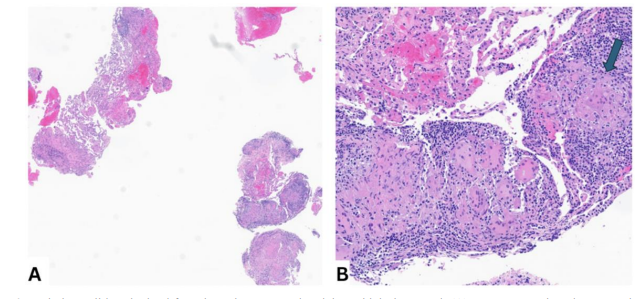
Figure 2: Pathology slides obtained from bronchoscopy and endobronchial ultrasound. (A) Low-power view demonstrating multiple well-formed, non-necrotizing granulomas within the lung parenchyma. (B) High-power view demonstrating multiple well-formed, non-necrotizing granulomas
Discussion
This case highlights the dual impact of immune checkpoint inhibitors in MSI-high cholangiocarcinoma: their remarkable anti-tumor efficacy and the potential for complex irAEs. In this patient, durvalumab therapy led to profound tumor regression, allowing discontinuation of cytotoxic chemotherapy and eventually a complete pathological response.
Cholangiocarcinoma is an aggressive malignancy with limited systemic options [5]. It is recognized as a “target-rich” malignancy, with next-generation sequencing identifying actionable alterations in up to 40-50% of patients, including FGFR2, IDH, BRAF V600E, and HER2 amplifications, forming a foundation for precision therapy [14]. Durvalumab, an anti-PD-L1 checkpoint inhibitor, became the standard frontline treatment for advanced biliary tract cancer after the TOPAZ-1 trial, which showed 24-month overall survival rates of 24.9% vs 10.4% with durvalumab plus gemcitabine/cisplatin versus chemotherapy alone, demonstrating statistically significant improvement in overall survival (HR 0.80; 95% CI 0.66-0.97) [5]. PD-1 and PD-L1 inhibitors exert their effects by modulating the tumor microenvironment through the regulation of regulatory T cells, tumor-associated macrophages, and myeloid-derived suppressor cells [7]. In addition, chemotherapy acts through a direct immunostimulatory mechanism, downregulating the immunosuppressive microenvironment and increasing tumor immunogenicity [15].
As the use of ICIs expanded, recognition of irAEs, particularly sarcoid-like reactions, has become critical; durvalumab-induced sarcoid-like reactions are driven by PD-L1 blockade, which disrupts the PD-1/PD-L1 inhibitory axis, causing unopposed CD4+ Th1 lymphocyte activation, excessive interferon-gamma and TNF-alpha production, and subsequent macrophage aggregation into non-caseating granulomas [16].
A key clinical challenge is distinguishing durvalumab-induced sarcoid-like reaction from true cholangiocarcinoma metastasis, as overlapping radiographic findings can lead to misclassification. Therefore, tissue biopsy with histopathologic confirmation and exclusion of infection are essential prior to any treatment modification [17]. Drug-induced sarcoid-like reaction is diagnosed based on radiographic findings of hilar/mediastinal lymphadenopathy and histopathology findings of non-caseating granulomas on biopsy [18]. There are a few case reports describing drug-induced sarcoid-like reaction after use of durvalumab (Table 1) [11-13]. Prospective data on durvalumab rechallenge after sarcoid-like reaction are lacking; however, case series suggest that patients with steroid-responsive disease and clear oncologic benefit may be cautiously rechallenged under close multidisciplinary monitoring, sometimes with prophylactic low-dose corticosteroids [19].
This case emphasizes the importance of biomarker-guided therapy, remarkable responses to immune checkpoint inhibitors, careful monitoring for immune-related adverse events, and multidisciplinary management to achieve optimal outcomes. Clinicians should maintain a high index of suspicion for drug-induced sarcoid-like reaction in patients receiving ICIs, even in rare malignancies, to avoid misinterpretation of imaging findings and ensure timely intervention.
|
Author (Year) |
Cancer Type |
Treatment |
Time to Onset |
Clinical/Imaging Findings |
Diagnostic Confirmation |
Management |
|
Sanderson et al. [11] |
NSCLC |
Lobectomy; cisplatin and vinorelbine; durvalumab |
6 months |
Bilateral hilar and mediastinal lymphadenopathy |
Biopsy-proven sarcoid-like reaction |
Corticosteroids; durvalumab discontinued |
|
Minami et al. [12] |
NSCLC |
Cisplatin and vinorelbine, durvalumab; Lobectomy radiation |
1.5 years |
Pulmonary nodules and mediastinal lymphadenopathy |
Histopathology from lobectomy consistent with sarcoid-like reaction |
Lobectomy |
|
Sobash et al. [13] |
NSCLC |
Carboplatin/ pemetrexed; radiation; durvalumab |
15 days |
Pneumonitis |
Tissue confirmation |
Observation or corticosteroids |
Table 1: Published Cases of Durvalumab-Associated Sarcoid-like Reaction
Acknowledgments
The authors would like to thank the patient and their family for consenting to the publication of this case. We also acknowledge the multidisciplinary teams in oncology, pathology, radiology, and pulmonology for their contributions to patient care.Funding
The authors received no specific funding for this work.Disclosures Disclaimers
Midhun Malla- Consulting: Incyte, Bristol-Myers Squibb, AstraZeneca, DoMoore, Ipsen, Exelixis, Elevar Therapeutics, Merus, Natera, Taiho Oncology Mehmet Akce- Research: Bristol-Myers Squibb-Ono Pharmaceutical (Inst), GSK (Inst), Genentech (Inst), Bayer (Inst), Relay (Inst), ProDa BioTech (Inst), Exelixis (Inst), AstraZeneca (Inst)
Consulting or advisory role: Eisai, Ipsen, Exelixis, GSK, QED, Isofol, Curio Science, AstraZeneca, Genentech, Incyte, Taiho, BMS, Partner Therapeutics, MJH Life Sciences.
The remaining authors have no relevant conflicts of interest to disclose. No disclaimers applicable.
References
- Saha, S. K., Zhu, A. X., Fuchs, C. S., & Brooks, G. A. (2016). Forty-year trends in cholangiocarcinoma incidence in the US: intrahepatic disease on the rise. The oncologist, 21(5), 594-599.
- Nakeeb, A., Pitt, H. A., Sohn, T. A., Coleman, J., Abrams,A., Piantadosi, S., ... & Cameron, J. L. (1996). Cholangiocarcinoma: a spectrum of intrahepatic, perihilar, and distal tumors. Annals of surgery, 224(4), 463-475.
- DeOliveira, M. L., Cunningham, S. C., Cameron, J. L., Kamangar, F., Winter, J. M., Lillemoe, K. D., ... & Schulick,R. D. (2007). Cholangiocarcinoma: thirty-one-year experience with 564 patients at a single institution. Annals of surgery, 245(5), 755-762.
- Khan, S. A., Thomas, H. C., Davidson, B. R., & Taylor-Robinson, S. D. (2005). Cholangiocarcinoma. The Lancet, 366(9493), 1303-1314.
- Oh, D. Y., He, A. R., Bouattour, M., Okusaka, T., Qin, S., Chen, L. T., ... & Valle, J. W. (2024). Durvalumab or placebo plus gemcitabine and cisplatin in participants with advanced biliary tract cancer (TOPAZ-1): updated overall survival from a randomised phase 3 study. The lancet Gastroenterology & hepatology, 9(8), 694-704.
- Eluri, M., Danner De Armas, A., Ross, J. S., Sharaf, R., Pavlick, D. C., Chatterjee, D., & Javle, M. M. (2023). Clinical and mutational profiles of microsatellite instability (MSI-H) in intrahepatic cholangiocarcinoma (iCCA). J Clin Oncol, 41(4_suppl), 607.
- Kelley, R. K., Ueno, M., Yoo, C., Finn, R. S., Furuse, J., Ren, Z., ... & Power, D. (2023). Pembrolizumab in combination with gemcitabine and cisplatin compared with gemcitabine and cisplatin alone for patients with advanced biliary tract cancer (KEYNOTE-966): a randomised, double-blind, placebo-controlled, phase 3 trial. The Lancet, 401(10391), 1853-1865.
- Goeppert, B., Roessler, S., Renner, M., Singer, S., Mehrabi, A., Vogel, M. N., ... & Kloor, M. (2019). Mismatch repair deficiency is a rare but putative therapeutically relevant finding in non-liver fluke associated cholangiocarcinoma: Immunotherapy. British journal of cancer, 120(1), 109-114.
- Wang, D. Y., Salem, J. E., Cohen, J. V., Chandra, S., Menzer, C., Ye, F., ... & Johnson, D. B. (2018). Fatal toxic effects associated with immune checkpoint inhibitors: a systematic review and meta-analysis. JAMA oncology, 4(12), 1721-1728.
- Ebinama, U., Sheshadri, A., Anand, K., & Swaminathan, I. (2023). Pulmonary immune-related adverse events of PD-1 versus PD-L1 checkpoint inhibitors: a retrospective review of pharmacovigilance. Journal of immunotherapy and precision oncology, 6(4), 177-184.
- Sanderson, E., Wimaleswaran, H., Senko, C., White, S., & McDonald, C. F. (2020). Durvalumab induced sarcoid-like pulmonary lymphadenopathy. Respirology case reports, 8(3), e00542.
- Minami, S., Yasuoka, H., Shoshihara, N., Ishida, D., & Sakamaki, Y. (2022). Sarcoid-like granulomatosis of the lung related to durvalumab after chemoradiation for pulmonary squamous cell carcinoma. Journal of Medical Cases, 14(1), 19-24.
- Sobash, P., Desikan, R., & Kakkera, K. (2020). Single-dose durvalumab induced sarcoidosis-like reaction with granuloma in non-small cell lung cancer. Chest, 158(4), A1518.
- Javle, M., Bekaii-Saab, T., Jain, A., Wang, Y., Kelley, R. K., Wang, K., ... & Ross, J. (2016). Biliary cancer: utility of next-generation sequencing for clinical management. Cancer, 122(24), 3838-3847.
- Galluzzi, L., Humeau, J., Buqué, A., Zitvogel, L., & Kroemer,G. (2020). Immunostimulation with chemotherapy in the era of immune checkpoint inhibitors. Nature reviews Clinical oncology, 17(12), 725-741.
- Capaccione, K. M., Valiplackal, J. P., Huang, A., Roa, T., Fruauff, A., Liou, C., ... & Salvatore, M. M. (2022). Checkpoint inhibitor immune-related adverse events: a multimodality pictorial review. Academic Radiology, 29(12), 1869-1884.
- Nykaza, I., Murciano-Goroff, Y. R., Desilets, A., Harada, G., Postow, M. A., Callahan, M. K., ... & Friedman, C. F. (2025). Sarcoid-like reactions in patients treated with checkpoint inhibitors for advanced solid tumors. The Oncologist, 30(5), oyaf017.
- Miedema, J., & Nunes, H. (2021). Drug-induced sarcoidosis-like reactions. Current opinion in pulmonary medicine, 27(5), 439-447.
- Schneider, B. J., Naidoo, J., Santomasso, B. D., Lacchetti, C., Adkins, S., Anadkat, M., ... & Bollin, K. (2021). Management of immune-related adverse events in patients treated with immune checkpoint inhibitor therapy: ASCO guideline update. Journal of clinical oncology, 39(36), 4073-4126.