

International Journal of Orthopaedics Research(IJOR)
ISSN: 2690-9189 | DOI: 10.33140/IJOR

Impact Factor: 1.6

Research Article - (2026) Volume 9, Issue 3
Comparative Study Between Short Proximal Femoral Nail and Long Proximal Femoral Nail in Management of Intertrochanteric Fractures
2Professor and Head of the Unit 2, Department of Orthopaedics, Smt. B.K. Shah Medial Institute and Research Center, Sumandeep Vidhyapeeth (Deemed to be University, India
32rd year Resident Doctor, Department of Orthopaedics Smt. B.K. Shah Medial Institute and Research Ce, Sumandeep Vidhyapeeth (Deemed to be University), India
Received Date: Jul 10, 2026 / Accepted Date: Aug 07, 2026 / Published Date: Sep 23, 2026
Copyright: ©2026 Pranavkumar Jagdishbhai Pandya, et al. This is an openaccess article distributed under the terms of the Creative Commons Attribution License, which permits unrestricted use, distribution, and reproduction in any medium, provided the original author and source are credited.
Citation: Pandya, P. J., Rawat, S., Gandhi, D. H. (2026). Comparative Study Between Short Proximal Femoral Nail and Long Proximal Femoral Nail in Management of Intertrochanteric Fractures. Int J Ortho Res, 9(3), 01-43
Abstract
Background: Intertrochanteric fractures are among the most common orthopedic injuries in the elderly population and are associated with significant morbidity and mortality. Proximal femoral nailing (PFN) has emerged as a preferred treatment modality because of its biomechanical advantages, minimally invasive approach, and ability to provide stable fixation. However, the choice between short proximal femoral nail and long proximal femoral nail remains controversial, with each implant having distinct advantages and limitations in terms of surgical parameters, biomechanical stability, and postoperative outcomes.
Aim: To evaluate and compare the surgical and functional outcomes of short proximal femoral nail and long proximal femoral nail in the management of intertrochanteric fractures.
Objectives: To assess the rate and time of fracture union in patients treated with short and long PFN and to analyze the complications associated with each implant type.
Methodology: A prospective observational study was conducted in the Department of Orthopaedics at SBKS Medical Institute and Research Centre, Dhiraj Hospital, Gujarat, India. Patients with intertrochanteric fractures were managed using either short PFN or long PFN depending on fracture characteristics and surgeon preference. Outcomes assessed included operative parameters, fracture union, functional recovery, and postoperative complications.
Background: Intertrochanteric fractures are among the most common orthopedic injuries in the elderly population and are associated with significant morbidity and mortality. Proximal femoral nailing (PFN) has emerged as a preferred treatment modality because of its biomechanical advantages, minimally invasive approach, and ability to provide stable fixation. However, the choice between short proximal femoral nail and long proximal femoral nail remains controversial, with each implant having distinct advantages and limitations in terms of surgical parameters, biomechanical stability, and postoperative outcomes.
Aim: To evaluate and compare the surgical and functional outcomes of short proximal femoral nail and long proximal femoral nail in the management of intertrochanteric fractures.
Objectives: To assess the rate and time of fracture union in patients treated with short and long PFN and to analyze the complications associated with each implant type.
Methodology: A prospective observational study was conducted in the Department of Orthopaedics at SBKS Medical Institute and Research Centre, Dhiraj Hospital, Gujarat, India. Patients with intertrochanteric fractures were managed using either short PFN or long PFN depending on fracture characteristics and surgeon preference. Outcomes assessed included operative parameters, fracture union, functional recovery, and postoperative complications.
Results: Short PFNs demonstrated advantages in terms of reduced operative time, lower blood loss, decreased fluoroscopy exposure, and ease of insertion. Long PFNs provided superior biomechanical stability, especially in unstable fractures, subtrochanteric extension, and osteoporotic bone, with reduced stress concentration and lower risk of peri-implant fractures. Functional outcomes and fracture union rates were generally comparable between the two implants, while implant selection was found to depend largely on fracture morphology, bone quality, patient comorbidities, and surgeon expertise.
Conclusion: Both short and long proximal femoral nails are effective treatment options for intertrochanteric fractures. Short PFN offers perioperative advantages such as shorter surgery duration and reduced blood loss, making it suitable for stable fractures and frail elderly patients. Long PFN provides enhanced biomechanical stability and is preferable in unstable fracture patterns, osteoporotic bone, and fractures with subtrochanteric extension. Individualized implant selection based on patient and fracture characteristics is essential for optimizing outcomes.
Keywords
Intertrochanteric Fracture, Proximal Femoral Nail, Short PFN, Long PFN, Intramedullary Fixation, Fracture Union, Harris Hip Score, Orthopedic Trauma
Abbreviations
PFN : Proximal Femoral Nail
PFNA : Proximal Femoral Nail Antirotation
DHS : Dynamic Hip Screw AO/OTA : Arbeitsgemeinschaft für Osteosynthesefragen /Orthopaedic Trauma Association
HHS : Harris Hip Score
PMS : Parker Mobility Score
TAD : Tip-Apex Distance
ZNN : Zimmer Natural Nail
PFNA II : Proximal Femoral Nail Antirotation II
SN : Short Nail LN : Long Nail
CMN : Cephalomedullary Nail
SF-36 : Short Form-36 Health Survey
SBKSMI&RC: SBKS Medical Institute and Research Centre
RCT : Randomized Controlled Trial COPD : Chronic Obstructive Pulmonary Disease
CT : Computed Tomography
DHS : Dynamic Hip Screw
PFNA 2 : Proximal Femoral Nail Antirotation 2
Introduction
Intertrochanteric fractures of the femur represent one of the most common and challenging injuries encountered in orthopedic practice, particularly among the elderly population. These fractures occur between the greater and lesser trochanters of the femur and are generally the result of low-energy trauma such as falls in elderly individuals with osteoporosis, although high-energy trauma in younger patients can also be a cause. The increasing incidence of intertrochanteric fractures is closely linked with demographic changes, including longer life expectancy and the rising prevalence of osteoporosis, making these injuries a significant public health concern worldwide. Their management requires not only effective stabilization to allow for early mobilization and functional recovery but also a careful consideration of the surgical technique and implant selection to minimize complications and optimize outcomes [1,2].
Intertrochanteric fractures constitute a major proportion of hip fractures globally and represent a significant public health burden, particularly in the geriatric population. The global incidence of hip fractures is estimated to exceed 1.6 million cases annually, and this number is projected to rise to over 6 million by 2050 due to increasing life expectancy and aging populations [2].
Among these, intertrochanteric fractures account for approximately 45–50% of all hip fractures. The incidence rate varies geographically, ranging from 150 to 300 per 100,000 population per year in developed countries, with a rapidly rising trend in developing nations like India due to demographic transition and increased prevalence of osteoporosis [3].
A marked gender difference is observed in the epidemiology of intertrochanteric fractures. These fractures are significantly more common in females, primarily due to postmenopausal osteoporosis. The female-to-male ratio ranges from 2:1 to 3:1, although recent studies suggest a gradual increase in incidence among elderly males due to improved life expectancy [3,4].
Age is a critical determinant, with the majority of cases occurring in individuals above 60 years, and the highest incidence seen in those over 75 years. In younger populations, these fractures are relatively rare and are typically associated with high-energy trauma, such as road traffic accidents or falls from height [3]. In the Indian context, hospital-based studies have reported a steady rise in intertrochanteric fractures, with increasing burden on healthcare systems. Sedentary lifestyle, nutritional deficiencies, and lack of early osteoporosis screening further contribute to this growing problem.
Over the years, a variety of fixation methods have been developed to treat intertrochanteric fractures, ranging from extramedullary devices such as dynamic hip screws to intramedullary devices such as proximal femoral nails. The evolution of surgical techniques has been driven by the understanding that early mobilization is crucial for reducing morbidity and mortality in these patients, as prolonged immobilization is associated with risks such as deep vein thrombosis, pneumonia, pressure sores, and loss of independence. Among the various implants available, proximal femoral nailing has emerged as a widely accepted modality of treatment because of its biomechanical advantages, minimally invasive nature, and ability to provide stable fixation even in osteoporotic bone [2,3].
Proximal femoral nails are intramedullary implants designed to act as load-sharing devices, reducing the lever arm and thereby decreasing the bending stresses on the implant when compared to extramedullary devices. Their central placement within the medullary canal provides improved stability, especially in unstable fracture patterns, and allows for earlier weight bearing. Within the category of proximal femoral nails, two main variants are commonly used in clinical practice: short proximal femoral nails and long proximal femoral nails. The choice between these two options remains a topic of ongoing debate among orthopedic surgeons, as each has distinct theoretical advantages and potential drawbacks [3,4].
The short proximal femoral nail is characterized by a shorter intramedullary stem, which reduces operative time, blood loss, and soft tissue dissection. Its design makes it easier to insert, particularly in elderly patients with poor physiological reserves, thereby reducing intraoperative complications. Short nails also require less fluoroscopy time, which benefits both the patient and surgical team by minimizing radiation exposure. In addition, their smaller size may be particularly suitable for patients with smaller femoral canals. However, concerns have been raised regarding the potential for peri-implant fractures at the distal tip of the nail, as the short implant may create a stress riser at the junction between the nail and the diaphyseal bone. This risk is believed to be higher in osteoporotic bone, where cortical support is reduced, and in patients with preexisting femoral deformities [4,5].
On the other hand, long proximal femoral nails extend further distally into the femoral shaft, distributing the load across a greater surface area and potentially reducing the risk of stress concentration at the distal end of the implant. By spanning a larger portion of the femur, long nails provide more stability, especially in cases with associated shaft fractures, subtrochanteric extension, or highly unstable intertrochanteric fracture patterns. They are particularly advantageous in patients with poor bone quality, as the longer construct reduces the risk of implant failure. Despite these biomechanical advantages, long nails are associated with certain challenges. Their insertion typically requires longer operative time and greater blood loss due to the need for more extensive reaming of the medullary canal. This may be a limiting factor in elderly patients with multiple comorbidities who may not tolerate prolonged anesthesia. Furthermore, technical difficulties such as malalignment, distal locking complications, and increased radiation exposure during the procedure are reported more frequently with long nails compared to short nails [5,6].
The decision regarding whether to use a short or long proximal femoral nail is influenced by multiple factors, including the fracture pattern, patient’s age, bone quality, comorbidities, surgeon’s expertise, and institutional resources. For stable intertrochanteric fractures, short nails are often considered sufficient, as they provide adequate stability with the added benefit of reduced surgical morbidity. For unstable fractures, especially those with subtrochanteric extension or reverse obliquity patterns, long nails are frequently preferred to achieve stronger fixation. However, recent studies and meta-analyses have demonstrated mixed outcomes when comparing short and long nails, with some reporting no significant differences in union rates, functional recovery, or complication rates, while others highlight specific contexts where one option is superior to the other [6,7].
From a biomechanical standpoint, the superiority of one implant over the other is not absolute but context-dependent. The short proximal femoral nail allows for quicker surgical procedures, reduced perioperative morbidity, and is often associated with fewer immediate complications. It is, therefore, particularly useful in elderly and frail patients where surgical efficiency and reduced anesthesia time are critical. Conversely, the long proximal femoral nail offers additional mechanical stability and reduces the risk of secondary fractures at the nail tip, making it more suitable for complex and unstable fracture patterns. Despite these theoretical considerations, real-world outcomes often depend on patient-specific variables and surgical expertise rather than the implant length alone [7,8].
The growing body of literature comparing short and long proximal femoral nails emphasizes the need for individualized treatment planning rather than a uniform approach. Randomized controlled trials, retrospective analyses, and systematic reviews have all contributed to this debate, highlighting the importance of considering not just radiological outcomes but also functional recovery, complication rates, and overall quality of life in the postoperative period. Functional independence and the ability to return to pre-injury levels of mobility are of paramount importance, especially in the elderly population, where maintaining autonomy can significantly affect long-term survival [8].
Another important dimension in this comparison lies in the economic implications of implant choice. Short nails are generally less expensive than long nails, and the reduced operative time and hospital stay associated with them may further decrease overall treatment costs. However, in cases where implant failure or peri-implant fracture occurs, the costs associated with revision surgery can offset these savings. Thus, the financial considerations must also be balanced against the potential risks and long-term benefits when deciding between short and long nails [8,9].
The aim of this study was to evaluate the comparative study between short proximal femoral nail and long proximal femoral nail in management of intertrochanteric fractures.
Aim & Objectives
AIM
• Surgical and functional outcomes of results of short proximal femoral nail and long proximal femoral nail
Objectives
• Primary: To assess the rate and time of union for short proximal femoral nail and long proximal femoral nail at our institute.
• Secondary: To analyze complications associated with it.
Review of Literature
Anatomical Considerations of the Proximal Femur
The proximal femur occupies a central role in weight-bearing biomechanics, locomotion, and fracture pathology. Its anatomical configuration—comprising the femoral head, neck, and trochanteric region—makes it uniquely adapted to withstand the high stresses of daily activity while simultaneously predisposing it to characteristic fracture patterns in elderly populations with osteoporosis or in younger individuals exposed to high-energy trauma. Understanding the structural, vascular, and morphological features of this region is vital not only for appreciating the pathophysiology of intertrochanteric fractures but also for selecting the appropriate fixation strategy, particularly in the context of short versus long proximal femoral nails (PFNs) [10].

Figure 1: Proximal Femoral
Proximal Femoral Anatomy
Proximal Femoral Anatomy
Femoral Head
The femoral head is a spherical structure covered with articular cartilage, articulating with the acetabulum to form the hip joint. It transmits compressive forces from the body to the femoral shaft. Approximately two-thirds of the head is contained within the acetabulum, enhancing stability while still permitting a wide range of motion. The head’s trabecular architecture is organized into compressive and tensile systems: the primary compressive trabeculae extend from the superior cortex of the femoral shaft into the weight-bearing portion of the head, while tensile trabeculae cross perpendicularly, resisting shear forces. These trabecular patterns directly influence fracture propagation and healing potential [11].
Femoral Neck
The femoral neck serves as a conduit between the head and shaft, angulated at approximately 125–135 degrees (neck-shaft angle). This angulation optimizes mechanical efficiency by reducing bending stresses on the shaft while maintaining mobility at the hip joint. The femoral neck is enclosed in a capsule and surrounded by synovial fluid, which facilitates motion but also limits the capacity for callus formation in intracapsular fractures. Importantly, the lack of periosteum around most of the femoral neck further compromises its healing ability, making fractures in this region particularly problematic [12].
Greater and Lesser Trochanters
The greater trochanter projects laterally and superiorly, serving as the attachment point for abductor and rotator muscles (gluteus medius, minimus, and short external rotators). The lesser trochanter, located posteromedially, serves as the insertion for the iliopsoas tendon. The functional significance of these bony prominences lies in their role in muscle lever arms, which profoundly influence gait and stability. Fractures that involve trochanteric regions disrupt muscular attachments, altering biomechanics and increasing the need for rigid fixation [13].
Significance of the Calcar Femorale and Medial Cortex in Load Transfer
Calcar Femorale
The calcar femorale is a dense vertical plate of bone that extends from the posteromedial aspect of the femoral shaft into the posteroinferior portion of the femoral neck. It is a crucial structural support, acting as an internal buttress that redistributes compressive loads from the femoral head into the shaft. The calcar provides resistance to varus collapse—a common complication in intertrochanteric fractures. When the calcar is disrupted, fracture stability diminishes, and fixation failure risk increases [13].
Biomechanically, the calcar femorale serves as a fulcrum against which intramedullary implants, particularly PFNs, gain purchase. Preservation of this structure during surgical fixation improves implant anchorage and reduces the risk of cut-out or excessive subsidence [14].
Medial Cortex
The medial cortex of the proximal femur, especially the inferomedial region near the lesser trochanter, plays an equally vital role in load transmission. It functions as the primary compressive trabecular pathway, channeling forces directly from the femoral head to the shaft. Intact medial cortical support ensures stable fixation, while cortical comminution predisposes to varus malalignment, implant failure, and non-union [14].
In clinical practice, the assessment of medial cortical integrity is a decisive factor in implant choice. Short PFNs may suffice for stable fractures with preserved medial cortex, whereas long PFNs are preferred when cortical compromise or subtrochanteric extension exists [15].
Vascular Anatomy Relevant to Fracture Healing and Implant Choice
Extracapsular Blood Supply
The proximal femur is richly supplied by an anastomotic vascular network. The extracapsular arterial ring, formed by branches of the medial and lateral femoral circumflex arteries, gives rise to ascending cervical branches that supply the femoral neck and head. The medial femoral circumflex artery, particularly its deep branch, is the most important source for the weight-bearing portion of the femoral head [15].
Retinacular Vessels
Ascending cervical branches traverse the retinacula along the femoral neck to reach the head. These vessels are vulnerable in intracapsular fractures but relatively preserved in extracapsular (intertrochanteric) fractures, which explains the lower incidence of avascular necrosis in the latter [16].
Nutrient and Metaphyseal Vessels
The nutrient artery supplies the femoral shaft and metaphysis, while metaphyseal vessels contribute to the trochanteric region. These vessels are important in fracture healing, as their disruption can delay callus formation [16].
Relevance to Implant Choice
The selection between short and long PFNs must consider vascular preservation. Intramedullary devices are generally favorable because they minimize disruption of periosteal blood supply compared to extramedullary implants. However, incorrect entry point or reaming can injure nutrient vessels. Moreover, preserving the trochanteric region and minimizing surgical trauma are critical for enhancing vascular integrity and promoting union [17].

Figure 2: Vascular Anatomy of Proximal Femoral
Anatomical Variations and Their Impact on Fracture Stability
Femoral Neck–Shaft Angle Variations
The normal neck–shaft angle ranges between 120–135 degrees, but variations exist across populations and with aging. A coxa vara configuration increases bending stresses on the implant and predisposes to medial cortical collapse. In such cases, a longer PFN may be advantageous for load distribution [17].
Femoral Anteversion and Torsional Anatomy
Femoral anteversion, defined as the forward angulation of the femoral neck relative to the shaft, varies widely. Excessive anteversion complicates nail insertion and may lead to malalignment. Long nails, with their greater intramedullary contact, may reduce torsional stresses but require careful insertion to avoid iatrogenic fractures [18].
Canal Morphology and Medullary Diameter
The proximal femur’s canal morphology influences implant length selection. Wide medullary canals accommodate both short and long nails, while narrow, stovepipe-shaped canals may pose technical difficulties for long PFN insertion. In osteoporotic bone, the reduced cortical thickness further impacts implant anchorage, making precise implant sizing crucial [18].
Age-Related Changes
With aging, trabecular bone loss, cortical thinning, and widening of the medullary canal occur. These changes reduce load-bearing capacity and alter fracture patterns. Short PFNs may provide adequate stabilization in elderly patients with low activity levels, while long PFNs are often chosen to prevent future periprosthetic fractures in osteoporotic bone [19,20].
Clinical Relevance of Anatomical Understanding in Implant Selection
Anatomical understanding of the proximal femur directly informs surgical decision-making. A fracture extending into the subtrochanteric region with compromised calcar and medial cortex often necessitates a long PFN for additional shaft stabilization. Conversely, stable intertrochanteric fractures with intact calcar support can be effectively managed with short PFNs, which reduce operative time, blood loss, and fluoroscopy exposure [21].
Furthermore, anatomical variations such as coxa vara, wide medullary canals, or excessive anteversion influence the surgeon’s choice of implant length and configuration. Recognizing these nuances reduces the likelihood of complications such as cut-out, implant breakage, and distal femoral shaft fractures [22].
Biomechanical Rationale of Intramedullary Fixation
The management of intertrochanteric fractures has undergone a paradigm shift over the past few decades, driven largely by biomechanical innovations in internal fixation. Historically, extramedullary devices such as the Dynamic Hip Screw (DHS) were considered the gold standard. However, advances in implant design, along with an improved understanding of fracture biomechanics, have positioned intramedullary devices—particularly the Proximal Femoral Nail (PFN)—as the treatment of choice in many clinical scenarios. The rationale for this preference stems from fundamental biomechanical principles that dictate stability, load transfer, and fracture healing [23].
The choice between short and long PFNs is not merely a surgical preference but a reflection of nuanced biomechanical considerations. While both designs share the core principle of intramedullary fixation, their performance diverges in terms of stress distribution, control of collapse, and protection against secondary fractures [24].
Load-Sharing vs. Load-Bearing Concepts
Principles of Load Transmission
In the native femur, physiological loading is transmitted from the femoral head through the neck and into the diaphysis, with the medial cortex and calcar femorale serving as primary load-bearing structures. Intertrochanteric fractures disrupt this pathway, necessitating external stabilization [24].
Extramedullary devices, such as the DHS, function as load-bearing implants, assuming much of the mechanical stress until bony union is achieved. This principle, while effective in stable fractures, places a significant mechanical burden on the implant, especially in osteoporotic bone or unstable fracture patterns. The consequence is implant fatigue, screw cut-out, or varus collapse if fracture healing is delayed [25].
Intramedullary nails, in contrast, operate on a load-sharing principle. Positioned within the medullary canal, they are aligned closer to the mechanical axis of the femur. This central alignment permits physiological transmission of compressive loads through both the implant and the surrounding bone, distributing stresses more evenly. By reducing the mechanical load borne solely by the implant, intramedullary nails minimize implant failure and promote biological fracture healing [25].
Implications for Short vs. Long PFN
Both short and long PFNs embody the load-sharing concept, but their performance differs in specific fracture scenarios. A short PFN, terminating above the isthmus, may concentrate stress at its distal tip, particularly in osteoporotic femora. This predisposes patients to secondary femoral shaft fractures distal to the nail, a complication well-documented in clinical literature. Conversely, a long PFN spans the entire femoral diaphysis, thereby dissipating forces more uniformly along the shaft. While this confers mechanical advantage in unstable fractures or osteoporotic bone, it comes at the cost of greater surgical dissection, longer operative time, and increased blood loss [26].

Figure 3: Schematic Two-Dimensional Representation of the Resultant Force of Weight Bearing, Showing its Relationship to the Internal Vertical and Horizontal Trabecular Columns at the Times of Full Extension and Full Flexion of the Hip Joint.
Biomechanical Stability of Intramedullary Nails vs. Extramedullary Devices
Lever Arm Mechanics
One of the most compelling biomechanical justifications for intramedullary fixation is the principle of shorter lever arms. Extramedullary devices, such as DHS, are anchored laterally on the femoral cortex, creating a longer lever arm between the implant and the body’s line of weight transmission. This longer moment arm amplifies the bending stresses on the implant, particularly in unstable fracture configurations [26].
Intramedullary nails, by virtue of their central placement within the medullary canal, significantly reduce the lever arm and therefore the bending moment acting on the construct. This confers superior resistance to varus forces and reduces the risk of implant failure [27].
Torsional and Axial Stability
Rotational control is critical in intertrochanteric fractures, especially those with comminution or posteromedial cortical disruption. Intramedullary nails are biomechanically superior in resisting rotational forces, owing to the combination of proximal lag screws and anti-rotation elements. Moreover, their intramedullary position aligns them with the weight-bearing axis, ensuring better axial stability under loading. Extramedullary devices, in contrast, are less effective in preventing torsional displacement, particularly in unstable or reverse oblique fractures [28].
Short vs. Long PFN in Stability
While both short and long PFNs provide enhanced stability compared to extramedullary devices, their relative performance depends on the fracture pattern. Short PFNs are often adequate for stable two-part intertrochanteric fractures, where the medial buttress is preserved. In unstable or subtrochanteric extensions, however, long PFNs demonstrate superior stability by controlling distal propagation of stresses and offering a longer working length for load distribution [29].
Figure 4: Proximal Femoral Nail (PFN)
Stress Distribution Differences Between Short and Long PFNs
Stress Concentration and Distal Femoral Fractures
Biomechanical studies using finite element analysis and cadaveric models consistently highlight differences in stress distribution between short and long PFNs. Short PFNs, due to their abrupt termination in the proximal femoral diaphysis, create a stress riser at the distal tip. Under repetitive loading, particularly in elderly osteoporotic patients, this stress concentration predisposes to femoral shaft fractures just below the nail. Long PFNs, by spanning the entire diaphysis, mitigate this phenomenon by distributing stresses along a greater surface area. This advantage explains their preference in cases with poor bone stock or high-energy unstable fractures [28].
Load Transfer and Implant Fatigue
Load transfer dynamics also differ between the two nail lengths. Short PFNs allow partial load transfer to the bone relatively early during healing, which can stimulate callus formation. However, the concentrated loading at the distal tip remains a concern. Long PFNs, by dissipating forces more uniformly, reduce the risk of implant fatigue but may delay early callus response due to reduced micromotion at the fracture site [29].
Clinical Relevance
The choice of nail length thus reflects a trade-off: while short PFNs offer ease of insertion and less operative morbidity, they carry a higher risk of stress-related complications; long PFNs provide superior biomechanical safety in complex fractures but at the expense of increased surgical demands [30].
Prevention of Varus Collapse and Rotational Instability
Varus Collapse
Varus collapse is a well-recognized complication of intertrochanteric fracture fixation, characterized by medialization of the femoral shaft and loss of neck-shaft angle. The biomechanical determinant of varus collapse lies in the integrity of the medial cortical buttress and the ability of the implant to resist bending stresses [30].
Intramedullary nails, particularly PFNs, offer superior protection against varus collapse compared to DHS because of their central alignment and shorter lever arm. By maintaining a vertical load path closer to the body’s mechanical axis, PFNs reduce bending moments and better preserve the femoral neck-shaft angle during healing [31].
Rotational Instability
Rotational instability arises from inadequate control of the proximal fragment, especially in multi-fragmentary fractures. PFNs address this through the use of dual proximal screws—a larger lag screw for load-bearing and a smaller anti-rotation screw for torsional control. This construct enhances rotational stability and reduces the risk of screw cut-out, one of the most common mechanical failures in intertrochanteric fracture fixation [31].
Short vs. Long PFN in Preventing Collapse and Instability
The role of nail length in preventing varus and rotational complications is complex. Short PFNs provide adequate control in stable fracture types but may fail in unstable patterns where medial cortical support is compromised. Long PFNs, by virtue of their extended diaphyseal fixation, not only resist varus deformity more effectively but also protect against secondary fractures that might exacerbate instability. Clinical studies have reported lower rates of varus collapse and screw cut-out with long PFNs in unstable fracture subtypes, further reinforcing their biomechanical superiority in selected cases [32].
Synthesis of Evidence
The biomechanical rationale for intramedullary fixation is well established, rooted in principles of load sharing, reduced lever arm mechanics, and superior control of rotational and axial forces. Within this framework, the distinction between short and long PFNs lies in their ability to manage stress distribution and prevent secondary complications [33].
Short PFNs, while advantageous for stable fractures and low surgical morbidity, are limited by stress concentration at the distal tip. Long PFNs, although technically more demanding, confer biomechanical superiority in unstable, osteoporotic, or high-energy fractures by distributing loads across the entire femoral shaft. Ultimately, the selection of implant length must be individualized, guided by fracture morphology, patient bone quality, and surgeon expertise [34].
Design Features of Short Vs. Long PFN
The management of intertrochanteric fractures has undergone a paradigm shift with the evolution of intramedullary fixation devices, particularly proximal femoral nails (PFNs). Both short and long PFNs have been developed with the goal of optimizing stability, reducing complications, and facilitating early mobilization in a vulnerable patient population. While their overarching purpose remains the same—achieving stable internal fixation—the design features of these implants differ significantly, influencing their biomechanical performance, surgical handling, and clinical outcomes [35].
Implant Geometry: Length, Diameter, and Proximal Angulation
Length Considerations
The most obvious distinction between short and long PFNs lies in their overall length. Short PFNs typically measure between 170–240 mm, designed to span only the proximal half of the femur. They are intended for fractures confined to the intertrochanteric region without significant subtrochanteric extension. Their compact length reduces operative time, minimizes soft tissue dissection, and decreases fluoroscopy exposure. However, their limited coverage of the femoral shaft concentrates stress at the nail tip, predisposing to distal femoral shaft fractures, particularly in osteoporotic bone [35]. Conversely, long PFNs extend the full length of the femoral canal, generally ranging from 300–460 mm. This longer geometry provides more uniform load distribution throughout the femur, reducing stress concentration at the implant terminus. It is particularly advantageous in unstable fractures, reverse obliquity patterns, or when there is subtrochanteric extension. Nevertheless, insertion of long PFNs is technically more demanding, requiring more extensive reaming, higher blood loss, and prolonged operative times. Thus, the balance between biomechanical safety and surgical morbidity often guides the choice of implant length [36].
Diameter and Canal Occupancy
Both short and long PFNs are available in varying diameters, usually between 9–12 mm. Nail diameter plays a critical role in providing rotational and axial stability. A snug fit within the femoral canal reduces micromotion and prevents toggling, but excessive mismatch between the implant and the canal diameter can increase the risk of intraoperative fracture. Long PFNs, spanning the entire shaft, demand more precise canal reaming to ensure proper fit and alignment. In contrast, short PFNs, being shorter, often tolerate mild mismatch better, though at the expense of stress concentration [36].
Proximal Angulation and Design
Both short and long PFNs share a common proximal geometry characterized by a mediolateral valgus angle (commonly 6°) to match the natural femoral neck-shaft angle. This angulation allows for optimal placement of proximal screws within the femoral head and neck. Some designs incorporate anatomical curvature along the shaft to mirror the femur’s anterior bowing, with long nails being more sensitive to mismatch due to their greater length. Improper accommodation of the femoral bow may result in anterior cortical impingement, malalignment, or intraoperative fracture when long PFNs are inserted, whereas short PFNs largely avoid this complication due to their limited extent [37].
Distal Locking Screw Configurations and Stress Modulation
Purpose of Distal Locking
Distal locking is critical for controlling axial and rotational stability, particularly in unstable fracture patterns. It prevents nail migration, telescoping, or rotation, and maintains alignment until fracture union. Both short and long PFNs are equipped with distal locking options, though their biomechanics differ due to implant length [37].
Figure 5: Distal Locking Screw Configurations
Configurations in Short PFNs
Short PFNs rely heavily on distal locking screws because their limited span amplifies stress at the nail tip. Most short designs provide one or two distal locking holes, with screws oriented in either static (transverse) or dynamic (oblique) modes. Static locking ensures rigid stabilization in multiple planes, reducing risk of distal fracture displacement, while dynamic options allow controlled axial compression at the fracture site, promoting healing. However, despite these provisions, stress risers at the distal end of short PFNs remain a recognized risk factor for subsequent femoral shaft fractures, especially in osteoporotic bone or after trivial trauma [38].
Configurations in Long PFNs
Long PFNs offer multiple distal locking options, generally spaced further apart along the shaft. This allows greater flexibility in tailoring stability to fracture morphology. Since long PFNs distribute stress more evenly, the distal locking screws play a secondary role compared to short PFNs. Moreover, multiple distal holes allow static and dynamic options, with surgeons often preferring dynamic locking in stable patterns to encourage axial loading and bone remodeling. The longer lever arm also reduces the mechanical stress concentration, translating to lower incidence of distal femoral fractures than short PFNs [38].
Biomechanical Implications
Biomechanical studies have demonstrated that distal locking in short PFNs significantly influences construct strength, while in long PFNs the implant itself provides sufficient resistance against axial and torsional forces. This explains the higher association of distal femoral shaft fractures with short nails. Additionally, advances such as advanced locking mechanisms (helical blades, angular stable screws) are being incorporated to enhance distal fixation and reduce stress risers [39].

Figure 6: Short PFNs
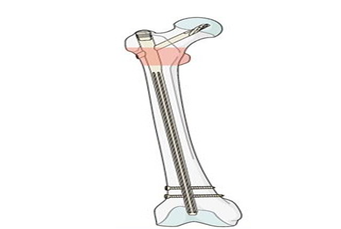
Figure 7: Long PFNs
Entry Point Considerations and Surgical Technique Variations
Anatomical Entry Points The standard entry point
for both short and long PFNs is the tip of the greater trochanter or slightly medial to it in the piriformis fossa, depending on the nail design. However, subtle differences in technique are magnified by implant length. Short PFNs, being compact, are more forgiving in entry point variability. A slightly off-center entry may still allow satisfactory alignment due to the short trajectory of the nail [39].
Long PFNs, in contrast, demand meticulous accuracy in entry point selection. A lateralized entry point risks varus malreduction, while a medialized or posterior entry increases the chance of cortical perforation. Given the length of the implant, even minor deviations translate into significant malalignment distally. Thus, proper fluoroscopic control and surgeon experience are critical for safe insertion [40].
Surgical Insertion Technique
Insertion of short PFNs generally requires less reaming, reduced operative time, and minimal fluoroscopy exposure. The procedure is relatively straightforward, making it suitable for emergency stabilization in elderly patients with multiple comorbidities. Long PFNs, however, necessitate extensive canal preparation. Reaming must match the femoral curvature, and improper technique risks cortical blowout, fracture propagation, or implant mismatch. Moreover, distal locking of long PFNs is technically more challenging, often requiring advanced targeting devices or freehand techniques under fluoroscopic guidance, which prolong operative duration [40].
Learning Curve and Technical Demands
Surgeons often find short PFNs easier to implant, with a shorter learning curve, whereas long PFNs demand greater surgical expertise. This difference has implications for outcomes in high-volume trauma centers where surgical turnover and resource availability influence implant choice. Moreover, in cases of polytrauma or hemodynamic instability, short PFNs may be favored due to shorter operative times [41]
Proximal Lag Screw and Anti-Rotation Screw: Stabilization Mechanics
Lag Screw Function
The proximal fixation component of PFNs typically consists of a large lag screw inserted into the femoral head, providing anchorage and transmitting load across the fracture site. Its primary role is to resist axial loading, prevent fracture collapse, and allow controlled sliding for impaction and union. Both short and long PFNs utilize this mechanism, with screw diameter and thread design optimized for purchase in osteoporotic bone [41].
Anti-Rotation Screw Function
In addition to the lag screw, many PFN designs incorporate a smaller anti-rotation screw positioned superiorly. This prevents rotational instability of the femoral head and neck fragment, a common cause of fixation failure. By sharing load with the lag screw, the anti-rotation screw enhances resistance against varus collapse and cut-out. The configuration is particularly advantageous in unstable intertrochanteric fractures, where rotational forces are pronounced [42].
Design Innovations
Several design innovations have refined proximal fixation. Some systems use helical blades instead of screws, which compact cancellous bone during insertion, enhancing purchase in osteoporotic femoral heads. The combination of lag screw and anti¬rotation screw provides a dual anchor system, reducing the risk of implant cut-out, one of the most dreaded complications of PFN fixation. Comparative studies suggest that these features, common to both short and long PFNs, contribute more to fixation success than implant length itself, underscoring the critical importance of proximal screw configuration [40].
Biomechanical Performance
Biomechanical testing has demonstrated that the dual-screw design provides superior torsional stability compared to single-screw systems. The anti-rotation screw not only prevents femoral head spin but also decreases the load borne by the lag screw, reducing risk of migration and cut-out. Thus, the proximal fixation mechanism is a cornerstone of PFN success, irrespective of nail length, but its biomechanical efficacy may be particularly relevant in osteoporotic patients where bone purchase is compromised [41,42].
Indications and Patient Selection
Intertrochanteric fractures of the femur represent one of the most common injuries in the elderly, particularly among individuals with osteoporosis and reduced mobility. These fractures, located between the greater and lesser trochanters of the proximal femur, carry significant morbidity and mortality due to their association with prolonged immobilization, thromboembolic risk, and impaired quality of life. Surgical fixation remains the cornerstone of management, with intramedullary devices such as the proximal femoral nail (PFN) increasingly replacing traditional extramedullary implants like the dynamic hip screw (DHS).
Within intramedullary devices, a critical distinction exists between short PFN and long PFN, each with unique biomechanical properties, advantages, and limitations. The choice between these two options is not merely technical but depends on the underlying fracture pattern, patient characteristics, and surgeon’s clinical judgment. Thus, a comprehensive understanding of indications and patient selection is central to achieving optimal outcomes [43,44].


Figure 8: AO/OTA Type
Indications for Short Proximal Femoral Nail
Short PFN in Stable Intertrochanteric Fractures
Stable intertrochanteric fractures, often classified under AO/OTA type 31-A1, typically demonstrate an intact posteromedial cortex and minimal comminution. In such cases, mechanical stability is inherent, and the implant serves mainly as a load-sharing device to maintain reduction during healing. The short PFN is particularly well-suited here due to: [43-45].
• Ease of insertion: Short PFNs require less reaming, reduce operative time, and minimize fluoroscopic exposure.
• Reduced surgical trauma: By limiting intramedullary reaming, they are beneficial in elderly patients with frailty, minimizing perioperative morbidity.
• Comparable biomechanical stability: In stable fractures, the shorter construct does not compromise fixation strength or healing potential.
Several comparative studies have demonstrated no significant difference in union rates or functional outcomes between short and long PFNs in stable fracture configurations, further reinforcing the appropriateness of short nails in this setting.
Short PFN in Select Unstable Intertrochanteric Fractures
While the traditional role of short PFNs has been limited to stable fractures, recent literature suggests that in select unstable fractures without subtrochanteric extension, short nails may still offer satisfactory outcomes. For instance, fractures with lateral wall involvement or moderate comminution, but without propagation into the subtrochanteric region, can be managed with short PFNs provided careful reduction and proper implant placement are achieved [44].
The key to success in these cases lies in the surgeon’s ability to restore medial support and avoid excessive stress concentration at the distal tip, which otherwise increases the risk of femoral shaft fractures. Thus, short PFNs may be cautiously applied in unstable patterns under strict intraoperative vigilance [45].
Indications for Long Proximal Femoral Nail
Long PFN in Subtrochanteric Extension
One of the clearest indications for the use of a long PFN is when the intertrochanteric fracture extends into the subtrochanteric region. Subtrochanteric extension compromises the proximal femur’s shorter constructs cannot adequately resist [45-47]. Long nails span a greater length of the femoral shaft, thereby:
• Dissipating stress more evenly along the bone-implant interface.
• Preventing secondary shaft fractures distal to the implant tip.
• Providing greater control in cases of long oblique or spiral fracture lines that cross into the subtrochanteric zone.
Long PFN in Osteoporotic Bone
In elderly patients with severe osteoporosis, fixation stability is often compromised due to poor bone quality. The insertion of a long PFN allows for improved distribution of forces across a larger segment of the femur, thereby reducing the risk of implant-related complications. Moreover, long nails reduce the incidence of peri-implant fractures, which are more common with short nails in osteoporotic bone [47].
Clinical series have highlighted that elderly osteoporotic patients treated with long PFNs demonstrate reduced incidence of postoperative femoral shaft fractures compared with those managed using short PFNs. Although operative time and blood loss may be higher, the overall benefit in terms of implant longevity and reduced revision rates supports their use [48].
Long PFN in Cases of High Comminution
Highly comminuted fractures, classified under AO/OTA type 31-A2 or A3, are inherently unstable. The disruption of medial cortical support and lateral wall integrity means that a short construct risks excessive stress concentration and mechanical failure. A long PFN provides additional leverage by spanning the entire femur, thereby neutralizing forces and aiding in the restoration of limb alignment [48].
Long implants are also advantageous in cases where reduction is difficult or when intraoperative stability remains questionable, as they provide additional fixation points along the shaft [48].
Patient Factors Influencing Implant Choice
Age and Physiological Reserve
Age plays a dual role: younger patients with better physiological reserve may tolerate longer operative times and slightly higher surgical morbidity associated with long PFNs, whereas elderly frail patients may benefit from shorter operative procedures achievable with short PFNs. However, in elderly patients with unstable fractures or poor bone quality, the biomechanical superiority of long PFNs may outweigh the risks of prolonged surgery [49].
Bone Quality
Bone mineral density remains a critical determinant. In osteopenic or osteoporotic bone, long PFNs are preferred to reduce the risk of implant cut-out and peri-implant fracture. In contrast, younger patients with good bone stock and stable fracture morphology are ideal candidates for short PFNs, which avoid unnecessary surgical trauma [50].
Comorbidities and Perioperative Risk
Patients with significant comorbidities—such as chronic obstructive pulmonary disease, ischemic heart disease, or renal insufficiency—may not tolerate prolonged anesthesia or high intraoperative blood loss. In such populations, short PFN is often the safer choice, provided the fracture pattern is stable or only moderately unstable [51].
Pre-Injury Mobility and Functional Status
The pre-fracture activity level and mobility status influence implant selection. Patients with high functional demands, particularly those who were ambulatory without aids, may benefit from the biomechanical security of a long PFN in unstable fracture patterns to ensure long-term durability. Conversely, in low-demand, bedridden, or nursing-home patients, short PFN may suffice, particularly when the primary goal is pain control and early mobilization rather than restoration of high-level function [52].
Balancing Surgical and Patient-Specific Factors
The decision between short and long PFN should never be made in isolation but rather as a balance between fracture characteristics and patient-specific considerations. The guiding principle is to minimize operative morbidity while maximizing mechanical stability [48-50]. For example:
• A frail elderly patient with a stable fracture may benefit from a short PFN due to shorter surgical duration.
• A physiologically fit elderly patient with an unstable, osteoporotic fracture is better served with a long PFN to prevent complications.
• In younger patients, implant selection often leans towards long PFNs in high-energy unstable patterns, while stable low-energy fractures can be reliably managed with short PFNs.
Evidence from Literature
Several comparative studies and meta-analyses have explored indications and outcomes [50-52]:
•Stable fractures: No significant difference in union or functional scores between short and long PFNs, with short PFNs demonstrating shorter operative times.
• Unstable and subtrochanteric fractures: Long PFNs consistently outperform short PFNs in terms of implant survival and reduced complications.
• Osteoporotic bone: Long PFNs reduce the risk of peri- implant fractures compared to short constructs.
• Patient comorbidity: Short PFNs demonstrate advantages in reducing intraoperative morbidity but require careful fracture pattern selection. This body of evidence underscores the importance of individualized treatment planning.
Intraoperative Parameters, Surgical Considerations amp; Postoperative Functional Outcomes
The management of intertrochanteric fractures has undergone a paradigm shift with the advent of intramedullary fixation devices, most notably the proximal femoral nail (PFN). The choice between short and long PFN not only influences long-term outcomes but also carries profound intraoperative implications. Surgical parameters such as operating time, blood loss, ease of insertion, and radiation exposure have a direct impact on perioperative morbidity, while technical challenges such as fracture reduction, distal locking, and canal preparation often determine the success of fixation. A comprehensive analysis of intraoperative parameters provides valuable insight into the relative strengths and limitations of short versus long PFN [53].
Operating Time and Ease of Insertion
Operating time is a critical determinant of perioperative morbidity, particularly in elderly patients with limited physiological reserve. Literature consistently demonstrates that short PFNs are associated with shorter operative durations compared to long PFNs. This difference arises primarily due to the reduced need for canal reaming and the relative ease of distal locking with shorter implants. The simplicity of insertion translates into less surgical fatigue, reduced anesthesia exposure, and lower risk of intraoperative complications in vulnerable patients [53].
Long PFNs, while biomechanically advantageous in certain fracture patterns, often prolong operating time due to technical demands. The need for precise canal preparation, careful alignment of the extended intramedullary device, and accurate distal locking contribute to longer surgical duration. Studies have reported an average difference of 15–25 minutes between short and long PFN procedures, though this varies by surgeon expertise and institutional protocols. The cumulative impact of this difference is sign ificant when considering large patient cohorts or resource-limited settings, where operating room efficiency is paramount [54].
Blood Loss and Fluoroscopy Exposure
Intraoperative blood loss is another parameter heavily influenced by implant choice. Short PFNs, with their limited requirement for canal preparation, are associated with lower blood loss. Reduced surgical dissection and reaming correlate with diminished intramedullary pressure changes and less marrow extrusion, thereby lowering the risk of fat embolism. Conversely, long PFNs often necessitate extensive reaming of the femoral canal, particularly in patients with narrow or osteoporotic femora, leading to increased blood loss. This may not always be clinically significant but can pose added risks in elderly patients with comorbid anemia or cardiovascular compromise [54].
Fluoroscopy exposure is closely linked with the complexity of insertion. The distal locking step in long PFNs requires more precise imaging and prolonged fluoroscopy use compared to short PFNs, where distal locking is technically less demanding. Prolonged radiation exposure carries occupational hazards for the surgical team and increases cumulative patient exposure, making this a non-trivial consideration in high-volume trauma centers. Technological advances in targeting devices have mitigated some of these challenges, yet a consistent difference in exposure between short and long PFNs persists in comparative studies [55].
Fracture Reduction and Technical Considerations
Achieving anatomic or near-anatomic reduction of intertrochanteric fractures is a prerequisite for successful fixation. Short PFNs allow relatively straightforward reduction due to minimal canal engagement and reduced torsional stress during insertion. The shorter lever arm reduces the likelihood of iatrogenic displacement during nail advancement, simplifying intraoperative handling. By contrast, long PFNs are more demanding in terms of reduction technique. The extended nail length increases the risk of malalignment, particularly in the sagittal or coronal plane, if reduction is not adequately maintained during insertion [55].
Another technical nuance involves the entry point. Both short and long PFNs require precise entry through the tip of the greater trochanter. However, long PFNs demand more stringent accuracy, as minor deviations at the entry point may translate into significant malalignment distally. This magnified effect underscores the importance of meticulous preoperative planning and intraoperative imaging when opting for a long implant [56].
Reaming Requirements and Canal Preparation
The need for intramedullary canal reaming distinguishes long PFNs from their shorter counterparts. While short PFNs can often be inserted with minimal or no reaming, long PFNs frequently require extensive reaming to accommodate their length and diameter. Reaming increases operating time and blood loss, and in osteoporotic bone, it may predispose to cortical perforation or propagation of microfractures. Moreover, reaming elevates intramedullary pressures, raising the theoretical risk of fat embolism syndrome, although the clinical significance remains debated [57].
On the other hand, reaming offers advantages in terms of nail fit and stability. A well-reamed canal reduces the risk of nail toggling and ensures more uniform load distribution along the femoral shaft. The balance between these risks and benefits continues to shape surgeon preference for long versus short nails in varying fracture contexts [58].
Distal Locking: Accuracy and Challenges
Distal locking represents one of the most technically challenging aspects of intramedullary fixation. In short PFNs, the distal locking holes are closer to the entry point, making them easier to access under fluoroscopic guidance. The reduced working length simplifies targeting and shortens fluoroscopy time. Conversely, distal locking in long PFNs is considerably more complex due to the increased distance from the proximal entry point and the potential for nail deformation during insertion. Targeting devices may lose accuracy over this extended length, necessitating free-hand techniques that are technically demanding and time-consuming [56].
Errors in distal locking may compromise implant stability, leading to mechanical failure or implant migration. Surgeons with less experience often find short PFN distal locking more manageable, which partly explains the preference for shorter implants in many centers, especially where surgical training opportunities are limited [57].

Figure 9: Proximal Lock-Related Complications. A: Proximal Screw Cut-Out; B: Z-Effect; C: Proximal Screw Back-Out; D: Proximal Screw Breakage
Reaming Requirements and Canal Preparation
The need for intramedullary canal reaming distinguishes long PFNs from their shorter counterparts. While short PFNs can often be inserted with minimal or no reaming, long PFNs frequently require extensive reaming to accommodate their length and diameter. Reaming increases operating time and blood loss, and in osteoporotic bone, it may predispose to cortical perforation or propagation of microfractures. Moreover, reaming elevates intramedullary pressures, raising the theoretical risk of fat embolism syndrome, although the clinical significance remains debated [57].
On the other hand, reaming offers advantages in terms of nail fit and stability. A well-reamed canal reduces the risk of nail toggling and ensures more uniform load distribution along the femoral shaft. The balance between these risks and benefits continues to shape surgeon preference for long versus short nails in varying fracture contexts [58]
Intraoperative Complications: Shaft Fracture, Malalignment, and Technical Errors
Intraoperative complications remain a concern with both implant types but are more pronounced with long PFNs. Periprosthetic femoral shaft fractures may occur during insertion of long nails, particularly in osteoporotic bone where excessive reaming or forceful advancement can propagate cortical cracks. Malalignment is another recognized complication, often stemming from subtle errors in entry point or canal preparation. Malreduction may compromise biomechanics, leading to impaired healing and functional deficits [58].
Short PFNs, while easier to insert, are not without risks. Their shorter working length increases stress concentration at the nail tip, occasionally resulting in intraoperative or early postoperative femoral shaft fractures just distal to the implant. Nonetheless, such events are less frequent intraoperatively compared to complications observed with long PFNs [59].
Comparative Perspectives: Short vs. Long PFN in Surgical Context Taken together, intraoperative considerations underscore a trade-off between the simplicity and efficiency of short PFN and the biomechanical comprehensiveness of long PFN. Short nails minimize operating time, blood loss, and fluoroscopy exposure, making them attractive in frail or high-risk patients. Long nails, while technically demanding, offer superior protection against distal femoral fractures and may provide biomechanical advantages in unstable or extended fracture patterns. Surgeon experience, institutional resources, and patient-specific anatomy ultimately determine the intraoperative choice [59].
Comparative Perspectives: Short vs. Long PFN in Surgical Context
A cornerstone of intertrochanteric fracture management is the ability to restore mobility and allow early weight-bearing. Short PFNs, due to their minimally invasive nature and reduced surgical trauma, often permit earlier mobilization. Reduced soft tissue disruption and lower perioperative morbidity translate into faster recovery trajectories. Patients may tolerate partial to full weight-bearing sooner, especially when fixation stability is adequate [57].
Long PFNs, by virtue of their extended intramedullary support, offer greater biomechanical stability in unstable fracture patterns. This enhanced stability may, in theory, facilitate immediate weight-bearing in selected cases. However, the increased surgical trauma associated with long nails can paradoxically delay early mobilization in frail patients. Thus, while both implants ultimately allow progressive mobilization, the time to initiate weight-bearing is often shorter with short PFNs in routine practice [58].
Postoperative Functional Outcomes
Early Mobilization and Weight-Bearing Potential
Functional outcomes following fixation are frequently assessed using standardized scales such as the Harris Hip Score (HHS) and the Parker Mobility Score (PMS). Studies consistently demonstrate comparable long-term functional outcomes between short and long PFNs, though short PFNs may show marginal advantages in the early postoperative phase. Patients treated with short nails often report higher early HHS due to reduced surgical morbidity and quicker mobilization, while differences tend to equalize at one year [59].
Parker Mobility Score, which evaluates independence and ambulation, similarly reflects early postoperative benefits with short PFNs. Long PFNs, however, maintain functional parity in the medium to long term, particularly in patients with unstable fractures where their biomechanical superiority translates into sustained mobility gains [60].
Rehabilitation Speed and Time to Return to Baseline Activity
Rehabilitation trajectories are a critical measure of postoperative success. Short PFNs often confer accelerated rehabilitation due to their lower surgical burden. Elderly patients, in particular, benefit from reduced hospital stays and earlier resumption of basic activities of daily living. Physical therapy milestones such as unassisted ambulation and stair climbing are often reached sooner with short PFNs [60].
Long PFNs, despite offering robust mechanical stability, may delay rehabilitation in the immediate postoperative period owing to increased thigh pain and soft tissue irritation. However, once the initial recovery phase is surpassed, rehabilitation outcomes are largely comparable between the two implant types. The choice of implant thus influences the pace rather than the ultimate extent of rehabilitation [61].
Long-Term Gait Patterns and Limb Function
Long-term gait analysis provides deeper insight into functional recovery beyond conventional scoring systems. Some studies suggest that short PFNs may be associated with a slightly higher incidence of limping or thigh discomfort due to stress concentration at the nail tip. Conversely, long PFNs distribute stress along the shaft more evenly, potentially mitigating such symptoms. Nonetheless, patient-reported outcomes indicate that most individuals achieve satisfactory limb function irrespective of implant type [61].
Persistent malalignment or limb length discrepancies, more commonly associated with long PFNs due to intraoperative technical challenges, can adversely affect gait and functional independence. Such complications highlight the importance of intraoperative precision in determining long-term outcomes [62].
Impact of Implant Design on Functional Independence
The design of the implant exerts subtle yet meaningful effects on postoperative independence. Short PFNs, with their lower insertional trauma, promote faster recovery of activities of daily living. Long PFNs, while initially burdensome, offer reassurance against future shaft fractures and implant-related complications, potentially preserving long-term independence. Ultimately, patient- specific factors such as baseline functional status, comorbidities, and social support networks modulate the relationship between implant design and postoperative independence [62].
Comparative Evidence from Literature on Short vs. Long PFN
Systematic reviews and meta-analyses consistently reveal no significant long-term differences in functional outcomes between short and long PFNs. The advantages of short PFNs are most apparent in the early postoperative period, with faster mobilization and shorter hospital stays. Long PFNs, though associated with longer surgeries and delayed early recovery, demonstrate equivalent functional recovery at six months to one year. These findings support the notion that both implants are viable, with implant selection best guided by fracture morphology and patient characteristics rather than functional outcome considerations alone [63].
Patient-Reported Outcomes and Quality of Life Measures
Beyond objective functional scales, patient-reported outcomes provide an essential perspective on recovery. Measures of pain, quality of life, and satisfaction often favor short PFNs in the early months, reflecting the lower perioperative burden. Over time, these differences diminish, with both implants yielding similar patient satisfaction rates. In unstable fractures, however, the durability of fixation with long PFNs contributes to sustained confidence in mobility and quality of life, highlighting the nuanced balance between surgical invasiveness and biomechanical security [60,61].
Synthesis: Functional Prognosis and Future Directions
Taken together, the postoperative functional outcomes of short and long PFNs converge over time, with early advantages favoring short nails and long-term outcomes remaining equivalent. The primary determinants of functional prognosis are less the implant itself than the quality of reduction, surgical expertise, and the patient’s baseline physiological status. Future research may focus on individualized algorithms that integrate fracture characteristics, bone quality, and patient comorbidities to optimize implant selection for both immediate and long-term functional success [62,63].
Past Studies
Hwang et al. discussed the role of the proximal femoral nail (PFN) and the proximal femoral nail anti-rotation (PFNA) as effective devices in the management of osteoporotic pertrochanteric fractures, highlighting their widespread clinical use and success [65]. Despite their efficacy, the authors emphasized the problem of geometrical mismatch between the natural antecurvation of the femur and currently available intramedullary nails, an aspect well-documented in earlier studies. However, they pointed out that the specific issue of mismatch between the femoral bowing and the PFN or PFNA during actual nailing procedures had not been sufficiently addressed in the literature. In their clinical experience, they encountered technical difficulties resulting from this mismatch between the medullary canal and the implants, which could complicate surgical management and potentially affect outcomes. The report elaborated on complications arising due to this mismatch, noting that such challenges may hinder proper nail insertion, alignment, and fixation. The authors further outlined certain technical pitfalls associated with the problem, suggesting that unrecognized mismatch could increase operative time, risk of iatrogenic damage, and even compromise fracture stabilization. To mitigate these risks, they offered technical tips and strategies aimed at avoiding or reducing the mismatch-related complications, thereby improving intraoperative ease and postoperative results. Their observations contributed to a deeper understanding of implant-femur compatibility, urging surgeons to anticipate possible mismatches and apply corrective measures during operative planning and execution [64].
De Bruijn et al. investigated the reliability of commonly used predictors for screw cutout following internal fixation of intertrochanteric hip fractures, including tip-apex distance (TAD), screw position in the femoral head, fracture reduction quality, and fracture classification systems [66]. The study evaluated all patients treated with either a dynamic hip screw or a gamma nail between January 2007 and June 2010 using a hip trauma database. The results demonstrated that TAD was a reliable predictor, with patients who experienced device cutout showing significantly higher TAD values. Interobserver agreement for screw position and fracture reduction was moderately reliable. After adjusting for TAD and screw position, A3 fractures were associated with a higher risk of cutout compared with A1 fractures. While poor fracture reduction correlated with cutout in univariate analysis, this association was not significant in multivariate analysis. Screw placement in central-inferior or anterior-inferior positions, after accounting for TAD and fracture type, was significantly protective against cutout. The study concluded that minimizing TAD and optimizing screw placement within the femoral head are critical to reducing the risk of cutout, highlighting the importance of precise surgical technique and careful preoperative planning in intertrochanteric fracture fixation [65].
Hoffmann et al. investigated the biomechanical performance of intramedullary nails with different lag screw designs in the fixation of unstable pertrochanteric fractures, aiming to determine whether proximal nail design influences stability and mechanical outcomes [67]. Using ten pairs of human cadaveric femora, they compared two short intramedullary nails: the Gamma3 nail without an interlocking lag screw and the Intertan nail with an interlocking lag screw. An unstable, multifragmentary pertrochanteric fracture model was created, and cyclic loading tests were conducted under conditions simulating single-leg stance, with progressive loading applied until failure. Parameters such as construct stiffness, failure load, cycles to failure, and fracture gap movement were measured. The results showed that the interlocking lag screw nail provided significantly greater initial stiffness, nearly 40% higher than the non-interlocking design, though this difference diminished with repeated loading. The interlocking construct also demonstrated a 13% higher failure load and survived 18% more cycles before failure, indicating enhanced endurance. Furthermore, femoral head rotation and varus collapse were markedly reduced—up to 84% lower—with the interlocking technique, though rotational instability gradually increased in both groups during prolonged testing. The trapezoidal design of the Intertan nail minimized toggling in the trochanteric region, improving stability and prolonging construct survival. The authors concluded that while the interlocking design experienced some decline in stability over time, the failure threshold corresponded to two to three months of limited activity, a period sufficient for clinical fracture healing, thereby supporting the biomechanical advantages of interlocking lag screw nails in unstable pertrochanteric fracture management [66,67].
Kleweno et al. conducted a retrospective cohort study to compare treatment failure rates between short and long cephalomedullary nails in patients over 65 years undergoing fixation of intertrochanteric hip fractures [68]. Data were collected from medical records and radiographs across three Level 1 trauma centers, including patients treated between January 2004 and December 2010. The primary outcome was postoperative treatment failure, defined as periprosthetic fracture or reoperation requiring nail removal or revision, including conversion to arthroplasty. Among 559 patients, the overall failure rate was 5.4%, with 5.9% in the short nail group (13 of 219) and 5.0% in the long nail group (17 of 340), a difference that was not statistically significant (P = 0.70). Periprosthetic fractures occurred in 2.0% of patients overall, with 2.7% after short nails and 1.5% after long nails (P = 0.35). Major reoperations requiring nail removal occurred in 19 patients, with 7 in the short nail group and 12 in the long nail group, for reasons including screw or helical blade cutout (16 cases), progressive arthritis (1), avascular necrosis of the femoral head (1), and symptomatic leg length discrepancy (1). The median follow-up for patients surviving at least one year postoperatively was 30 months, and overall one-year mortality was 25% (175 of 698 patients). The study concluded that contemporary short and long cephalomedullary nails provide similar treatment failure rates in elderly patients, supporting the use of either implant without significant differences in outcomes or complications [68].
Vaughn et al. examined the complication profiles of short versus long cephalomedullary nails in the treatment of intertrochanteric femur fractures, given the ongoing debate regarding the optimal implant choice [69]. With hip fractures becoming increasingly prevalent and associated with high morbidity, mortality, and healthcare costs, the study aimed to compare hardware-related complications and failure rates between the two devices. A retrospective review of cases treated between 2006 and 2011 identified 256 patients with AO class 31.1–32.3 fractures, of which 60 were managed with short nails and 196 with long nails. Radiographs and clinical records were analyzed for catastrophic failures, hardware complications, and fixation-related issues. The findings showed no statistically significant difference in overall catastrophic failure or hardware complication rates between the two groups, with a 3.1% overall catastrophic failure rate. In detail, short nails had a 5% failure rate compared with 2.6% for long nails, though this was not statistically significant (p = 0.191). Secondary femoral fractures occurred in 3.33% of the short-nail group but were absent in the long-nail group, a difference that approached statistical significance (p = 0.054). Proximal fixation failures were rare and nearly equivalent, with 1.67% in the short-nail group and 2.0% in the long-nail group (p = 0.406). Based on these results, the authors concluded that both short and long cephalomedullary nails demonstrate similar overall failure rates, suggesting comparable effectiveness. However, they emphasized that short nails may carry a slightly higher risk of secondary femoral fractures, a factor that warrants consideration when selecting implant length for intertrochanteric fracture management [69].
Hoskins et al. examined the impact of cerclage wire use on fracture reduction and clinical outcomes in subtrochanteric femur fractures, which present significant challenges due to complex anatomy and biomechanical stresses [70]. The study was a seven-year retrospective review conducted at a Level 1 trauma center, excluding pathological, bisphosphonate-associated, and segmental fractures. A total of 134 cases met inclusion criteria, and patients were analyzed based on the method of reduction: closed reduction (51.9%, n = 70), open reduction without cerclage wire (33.3%, n = 45), and open reduction with cerclage wire (14.8%, n = 20). The primary outcome was a composite of major complications, including return to theatre for fixation failure, nonunion, or implant failure. Secondary outcomes included fracture displacement, angulation, and overall quality of reduction. Results demonstrated a total major complication rate of 9.7%, with no complications in cases where cerclage wire was used, compared to an 11.4% complication rate when cerclage wire was not applied. Use of cerclage wire significantly improved fracture displacement (3.2 mm vs. 8.8 mm), angulation, and quality of reduction (p < 0.05), and fractures with greater displacement and distraction were associated with higher rates of return to theatre (p < 0.05). The authors concluded that achieving anatomical reduction is essential for successful subtrochanteric fracture management. While some fractures can be treated with indirect closed reduction, open reduction should be performed when necessary, and the use of cerclage wire is recommended when fracture patterns allow, as it enhances reduction quality without increasing major complications [70].
Shin et al. conducted a prospective randomized trial to compare the clinical efficacy and safety of two cephalomedullary implants, the Zimmer Natural Nail (ZNN) and the Proximal Femoral Nail Antirotation II (PFNA II), in the treatment of elderly patients with intertrochanteric hip fractures [71,72]. Recognizing that despite the mechanical advantages of cephalomedullary implants over extramedullary devices, complication and implant failure rates remain high in osteoporotic elderly patients, the study aimed to determine whether newer implant designs could reduce these risks. A total of 353 patients, with a mean age of 77 years and a mean follow-up of 12.3 months, were randomized to receive either the ZNN (n = 172, group I) or PFNA II (n = 181, group II) between 2011 and 2014. The primary outcome was the Harris Hip Score (HHS), while secondary outcomes included operation time, fluoroscopy time, lateral hip pain, walking ability, reoperation rates, and the incidence of lag screw cut-out, analyzed with respect to implant position and fracture reduction quality. The findings showed no significant differences between the groups in HHS, walking ability, or reoperation rates, indicating that both implants provided similar functional recovery. However, group I (ZNN) was associated with longer operation and fluoroscopy times and a higher incidence of lateral hip pain compared with group II (PFNA II). Cut-out rates were comparable between the groups and were strongly associated with the tip-apex distance (TAD) of the lag screw rather than the reduction quality or Cleveland zone classification. The authors concluded that both ZNN and PFNA II are effective options for managing intertrochanteric fractures in elderly patients, but PFNA II offered advantages in reducing operative complexity and lateral hip pain, while optimal lag screw positioning, as indicated by TAD, remained the critical factor in preventing cut-out [71].
Shin et al. conducted a prospective randomized trial to compare the clinical efficacy and safety of two cephalomedullary implants, the Zimmer Natural Nail (ZNN) and the Proximal Femoral Nail Antirotation II (PFNA II), in the treatment of elderly patients with intertrochanteric hip fractures [71,72]. Recognizing that despite the mechanical advantages of cephalomedullary implants over extramedullary devices, complication and implant failure rates remain high in osteoporotic elderly patients, the study aimed to determine whether newer implant designs could reduce these risks. A total of 353 patients, with a mean age of 77 years and a mean follow-up of 12.3 months, were randomized to receive either the ZNN (n = 172, group I) or PFNA II (n = 181, group II) between 2011 and 2014. The primary outcome was the Harris Hip Score (HHS), while secondary outcomes included operation time, fluoroscopy time, lateral hip pain, walking ability, reoperation rates, and the incidence of lag screw cut-out, analyzed with respect to implant position and fracture reduction quality. The findings showed no significant differences between the groups in HHS, walking ability, or reoperation rates, indicating that both implants provided similar functional recovery. However, group I (ZNN) was associated with longer operation and fluoroscopy times and a higher incidence of lateral hip pain compared with group II (PFNA II). Cut-out rates were comparable between the groups and were strongly associated with the tip-apex distance (TAD) of the lag screw rather than the reduction quality or Cleveland zone classification. The authors concluded that both ZNN and PFNA II are effective options for managing intertrochanteric fractures in elderly patients, but PFNA II offered advantages in reducing operative complexity and lateral hip pain, while optimal lag screw positioning, as indicated by TAD, remained the critical factor in preventing cut-out [72].
Shannon et al. conducted a randomized prospective study to compare the functional and clinical outcomes of patients with pertrochanteric hip fractures treated using either short or long cephalomedullary nails [73]. The investigation took place at the Mayo Clinic’s Level 1 Trauma Center, where 220 patients with intertrochanteric fractures were randomized into short nail (SN) and long nail (LN) cohorts. Of these, 168 patients (SN = 80; LN = 88) completed the minimum follow-up, with a mean duration of 13.9 months, while 52 patients were excluded for not meeting the three-month follow-up requirement. Baseline demographics between the groups were comparable, ensuring balanced cohorts. The primary outcomes were functional scores assessed by the Short Form-36 (SF-36) and Harris Hip Score (HHS) at three months. Secondary outcomes included implant failure, peri-implant fracture, mortality, operative time, estimated blood loss, and need for reoperation. Results showed that functional outcomes were equivalent between groups, with no significant differences in SF-36 scores and only clinically insignificant differences in HHS. Operative time was shorter in the short nail group, while blood loss, tip-to-apex distance, and subtrochanteric fracture extension showed no significant group differences. Importantly, there was no observed difference in implant cutout, deep infection, or peri-implant fracture rates between SN and LN cohorts. The authors concluded that both short and long cephalomedullary nails provide comparable functional and clinical outcomes in the treatment of pertrochanteric hip fractures. Furthermore, short nails offered the advantage of shorter operative times without compromising fixation stability, even in cases with up to 3 cm subtrochanteric fracture extension, thereby supporting their effectiveness as a reliable option [73].
Rahman MA et al. conducted a prospective randomized comparative study to evaluate the functional and radiological outcomes of short versus long proximal femoral nail (PFN) in the management of intertrochanteric fractures [74]. The study included 30 skeletally mature patients with fresh intertrochanteric fractures (AO/OTA 31-A1, A2, and A3), divided equally into two groups treated with short and long PFN. Functional outcomes were assessed using the Harris Hip Score. The results demonstrated that the mean duration of surgery was significantly shorter in the short PFN group (68.6 ± 6.62 minutes) compared to the long PFN group (78.6 ± 7.35 minutes). However, fracture union time was comparable between both groups (approximately 15.7 weeks), and functional outcomes were similar, with mean Harris Hip Scores of 81.0 ± 11.62 in the short PFN group and 80.3 ± 10.83 in the long PFN group. Complication rates were also comparable, including isolated cases of implant failure, screw back-out, non-union, and superficial wound infection in both groups. The authors concluded that both short and long PFN are effective implants for the management of intertrochanteric fractures, with the advantage of reduced operative time when using short PFN, while maintaining similar clinical and radiological outcomes [74].
Hantouly AT et al. performed a systematic review and meta-analysis of randomized controlled trials to evaluate the impact of nail length in the management of intertrochanteric fractures [75]. The analysis included six RCTs comprising a total of 620 patients, with approximately half treated using short nails and the remainder with long nails. The findings demonstrated that short proximal femoral nails were associated with significantly reduced operative time and lower intraoperative blood loss compared to long nails. However, no significant differences were observed between short and long nails in terms of functional outcomes (Harris Hip Score), length of hospital stay, complication rates, reoperation rates, or one-year mortality. The authors concluded that while nail length does not significantly influence long-term clinical and functional outcomes, short nails offer perioperative advantages such as shorter surgical duration and reduced blood loss, making them a favorable option in suitable cases of intertrochanteric fractures [75].
Schneider F et al. conducted a retrospective study to evaluate the clinical and radiological outcomes of proximal femoral fractures treated with Proximal Femoral Nail Antirotation (PFNA), with a particular focus on complications and reoperations, and to compare outcomes with and without cement augmentation [76]. The study included 264 patients with a mean age of 78.8 years, the majority being females, and most fractures classified as AO/OTA 31A1. The findings demonstrated that fracture healing was achieved in 99.6% of patients within three months, indicating excellent union rates. Implant positioning was rated as good in 84.1% of cases, and the overall reoperation rate was 6.8%. Importantly, the use of cement augmentation was associated with a significantly lower reoperation rate (2.3% vs. 9.0%, p = 0.04), and notably, no cases of cut-out or cut-through were observed in the augmented group. The duration of surgery was comparable between augmented and non-augmented groups. The authors concluded that PFNA provides excellent clinical and radiological outcomes, and cement augmentation further enhances implant stability and reduces postoperative complications, making it a valuable adjunct in the management of proximal femoral fractures [76].
Zhang S et al. conducted a meta-analysis of randomized controlled trials to compare the efficacy and safety of short versus long cephalomedullary nails (CMN) in the management of intertrochanteric femur fractures [77]. The analysis included seven studies comprising 658 patients. The results demonstrated that short CMN were associated with significantly reduced operative time and lower intraoperative blood loss compared to long CMN. Additionally, the tip–apex distance was significantly lower in the short CMN group, suggesting potentially improved implant positioning. However, no significant differences were observed between the two groups in terms of functional outcomes (Harris Hip Score), overall complication rates, reoperation rates, peri-implant fractures, length of hospital stay, or one-year mortality. The authors concluded that while short cephalomedullary nails offer intraoperative advantages, both short and long nails provide comparable clinical and functional outcomes, indicating that implant selection can be tailored based on surgeon preference and patient-specific factors [77].
Prabhat V et al. conducted a randomized trial to compare the radiological and functional outcomes of Proximal Femoral Nailing (PFN) and Proximal Femoral Nailing Antirotation 2 (PFNA 2) in the management of unstable intertrochanteric femur fractures [78]. The study included 76 patients treated over a one-year period. Functional outcomes were assessed using the Harris Hip Score. The results demonstrated that both PFN and PFNA 2 achieved comparable functional outcomes, with the majority of patients in both groups showing excellent to good results. Fracture union rates, radiological outcomes, postoperative complications, and recovery profiles were also similar between the two groups, indicating no significant superiority of one implant over the other in these aspects. However, PFNA 2 was associated with significantly reduced operative time and lower intraoperative blood loss, likely due to the helical blade design that allows better cancellous bone compaction and ease of insertion. The authors concluded that while both PFN and PFNA 2 are effective and reliable implants for unstable intertrochanteric fractures, PFNA 2 offers intraoperative advantages without compromising clinical outcomes [78].
Yu X et al. evaluated the clinical efficacy of Proximal Femoral Nail Antirotation (PFNA) compared to conservative management in super-elderly patients (aged >90 years) with intertrochanteric fractures. In this study, 112 patients were included and categorized into surgical (PFNA) and conservative (skeletal traction) groups, with propensity score matching applied to minimize confounding fractures [79]. The findings demonstrated that the PFNA group experienced significantly faster pain relief and shorter fracture healing time compared to the conservative group. Additionally, functional outcomes, as assessed by Harris Hip Score (HHS) and SF-36, were significantly better in the surgical group at final follow-up. The incidence of complications was lower, and importantly, the one-year survival rate was significantly higher in patients treated with PFNA (p < 0.05). The authors concluded that PFNA fixation provides superior clinical outcomes, improved functional recovery, reduced complications, and better survival rates compared to conservative treatment in super-elderly patients with intertrochanteric fractures, particularly in those with acceptable baseline health status [79].
Kodirov et al. highlighted that fractures of the proximal femur, including intertrochanteric fractures, represent a significant challenge in orthopedic practice, particularly among the elderly due to their high association with morbidity, disability, and mortality mortality [80]. The authors emphasized that early diagnosis using radiography and computed tomography, along with prompt hospitalization, plays a crucial role in improving outcomes. Surgical management has emerged as the gold standard for most proximal femoral fractures, with commonly employed techniques including dynamic hip screws (DHS), intramedullary fixation using proximal femoral nails (PFN) and Gamma nails, and hip arthroplasty in selected cases. Minimally invasive surgical approaches have gained importance due to their advantages in reducing operative trauma, blood loss, and hospital stay. Additionally, a multidisciplinary approach involving optimized anesthesia, thromboprophylaxis, infection control, and early rehabilitation significantly enhances functional recovery and overall prognosis. Early mobilization combined with physiotherapy and osteoporosis management further contributes to improved quality of life and reduction in recurrent fracture risk [80].
Methodology
Study Design
A prospective observational study design was employed. This design was selected as it allowed for the observation and comparison of outcomes in patients who received either a short or long proximal femoral nail (PFN) based on the attending surgeon's decision and the specific fracture characteristics, without the investigator interfering with the standard treatment protocol. Data were collected over a defined period from a consecutively enrolled cohort of patients meeting the study criteria.
Study Setting
The study was conducted at the Department of Orthopaedics, SBKS Medical Institute and Research Centre (SBKSMI&RC), Dhiraj Hospital, a tertiary care teaching hospital affiliated with Sumandeep Vidyapeeth, Piparia, Vadodara, Gujarat, India. The department was equipped with modern operating theatres, a dedicated orthopaedic trauma unit, a digital radiography system, and a physiotherapy department, providing a comprehensive setup for managing fracture cases and conducting clinical research.
Study Duration
The study duration encompassed the period from the approval of the synopsis by the institutional ethics committee to the completion of follow-up for the last enrolled patient. The patient recruitment phase lasted for a specified period until the target sample size was achieved. Each enrolled patient was then followed up for a minimum of 24 weeks (approximately 6 months) post-operatively to assess both radiological union and functional outcomes.
Participants - Inclusion and Exclusion Criteria
The selection of participants was governed by strict inclusion and exclusion criteria to ensure a homogeneous and relevant study population.
• Inclusion Criteria: o All patients aged 18 years and above who presented with a closed intertrochanteric femur fracture. o Patients who were willing to participate in the study and provided written informed consent. o Patients who were medically fit to undergo surgical intervention under anesthesia.
• Exclusion Criteria: o Patients with open intertrochanteric fractures. o Patients with associated ipsilateral sub-trochanteric or femoral shaft fractures. o Patients who declined to participate in the research program. o Patients who were lost to follow-up before completing the minimum two-month post-operative assessment.
Study Sampling
A purposive sampling technique was utilized. This non-probability sampling method was chosen because the study required specific types of participants—those with intertrochanteric fractures undergoing surgery with a specific implant (PFN). Every consecutive patient who met the inclusion criteria and was admitted to the department during the study period was invited to participate until the desired sample size was reached.
Study Sample Size
The initial planned sample size was 30 patients. It was decided that this number could be increased if necessary, depending on the patient inflow during the study period and to enhance the power of the study. The sample was distributed between the two study groups (Short PFN and Long PFN) based on surgical indications.
Study Groups
The participants were divided into two groups based on the implant used for their surgical fixation:
• Group A (Short PFN Group): This group consisted of patients whose intertrochanteric fractures were managed with a Short Proximal Femoral Nail.
• Group B (Long PFN Group): This group consisted of patients whose intertrochanteric fractures were managed with a Long Proximal Femoral Nail.
The assignment to either group was not randomized but was determined by the operating surgeon based on factors such as fracture pattern, bone quality, and anatomical considerations.
Study Parameters
The study parameters were categorized into primary and secondary outcomes to systematically evaluate the efficacy and safety of the two implants.
• Primary Outcome Parameters:
o The rate of radiological union, confirmed by the presence of solid, continuous callus formation on X-ray.
o The mean time taken to achieve radiological union, measured in weeks.
• Secondary Outcome Parameters: o Intra-operative details: duration of surgery and any intra-operative complications.
o Post-operative complications: including but not limited to surgical site infection, implant-related pain (e.g., anterior thigh pain), peri-implant fracture, and limb shortening.
o Functional outcome: assessed using the Modified Harris Hip Score (HHS) at each follow-up visit, with a final assessment at 24 weeks. o Post-operative hospital stay: recorded as the number of days from surgery to discharge.
Study Procedure
The study procedure was standardized for all participants and involved pre-operative, operative, and post-operative phases.
• Pre-operative Management: Upon admission, all patients were managed with skin traction applied to the affected limb using a weight of 3-6 kg. A thorough pre-anesthetic check-up was performed, including necessary laboratory investigations and radiological assessment (Antero-posterior and Lateral view X-rays of the affected hip). The nature of the study, the surgical procedure, and the follow-up protocol were explained in detail to the patients, and written informed consent was obtained.
• Operative Management: All surgeries were performed under regional or general anesthesia by experienced orthopaedic surgeons. The choice between a short or long PFN was made by the surgeon. A standard surgical technique for PFN insertion, as per the implant manufacturer's guidelines and institutional protocol, was followed for all cases.
• Post-operative Management: Routine post-operative care was administered, including antibiotics and analgesics. Physiotherapy for quadriceps strengthening and graduated weight-bearing was initiated as per the patient's pain tolerance and fracture stability. Patients were discharged upon achieving mobilization goals and having a satisfactory wound condition.
Study Data Collection
Data collection was a continuous process from admission until the final follow-up. A structured proforma/questionnaire was used to record all relevant data. This included:
• Demographic data: patient name, age, sex, occupation, etc.
• Pre-operative data: medical history, clinical findings, and radiological images.
• Intra-operative data: type of implant used, operative time, and any complications.
• Post-operative data: wound healing status, hospital stay, and complications at each follow-up.
• Follow-up data: Patients were scheduled for clinical and radiological evaluation at 2 weeks, 4 weeks, 6 weeks, 12 weeks, and 24 weeks. At each visit, a clinical assessment was performed, and a fresh set of X-rays (AP and Lateral views) was obtained. The functional outcome was quantitatively measured using the Modified Harris Hip Score at each of these intervals.
Data Analysis
The collected data were compiled, organized, and analyzed using appropriate statistical software. Descriptive statistics (mean, standard deviation, frequency, percentage) were used to summarize the demographic and clinical characteristics of the participants. For comparative analysis between Group A (Short PFN) and Group B (Long PFN), inferential statistical tests were applied. Continuous variables like operative time, time to union, and Harris Hip Scores were compared using the Student's t-test or Mann-Whitney U test, depending on the normality of the data. Categorical variables like union rate, complication rates, and proportion of excellent/good outcomes were compared using the Chi-square test or Fisher's exact test. A p-value of less than 0.05 was considered statistically significant.
Ethical Considerations
The study was conducted after obtaining formal approval from the Institutional Ethics Committee (IEC) of Sumandeep Vidyapeeth. The fundamental principles of bioethics—autonomy, beneficence, non-maleficence, and justice—were strictly adhered to throughout the study. Written informed consent was obtained from every participant after a detailed explanation of the study's purpose, procedures, potential risks, and benefits in a language they understood. A Participant Information Sheet (PIS) was provided to them. The anonymity and confidentiality of all participant data were rigorously maintained. Participants were explicitly informed of their right to withdraw from the study at any point without providing a reason and without any prejudice to their standard medical care. No additional financial burden was imposed on the patients for the purpose of this research. Any subsequent amendments to the study protocol were communicated to the IEC for approval before implementation.
Result and Analysis
|
Age Group (years) |
Long PFN (n=14) |
Short PFN (n=15) |
Total (n=29) |
|
45–60 |
6 |
8 |
14 |
|
61–75 |
8 |
7 |
15 |
|
Total |
14 |
15 |
29 |
|
Chi-square test: χ² = 0.318, df = 1 p-value = 0.573 |
|||
Table 1: Association Between Age Group and Implant Type
There was no statistically significant association between age group and the type of implant used (χ² = 0.318, p = 0.573). Patients in both the Long PFN and Short PFN groups were distributed similarly across the two age categories (45–60 years and 61–75 years). This indicates that both groups were comparable in terms of age distribution, and age was not a confounding factor influencing implant selection.
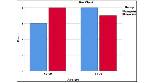
Figure 10: Association Between Age Group and Implant Type
|
Sex |
Long PFN (n=15) |
Short PFN (n=15) |
Total (n=30) |
|
Female |
8 |
7 |
15 |
|
Male |
7 |
8 |
15 |
|
Total |
15 |
15 |
30 |
|
p-value (Chi-square): 0.715 |
|||
Table 2: Association Between Sex and Type of Implant
Interpretation:
There was no statistically significant association between sex distribution and implant type (p = 0.715). Both groups were comparable in gender composition.
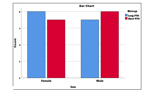
Figure 11: Association Between Sex and Type of Implant
|
Occupation |
Long PFN (n=15) |
Short PFN (n=15) |
Total (n=30) |
|
Farmer |
3 |
3 |
6 |
|
Housewife |
3 |
3 |
6 |
|
Labour |
3 |
3 |
6 |
|
Office |
3 |
3 |
6 |
|
Retired |
3 |
3 |
6 |
|
Total |
15 |
15 |
30 |
|
p-value (Chi-square): 1.000 |
|||
Table 3: Association Between Occupation and Type of Implant
Interpretation:
No significant association was found between occupation category and implant group (p = 1.000), indicating equal occupational distribution across groups.
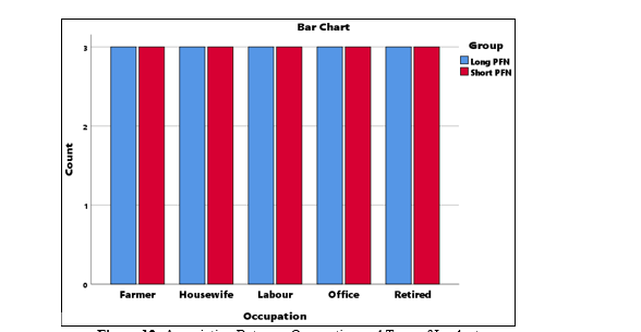
Figure 12: Association Between Occupation and Type of Implant
|
SES |
Long PFN (n=15) |
Short PFN (n=15) |
Total (n=30) |
|
High |
3 |
3 |
6 |
|
Low |
6 |
6 |
12 |
|
Middle |
6 |
6 |
12 |
|
Total |
15 |
15 |
30 |
|
p-value (Chi-square): |
|||
Table 4: Association Between Socioeconomic Status (SES) and Type of Implant
Interpretation:
SES distribution did not differ significantly between groups (p = 1.000). Both implants were used similarly across socioeconomic classes.
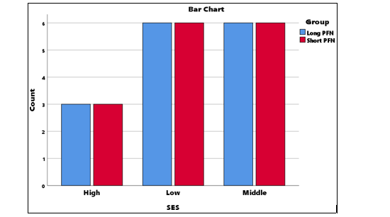
Figure 13: Association Between Socioeconomic Status (SES) and Type of Implant
|
Side |
Long PFN (n=15) |
Short PFN (n=15) |
Total (n=30) |
|
Left |
8 |
7 |
15 |
|
Right |
7 |
8 |
15 |
|
Total |
15 |
15 |
30 |
|
p-value (Chi-square): 0.715 |
|||
Table 5: Association Between Side of Injury and Type of Implant
Interpretation:
No significant association was seen between the side of fracture and implant used (p = 0.715). Both groups had an almost identical left–right distribution.
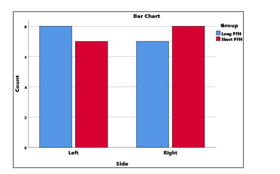
Figure 14: Association Between Side of Injury and Type of Implant
|
Mode of Injury |
Long PFN (n=15) |
Short PFN (n=15) |
Total (n=30) |
|
RTA |
5 |
0 |
5 |
|
Slip & Fall |
10 |
15 |
25 |
|
Total |
15 |
15 |
30 |
|
p-value (Chi-square): 0.014 |
|||
Table 6: Association Between Mode of Injury and Type of Implant
Interpretation:
A statistically significant association was observed between mode of injury and implant type (p = 0.014). RTA cases occurred exclusively in the Long PFN group, while Slip & Fall was more common in Short PFN.
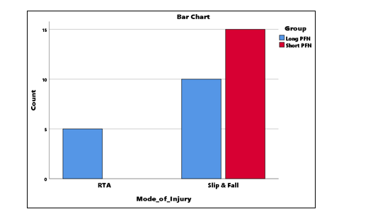
Figure 15: Association Between Mode of Injury and Type of Implant
|
Comorbidity |
Long PFN (n=15) |
Short PFN (n=15) |
Total (n=30) |
|
Both |
3 |
3 |
6 |
|
DM |
3 |
3 |
6 |
|
HTN |
3 |
3 |
6 |
|
None |
6 |
6 |
12 |
|
Total |
15 |
15 |
30 |
|
p-value (Chi-square): 1.000 |
|||
Table 7: Association Between Comorbidity and Type of Implant
Interpretation:
There was no statistically significant difference in comorbidity distribution between Short PFN and Long PFN groups (p = 1.000), showing that both groups were clinically comparable at baseline.
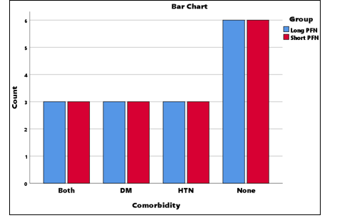
Figure 16: Association Between Comorbidity and Type of Implant
|
Evans Classification |
Long PFN (n=15) |
Short PFN (n=15) |
Total (n=30) |
p-value (Fisher’s Exact) |
|
Stable fracture |
9 |
11 |
20 |
0.842 |
|
Unstable fracture |
4 |
3 |
7 |
|
|
Reverse obliquity |
2 |
1 |
3 |
|
|
Total |
15 |
15 |
30 |
Table 8: Distribution of Fracture Patterns According to Evans Classification
There was no statistically significant association between Evans fracture classification and implant type according to Fisher’s Exact test (p = 0.842). Stable fractures were the predominant fracture pattern in both groups.
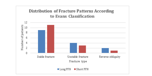
Figure 17: Distribution of Fracture Patterns According to Evans Classification
|
Variable |
Long PFN (Mean ± SD) |
Short PFN (Mean ± SD) |
t-value |
p-value |
95% CI of Difference |
|
Age (years) |
64.13 ± 8.85 |
59.27 ± 8.31 |
1.553 |
0.132 |
–1.55 to 11.28 |
|
Statistics |
— |
— |
Table 9: Comparison of Age Between Groups
Interpretation:
No significant age difference was noted between the groups, indicating both groups were comparable at baseline.
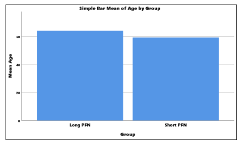
Figure 18: Comparison of Age Between Groups
|
Variable |
Long PFN (Mean ± SD) |
Short PFN (Mean ± SD) |
t-value |
p-value |
95% CI |
|
Operative Time (min) |
56.00 ± 1.69 |
51.40 ± 2.10 |
6.613 |
<0.001* |
3.17 to 6.03 |
|
Statistics |
— |
— |
Table 10: Comparison of Operative Time Between Groups
Interpretation:
PFN. Long PFN required significantly more operative time than Short PFN.
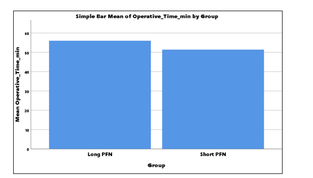
Figure 19: Comparison of Operative Time Between Groups
|
Variable |
Long PFN (Mean ± SD) |
Short PFN (Mean ± SD) |
t-value |
p-value |
95% CI |
|
Blood Loss (ml) |
100.80 ± 2.37 |
93.27 ± 2.66 |
8.198 |
<0.001* |
5.65 to 9.41 |
|
Statistics |
— |
— |
|
Table 11: Comparison of Blood Loss Between Groups
Interpretation:
Blood loss was significantly higher in the Long PFN group.
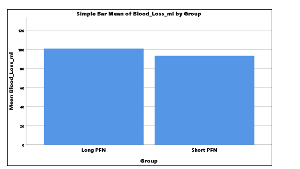
Figure 20: Comparison of Blood Loss Between Groups
|
Variable |
Long PFN (Mean ± SD) |
Short PFN (Mean ± SD) |
t-value |
p-value |
95% CI |
|
Union Time (weeks) |
16.47 ± 0.52 |
14.73 ± 0.59 |
8.532 |
<0.001* |
1.32 to 2.15 |
|
Statistics |
— |
— |
Table 12: Comparison of Union Time Between Groups
Interpretation:
Union occurred significantly earlier with Short PFN.
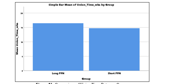
Figure 21: Comparison of Union Time Between Groups
|
Follow-up |
Long PFN (Mean ± SD) |
Short PFN (Mean ± SD) |
t-value |
p-value |
95% CI of Difference |
|
4 weeks |
84.47 ± 2.07 |
74.27 ± 2.22 |
13.031 |
<0.001* |
8.60 to 11.80 |
|
6 weeks |
88.47 ± 2.07 |
78.27 ± 2.22 |
13.031 |
<0.001* |
8.59 to 11.80 |
|
12 weeks |
90.47 ± 2.07 |
81.27 ± 2.22 |
11.753 |
<0.001* |
7.60 to 10.80 |
|
24 weeks |
92.47 ± 2.07 |
84.27 ± 2.22 |
10.476 |
<0.001* |
6.60 to 9.80 |
Table 13: Comparison of Functional Outcome (HHS) at Different Follow-ups
Functional outcomes, as assessed by Harris Hip Score, were significantly higher in the Long PFN group at all follow-up intervals (4, 6, 12, and 24 weeks; p < 0.001 for all comparisons). The results demonstrate a consistent and sustained functional advantage of Long PFN over Short PFN throughout the postoperative recovery period.
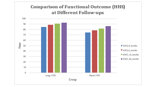
Figure 22: Comparison of Functional Outcome (HHS) at Different Follow-ups
|
Follow-up |
Callus Formation |
Long PFN (n=15) |
Short PFN (n=15) |
Total (n=30) |
p-value (Fisher’s Exact) |
|
|
|
|
|
|
|
|
4 weeks |
Absent |
5 |
8 |
13 |
0.472 |
|
Minimal |
8 |
6 |
14 |
||
|
Moderate |
2 |
1 |
3 |
||
|
Total |
15 |
15 |
30 |
||
|
6 weeks |
Minimal |
4 |
7 |
11 |
0.533 |
|
Moderate |
9 |
6 |
15 |
||
|
Good |
2 |
2 |
4 |
||
|
Total |
15 |
15 |
30 |
||
|
12 weeks |
Moderate |
3 |
6 |
9 |
0.421 |
|
Good |
10 |
7 |
17 |
||
|
Complete |
2 |
2 |
4 |
||
|
Total |
15 |
15 |
30 |
||
|
24 weeks |
Good |
2 |
4 |
6 |
0.648 |
|
Complete |
13 |
11 |
24 |
||
|
Total |
15 |
15 |
30 |
|
Table 14: Progression of Callus Formation
There was no statistically significant difference in callus formation between the two groups at any follow-up (all p > 0.05). Both groups showed progressive healing, with comparable outcomes by 24 weeks.
|
HHS Outcome |
Long PFN (n=15) |
Short PFN (n=15) |
Total (n=30) |
p-value |
|
Good |
1 |
13 |
14 |
<0.001* |
|
Excellent |
14 |
2 |
16 |
|
|
Total |
15 |
15 |
30 |
Table 15: Association Between Final Functional Outcome (HHS at 24 Weeks) and Implant Type
There was a highly significant association between implant type and final functional outcome (p < 0.001). A significantly higher proportion of patients in the Long PFN group achieved excellent outcomes, whereas most patients in the Short PFN group had only good outcomes, indicating superior functional results with Long PFN.
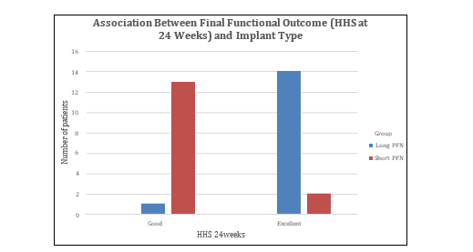
Figure 23: Association Between Final Functional Outcome (HHS at 24 Weeks) and Implant Type
|
Complication |
Long PFN (n=15) |
Short PFN (n=15) |
Total (n=30) |
|
Anterior thigh pain |
0 |
2 |
2 |
|
Implant failure |
0 |
1 |
1 |
|
None |
13 |
10 |
23 |
|
Persistent hip pain |
1 |
1 |
2 |
|
Superficial infection |
1 |
1 |
2 |
|
Total |
15 |
15 |
30 |
|
Chi-square (χ²) value 3.391, p = 0.495 |
|||
Table 16: Association Between Complications and Implant Type
There was no statistically significant association between type of implant and the occurrence of post-operative complications (p = 0.495). Although minor complications were slightly more common in the Short PFN group, the difference was not statistically meaningful. Both implants demonstrated a comparable safety profile.
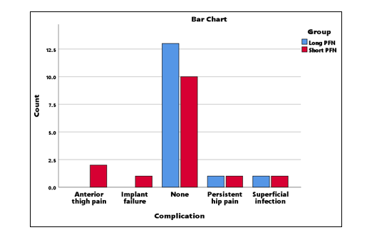
Figure 24: Association Between Complications and Implant Type
|
Paired Comparison |
Mean at Early Follow-up |
Mean at 24 Weeks |
Mean Difference |
t-value |
p-value |
95% CI of Difference |
|
HHS 4 weeks vs HHS 24 weeks |
79.37 ± 5.60 |
88.37 ± 4.67 |
−9.00 |
−48.466 |
<0.001* |
−9.38 to −8.62 |
|
HHS 6 weeks vs HHS 24 weeks |
83.37 ± 5.60 |
88.37 ± 4.67 |
−5.00 |
−26.926 |
<0.001* |
−5.38 to −4.62 |
|
HHS 12 weeks vs HHS 24 weeks |
85.87 ± 5.13 |
88.37 ± 4.67 |
−2.50 |
−26.926 |
<0.001* |
−2.69 to −2.31 |
Table 17: Paired Comparison of Functional Outcome (HHS) Across Follow-ups
There was a statistically significant improvement in functional outcome scores from early follow-up (4, 6, and 12 weeks) to final follow-up at 24 weeks (all p < 0.001). This indicates progressive recovery and improvement in hip function over time.
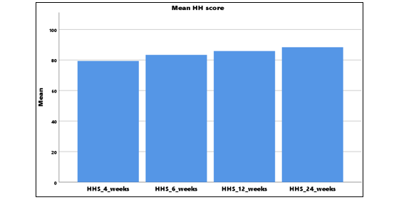
Figure 25: Mean HHS Across Follow-ups
Discussion
The aim of the present study was to compare the clinical, operative, radiological, and functional outcomes of Short Proximal Femoral Nail and Long Proximal Femoral Nail in the management of intertrochanteric fractures. The study was designed to evaluate whether implant length influences important treatment parameters such as operative time, intraoperative blood loss, fracture union time, callus formation, Harris Hip Score, final functional outcome, and postoperative complications. Intertrochanteric fractures are common proximal femoral fractures, especially in elderly individuals, and are associated with pain, immobility, loss of independence, morbidity, and increased healthcare burden. Stable internal fixation and early mobilization are essential for reducing complications and restoring functional ability. Proximal femoral nailing is widely used because it provides intramedullary fixation with good biomechanical stability; however, the choice between Short PFN and Long PFN remains clinically relevant. Short PFN may offer advantages such as shorter operative duration, reduced blood loss, easier insertion, and less surgical morbidity, whereas Long PFN may provide greater femoral support, improved load sharing, and better protection in unstable or high-energy fracture patterns. The significance of this study lies in its attempt to provide comparative evidence regarding these two commonly used implants in intertrochanteric fracture fixation. By assessing both perioperative and postoperative outcomes, the study helps identify the relative benefits and limitations of each implant. The findings may assist orthopaedic surgeons in selecting the most appropriate implant based on patient age, fracture pattern, mechanism of injury, bone quality, operative risk, and expected functional demand. The study is also significant because it emphasizes that implant selection should not be based only on surgeon preference, but should be individualized to achieve safe fixation, early union, reduced complications, and optimal hip function. Thus, this study contributes clinically useful information for improving decision-making and outcomes in patients with intertrochanteric fractures.
Age Distribution Age distribution
in the present study showed no statistically significant association with implant type, indicating baseline comparability between the Long PFN and Short PFN groups. In the 45–60 years age group, 6 patients were treated with Long PFN and 8 patients with Short PFN, while in the 61–75 years age group, 8 patients were treated with Long PFN and 7 patients with Short PFN. The Chi-square test showed χ² = 0.318 with p = 0.573, confirming that age group did not significantly influence implant selection. Mean age comparison also showed no significant difference, with mean age of 64.13 ± 8.85 years in the Long PFN group and 59.27 ± 8.31 years in the Short PFN group, with t = 1.553, p = 0.132, and 95% CI from −1.55 to 11.28. This indicates that both groups were comparable in terms of age, allowing valid comparison of operative and functional outcomes. Kleweno et al. studied patients older than 65 years and reported similar treatment failure rates between short and long cephalomedullary nails, with overall failure of 5.4%, 5.9% in the short nail group and 5.0% in the long nail group (p = 0.70) [68]. Shin et al. studied elderly patients with mean age of 77 years and found no significant difference in Harris Hip Score, walking ability, or reoperation rates between ZNN and PFNA II groups [71]. Schneider et al. included 264 patients with mean age of 78.8 years and reported fracture healing in 99.6% within three months after PFNA fixation [76]. Thus, age comparability in the present study strengthens the interpretation that observed differences between Long PFN and Short PFN were more likely related to implant-related and surgical factors rather than age-related bias.
Sex Distribution
Sex distribution was comparable between the two implant groups in the present study. Among females, 8 patients were treated with Long PFN and 7 patients with Short PFN, while among males, 7 patients were treated with Long PFN and 8 patients with Short PFN. The Chi-square test showed p = 0.715, indicating no statistically significant association between sex and type of implant. This finding suggests that both groups were well matched regarding gender composition, and sex was unlikely to confound the comparison of operative time, blood loss, union time, functional outcome, or complications. This is relevant because intertrochanteric fractures are often influenced by sex-related differences in osteoporosis, bone quality, fall risk, and postoperative recovery potential. Shannon et al. conducted a randomized prospective study involving 220 patients with pertrochanteric fractures and reported comparable baseline demographics between short nail and long nail cohorts before comparing clinical and functional outcomes [73]. Similarly, Hantouly et al., in a meta-analysis of six randomized controlled trials comprising 620 patients, compared short and long nails across balanced study groups and found that short nails reduced operative time and blood loss, while functional outcomes and complications remained comparable [75]. Zhang et al. also analyzed seven randomized studies involving 658 patients and found no significant differences between short and long cephalomedullary nails in Harris Hip Score, complication rates, reoperation rates, peri-implant fractures, hospital stay, or one-year mortality [77]. In the present study, the balanced male–female distribution supports reliable interpretation of implant-related outcomes. Therefore, the observed superiority of Short PFN in operative parameters and Long PFN in functional outcomes cannot be attributed to unequal sex distribution.
Occupation
Occupation was equally distributed between the Long PFN and Short PFN groups in the present study. Farmers, housewives, labourers, office workers, and retired individuals were represented equally, with 3 patients in each occupational category in both implant groups. The Chi-square test showed p = 1.000, confirming no statistically significant association between occupation and implant type. This indicates complete occupational comparability between the groups. Occupational background is important in intertrochanteric fracture studies because pre-injury activity level, physical workload, mobility demand, and rehabilitation expectations may affect postoperative recovery and functional outcome. Labourers and farmers may require higher functional restoration for return to routine activities, whereas retired or less physically active patients may have different rehabilitation goals. Rahman et al. conducted a prospective randomized comparative study of 30 patients with intertrochanteric fractures divided equally into short PFN and long PFN groups and assessed functional outcome using Harris Hip Score, reporting comparable functional scores of 81.0 ± 11.62 in the short PFN group and 80.3 ± 10.83 in the long PFN group [74]. Shannon et al. also emphasized comparable baseline characteristics in short and long nail cohorts before reporting equivalent functional outcomes [73]. Kodirov et al. highlighted that early mobilization, physiotherapy, osteoporosis management, and multidisciplinary care are essential for improving functional recovery and quality of life after proximal femoral fractures [80]. In the present study, equal occupational distribution eliminates occupational activity level as a major bias. Therefore, the significantly higher Harris Hip Score in the Long PFN group and shorter operative time with Short PFN can be interpreted as implant-related findings rather than consequences of occupational imbalance.
Socioeconomic Status
Socioeconomic status was not significantly associated with implant type in the present study. High socioeconomic status was present in 3 patients each in the Long PFN and Short PFN groups, while low socioeconomic status was present in 6 patients each and middle socioeconomic status in 6 patients each. The Chi-square test showed p = 1.000, indicating identical socioeconomic distribution between both groups. This finding is important because socioeconomic status may influence time to hospital presentation, nutritional status, access to rehabilitation, compliance with follow¬up, affordability of investigations, and general postoperative care. Equal distribution of socioeconomic status ensures that outcome comparison between Long PFN and Short PFN was not biased by differences in social or economic background. Kodirov et al. emphasized that prompt hospitalization, early diagnosis using radiography and computed tomography, optimized anesthesia, infection control, thromboprophylaxis, early rehabilitation, and osteoporosis management are essential components of successful proximal femoral fracture care [80]. Yu et al. demonstrated that PFNA fixation in super-elderly patients produced faster pain relief, shorter fracture healing time, better HHS and SF-36 scores, fewer complications, and higher one-year survival compared with conservative treatment (p < 0.05), emphasizing the importance of appropriate treatment access and postoperative care [79]. In the present study, socioeconomic comparability supports fair comparison of both implants. Therefore, the significantly shorter operative time and lower blood loss in the Short PFN group, and the significantly better functional outcome in the Long PFN group, were unlikely to be due to differences in socioeconomic status. This strengthens the internal validity of the study findings.
Side of Injury
The side of injury showed no statistically significant association with implant type in the present study. Left-sided fractures were seen in 8 patients in the Long PFN group and 7 patients in the Short PFN group, while right-sided fractures were seen in 7 patients in the Long PFN group and 8 patients in the Short PFN group. The Chi-square test showed p = 0.715, indicating that fracture laterality was almost equally distributed between both implant groups. This comparability is relevant because surgical positioning, surgeon handling, limb dominance, rehabilitation comfort, and gait recovery may be influenced by the side involved, although laterality itself is not usually a major determinant of union or implant failure. Shannon et al. randomized patients with pertrochanteric fractures into short nail and long nail cohorts and reported comparable baseline characteristics before comparing functional and clinical outcomes [73]. Kleweno et al. compared short and long cephalomedullary nails in elderly patients and found no significant difference in treatment failure, with 5.9% failure in the short nail group and 5.0% in the long nail group (p = 0.70) [68]. Vaughn et al. also reported no statistically significant difference in catastrophic failure or hardware complications between short and long nails, although secondary femoral fractures occurred in 3.33% of short nail cases and none of the long nail cases, approaching significance (p = 0.054) [69]. In the present study, equal left–right distribution supports unbiased interpretation of outcomes. Therefore, differences in operative time, blood loss, union time, and Harris Hip Score were unlikely to be influenced by fracture side.
Mode of Injury
Mode of injury showed a statistically significant association with implant type in the present study. Road traffic accident was present in 5 patients, all of whom were treated with Long PFN, whereas slip and fall injury was present in 25 patients, including 10 patients in the Long PFN group and all 15 patients in the Short PFN group. The Chi-square test showed p = 0.014, indicating a significant difference in injury mechanism between the two groups. This suggests that Long PFN was preferred in patients with high-energy trauma, while Short PFN was more commonly used for low-energy slip and fall injuries. This finding is clinically important because high-energy injuries may be associated with greater fracture instability, comminution, subtrochanteric extension, and higher mechanical demand on fixation. Hwang et al. described technical problems related to mismatch between femoral bowing and PFN/PFNA, noting that such mismatch may hinder nail insertion, affect alignment, increase operative time, and compromise stabilization [64]. Hoffmann et al. demonstrated that implant design can influence fixation stability, with interlocking lag screw nails showing nearly 40% higher initial stiffness, 13% higher failure load, 18% more cycles before failure, and up to 84% lower femoral head rotation and varus collapse compared with non-interlocking designs [67]. Shannon et al. reported that short nails could be effective even with up to 3 cm subtrochanteric fracture extension, without increased cutout, infection, or peri-implant fracture [73]. In the present study, selection of Long PFN for all RTA cases suggests a possible preference for longer fixation in more demanding fracture situations.
Comorbidity
Comorbidity distribution was identical between the Long PFN and Short PFN groups in the present study. Diabetes mellitus was present in 3 patients in each group, hypertension in 3 patients in each group, both diabetes mellitus and hypertension in 3 patients in each group, and no comorbidity in 6 patients in each group. The Chi-square test showed p = 1.000, confirming no statistically significant difference in baseline comorbidity status. This finding is important because diabetes mellitus may affect wound healing, infection risk, and bone union, while hypertension and other systemic conditions may increase perioperative risk and delay rehabilitation. Equal comorbidity distribution therefore strengthens the comparison between implants. Kleweno et al. studied elderly intertrochanteric fracture patients and reported one-year mortality of 25% among 698 patients, reflecting the systemic vulnerability of this fracture population [68]. Shin et al. studied elderly patients with mean age of 77 years and found that both ZNN and PFNA II produced similar Harris Hip Score, walking ability, and reoperation rates, although ZNN had longer operation and fluoroscopy times and more lateral hip pain [71]. Yu et al. found that PFNA fixation in patients aged more than 90 years produced faster pain relief, shorter healing time, better functional scores, lower complications, and higher one-year survival than conservative management (p < 0.05) [79]. In the present study, equal distribution of diabetes mellitus, hypertension, combined comorbidity, and absence of comorbidity suggests that the observed differences in operative time, blood loss, union, and functional outcomes were not due to unequal medical risk. Therefore, comorbidity did not appear to confound implant comparison.
Operative Time
Operative time was significantly shorter in the Short PFN group compared with the Long PFN group. The mean operative time was 56.00 ± 1.69 minutes in the Long PFN group and 51.40 ± 2.10 minutes in the Short PFN group, with t = 6.613, p < 0.001, and 95% CI from 3.17 to 6.03. This indicates that Short PFN required significantly less surgical time. The shorter operative duration may be due to easier nail insertion, shorter implant length, reduced canal instrumentation, less technical difficulty during distal locking, and reduced intraoperative manipulation. Rahman et al. reported similar findings in a prospective randomized study of 30 patients, where mean surgery duration was significantly shorter with short PFN at 68.6 ± 6.62 minutes compared with 78.6 ± 7.35 minutes for long PFN [74]. Shannon et al. also found shorter operative time in the short nail group, while functional outcomes, blood loss, tip-apex distance, implant failure, deep infection, and peri-implant fracture were not significantly different between short and long nails [73]. Hantouly et al., in a meta-analysis of six randomized controlled trials involving 620 patients, concluded that short nails significantly reduced operative time compared with long nails [75]. Zhang et al. also reported significantly reduced operative time with short cephalomedullary nails in seven randomized studies comprising 658 patients [77]. Hwang et al. emphasized that implant-femur mismatch may increase technical difficulty and operative time during PFN/PFNA insertion [64]. Therefore, the present study is consistent with previous evidence and supports Short PFN as a surgically efficient implant with reduced operative duration.
Blood Loss
Blood loss was significantly lower in the Short PFN group compared with the Long PFN group in the present study. The mean blood loss was 100.80 ± 2.37 ml in the Long PFN group and 93.27 ± 2.66 ml in the Short PFN group, with t = 8.198, p < 0.001, and 95% CI from 5.65 to 9.41. This result indicates that Long PFN was associated with greater intraoperative blood loss. The difference may be explained by longer operative time, increased intramedullary manipulation, longer implant passage, greater technical requirements, and distal locking demands. Hantouly et al. reported similar findings in a systematic review and meta-analysis of six randomized controlled trials including 620 patients, where short proximal femoral nails were associated with significantly lower intraoperative blood loss compared with long nails [75]. Zhang et al. also found that short cephalomedullary nails were associated with significantly reduced intraoperative blood loss compared with long nails in a meta-analysis of seven studies involving 658 patients [77]. Prabhat et al. demonstrated that PFNA 2 was associated with significantly lower intraoperative blood loss compared with PFN, likely because of easier insertion and helical blade design, while functional and radiological outcomes were comparable [78]. Kodirov et al. emphasized that minimally invasive surgical approaches in proximal femoral fracture management reduce operative trauma, blood loss, and hospital stay [80]. In the present study, Short PFN showed a clear perioperative advantage in reducing blood loss. However, despite higher blood loss, Long PFN demonstrated superior Harris Hip Score at all follow-up intervals, showing that intraoperative efficiency and final functional outcome may differ between implant groups.
Union Time
Union time was significantly earlier in the Short PFN group compared with the Long PFN group. The mean union time was 16.47 ± 0.52 weeks in the Long PFN group and 14.73 ± 0.59 weeks in the Short PFN group, with t = 8.532, p < 0.001, and 95% CI from 1.32 to 2.15. This suggests faster fracture union in patients treated with Short PFN. However, this finding should be interpreted with caution because mode of injury differed significantly between groups. All road traffic accident cases were managed with Long PFN, while all Short PFN patients had slip and fall injury. High-energy trauma may be associated with greater comminution and biological disruption, which can delay union. Rahman et al. reported comparable union time between short and long PFN groups, approximately 15.7 weeks, despite shorter operative time in the short PFN group [74]. Schneider et al. reported excellent radiological healing with PFNA fixation, with fracture healing achieved in 99.6% of 264 patients within three months [76]. Yu et al. found that PFNA fixation in super-elderly patients resulted in shorter fracture healing time compared with conservative skeletal traction [79]. Hoffmann et al. demonstrated that improved implant stability can reduce femoral head rotation and varus collapse, supporting the importance of mechanical stability during fracture healing [67]. In the present study, Short PFN was associated with earlier mean union, but both implants achieved satisfactory healing. The difference in union time may reflect both implant characteristics and injury severity distribution.
Callus Formation
Callus formation progressed in both groups over time, with no statistically significant difference between Long PFN and Short PFN at any follow-up. At 4 weeks, callus was absent in 5 Long PFN and 8 Short PFN patients, minimal in 8 Long PFN and 6 Short PFN patients, and moderate in 2 Long PFN and 1 Short PFN patient, with p = 0.472. At 6 weeks, minimal callus was seen in 4 Long PFN and 7 Short PFN patients, moderate callus in 9 Long PFN and 6 Short PFN patients, and good callus in 2 patients each, with p = 0.533. At 12 weeks, moderate callus was seen in 3 Long PFN and 6 Short PFN patients, good callus in 10 Long PFN and 7 Short PFN patients, and complete callus in 2 patients each, with p = 0.421. At 24 weeks, complete callus formation was noted in 13 Long PFN and 11 Short PFN patients, with p = 0.648. These findings indicate comparable radiological progression between both implants. Rahman et al. reported comparable radiological outcomes between short and long PFN groups, with union time of approximately 15.7 weeks in both groups [74]. Schneider et al. reported fracture healing in 99.6% of patients treated with PFNA within three months, supporting the effectiveness of cephalomedullary fixation in proximal femoral fractures [76]. Hoskins et al. emphasized the importance of anatomical reduction, showing that cerclage wire improved displacement from 8.8 mm to 3.2 mm and improved reduction quality without increasing major complications [70]. Therefore, in the present study, comparable callus formation suggests that both Short PFN and Long PFN provided adequate biological and mechanical conditions for fracture healing.
Functional Outcome at Follow-up
Functional outcome assessed by Harris Hip Score was significantly better in the Long PFN group at all follow-up intervals. At 4 weeks, the mean HHS was 84.47 ± 2.07 in the Long PFN group and 74.27 ± 2.22 in the Short PFN group, with p < 0.001. At 6 weeks, mean HHS was 88.47 ± 2.07 in Long PFN and 78.27 ± 2.22 in Short PFN, with p < 0.001. At 12 weeks, mean HHS was 90.47 ± 2.07 in Long PFN and 81.27 ± 2.22 in Short PFN, with p < 0.001. At 24 weeks, mean HHS was 92.47 ± 2.07 in Long PFN and 84.27 ± 2.22 in Short PFN, with p < 0.001. This demonstrates a consistent functional advantage with Long PFN. In contrast, Rahman et al. reported similar functional outcomes, with mean HHS of 81.0 ± 11.62 in the short PFN group and 80.3 ± 10.83 in the long PFN group [74]. Shannon et al. found no significant difference in SF-36 scores and only clinically insignificant differences in HHS between short and long nail groups [73]. Hantouly et al. and Zhang et al. also reported no significant difference in Harris Hip Score between short and long nails [75,77]. Shin et al. found similar HHS between ZNN and PFNA II groups [71]. Therefore, the present study differs from several previous studies by demonstrating superior functional recovery with Long PFN. This may be related to better mechanical stability, fracture pattern selection, rehabilitation differences, or sample-specific characteristics.
Final Functional Outcome
Final functional outcome at 24 weeks showed a highly significant association with implant type. In the Long PFN group, 14 patients achieved excellent outcome and 1 patient achieved good outcome, whereas in the Short PFN group, only 2 patients achieved excellent outcome and 13 patients achieved good outcome. The association was statistically significant with p < 0.001. This indicates that excellent functional recovery was much more common among patients treated with Long PFN. Although Short PFN produced good outcomes in most patients, Long PFN showed clear superiority in the final functional category. Rahman et al. reported comparable mean Harris Hip Score between short PFN and long PFN groups, with scores of 81.0 ± 11.62 and 80.3 ± 10.83, respectively [74]. Hantouly et al., in a meta-analysis of six randomized trials involving 620 patients, found no significant difference between short and long nails in Harris Hip Score, complication rates, reoperation rates, hospital stay, or one-year mortality [75]. Zhang et al. also concluded that functional outcomes were comparable between short and long cephalomedullary nails in seven studies including 658 patients [77]. In contrast, the present study showed a distinct functional advantage with Long PFN. Hoffmann et al. demonstrated that improved nail design and stability can reduce femoral head rotation and varus collapse by up to 84%, supporting the role of mechanical stability in better function [67]. Therefore, the present findings suggest that Long PFN may provide better final functional restoration, possibly due to greater biomechanical support and longer femoral stabilization.
Progressive Improvement in Harris Hip Score
The present study showed statistically significant progressive improvement in Harris Hip Score from early follow-up to final follow-up at 24 weeks. Mean HHS improved from 79.37 ± 5.60 at 4 weeks to 88.37 ± 4.67 at 24 weeks, with mean difference of −9.00, t = −48.466, p < 0.001, and 95% CI from −9.38 to −8.62. From 6 weeks to 24 weeks, HHS improved from 83.37 ± 5.60 to 88.37 ± 4.67, with mean difference of −5.00, t = −26.926, p < 0.001, and 95% CI from −5.38 to −4.62. From 12 weeks to 24 weeks, HHS improved from 85.87 ± 5.13 to 88.37 ± 4.67, with mean difference of −2.50, t = −26.926, p < 0.001, and 95% CI from −2.69 to −2.31. This shows continuous recovery of hip function over time. Shin et al. reported that both ZNN and PFNA II achieved similar Harris Hip Score and walking ability in elderly intertrochanteric fracture patients [71]. Prabhat et al. found that both PFN and PFNA 2 achieved comparable functional outcomes, with most patients having excellent to good results [78]. Yu et al. showed significantly better HHS and SF-36 outcomes after PFNA compared with conservative management in super-elderly patients [79]. Kodirov et al. emphasized early mobilization, physiotherapy, osteoporosis management, and multidisciplinary care for improved functional recovery [80]. Therefore, progressive HHS improvement in the present study reflects successful fracture fixation, rehabilitation, pain reduction, and gradual restoration of mobility after PFN fixation.
Complications
Postoperative complications were not significantly associated with implant type in the present study. In the Long PFN group, 13 patients had no complications, 1 patient had persistent hip pain, and 1 patient had superficial infection. In the Short PFN group, 10 patients had no complications, 2 patients had anterior thigh pain, 1 patient had implant failure, 1 patient had persistent hip pain, and 1 patient had superficial infection. The Chi-square value was 3.391 with p = 0.495, indicating no statistically significant difference in complication rates between the groups. Although complications were numerically more frequent in the Short PFN group, the difference was not statistically meaningful. Kleweno et al. reported similar treatment failure rates between short and long cephalomedullary nails, with 5.9% failure in short nails and 5.0% in long nails (p = 0.70), and periprosthetic fractures in 2.7% after short nails and 1.5% after long nails (p = 0.35) [68]. Vaughn et al. also found no significant difference in catastrophic failure or hardware complications, with overall catastrophic failure of 3.1%, short nail failure of 5%, and long nail failure of 2.6% (p = 0.191) [69]. However, secondary femoral fractures occurred in 3.33% of short nail cases and none of the long nail cases, approaching significance (p = 0.054) [69]. Schneider et al. reported an overall reoperation rate of 6.8%, reduced from 9.0% to 2.3% with cement augmentation (p = 0.04) [76]. Thus, the present study supports comparable safety of both implants, although Short PFN showed slightly more implant-related symptoms.
Overall Comparison Between Short PFN
and Long PFN Overall comparison showed that Short PFN and Long PFN had different advantages in the management of intertrochanteric fractures. Short PFN was superior in operative parameters, with significantly shorter operative time of 51.40 ± 2.10 minutes compared with 56.00 ± 1.69 minutes for Long PFN (p < 0.001), and significantly lower blood loss of 93.27 ± 2.66 ml compared with 100.80 ± 2.37 ml for Long PFN (p < 0.001). Short PFN also had significantly earlier union, with mean union time of 14.73 ± 0.59 weeks compared with 16.47 ± 0.52 weeks for Long PFN (p < 0.001). However, Long PFN showed better functional outcome, with higher HHS at 4, 6, 12, and 24 weeks. At final follow-up, HHS was 92.47 ± 2.07 in the Long PFN group and 84.27 ± 2.22 in the Short PFN group (p < 0.001), and excellent outcome was seen in 14 Long PFN patients compared with 2 Short PFN patients (p < 0.001). Hantouly et al. and Zhang et al. found that short nails reduced operative time and blood loss, but showed no significant difference in HHS, complications, reoperations, or mortality [75,77]. Rahman et al. reported shorter surgery with short PFN but similar HHS between short and long PFN groups [74]. In contrast, the present study found superior functional outcomes with Long PFN. Therefore, Short PFN may be preferred when reduced surgical duration and blood loss are priorities, while Long PFN may be considered when superior functional recovery and greater mechanical support are desired.
Strength of the Study
The strength of the present study lies in its direct comparison between Short Proximal Femoral Nail and Long Proximal Femoral Nail in the management of intertrochanteric fractures using multiple clinically relevant parameters. The study evaluated demographic variables, baseline comparability, operative characteristics, radiological healing, functional outcome, and postoperative complications, allowing a comprehensive assessment of both implants. The two groups were comparable in most baseline characteristics, including sex, occupation, socioeconomic status, side of injury, and comorbidity distribution, thereby reducing the possibility of major confounding from these variables. The use of objective operative parameters such as operative time and blood loss provided measurable intraoperative comparison between the two implants. Radiological healing was assessed through union time and serial callus formation at 4, 6, 12, and 24 weeks, enabling evaluation of fracture healing progression over time. Functional outcome was assessed using Harris Hip Score at multiple follow¬up intervals, which allowed not only final outcome comparison but also observation of progressive functional recovery. The study also included analysis of final functional grading and postoperative complications, making the evaluation clinically meaningful. Another important strength was the equal sample distribution in most tables, with 15 patients in each implant group, which allowed balanced comparison. The inclusion of both surgical efficiency outcomes and patient-centred functional outcomes provided a broader understanding of implant performance. The finding that Short PFN was associated with shorter operative time, lower blood loss, and earlier union, while Long PFN showed better functional outcome, gives practical value for clinical decision-making. Therefore, the study provides useful evidence for individualized implant selection in intertrochanteric fracture management.
Implications
The findings of the present study have important clinical implications for the surgical management of intertrochanteric fractures. Short PFN demonstrated advantages in operative efficiency, as it was associated with significantly shorter operative time, lower intraoperative blood loss, and earlier union compared with Long PFN. These findings suggest that Short PFN may be particularly useful in patients where minimizing operative duration and surgical blood loss is important, such as elderly patients, medically fragile individuals, or patients with limited physiological reserve. On the other hand, Long PFN showed significantly better Harris Hip Score at all follow-up intervals and a higher proportion of excellent final functional outcomes, suggesting that it may provide better functional recovery in selected cases. This implies that Long PFN may be preferred where greater mechanical support, longer femoral stabilization, and improved postoperative function are desired. The study also showed that most baseline demographic and clinical variables were comparable between the two groups, supporting the interpretation that observed differences were related mainly to implant performance and surgical factors. The absence of a significant difference in complication rates suggests that both implants can be considered safe when appropriately selected and properly inserted. The significant association between mode of injury and implant type indicates that fracture mechanism may influence implant selection, with Long PFN being more frequently used in high-energy trauma cases. Thus, the study supports individualized decision-making rather than universal preference for one implant. Implant selection should consider patient age, fracture pattern, injury mechanism, bone quality, surgical risk, and expected functional demand. These implications can guide orthopaedic surgeons in balancing operative simplicity with long¬term functional benefit.
Recommendations
Based on the findings of the present study, both Short PFN and Long PFN can be recommended as effective treatment options for intertrochanteric fractures, provided that implant selection is individualized according to patient and fracture characteristics. Short PFN may be recommended for patients with relatively stable fracture patterns, low-energy slip and fall injuries, and those in whom reduced operative time and lower blood loss are desirable. Its shorter operative duration and lesser blood loss make it suitable for elderly or medically compromised patients who may not tolerate prolonged surgery. Long PFN may be recommended in patients with high-energy trauma, suspected unstable fracture configuration, subtrochanteric extension, poor bone quality, or situations where greater mechanical stability and longer femoral support are required. Since Long PFN showed better Harris Hip Score and a higher proportion of excellent outcomes at final follow-up, it may be preferred in patients with higher functional expectations or fracture patterns requiring stronger biomechanical support. Careful preoperative planning should be performed in all patients, including proper radiological assessment of fracture type, femoral morphology, and implant suitability. Intraoperative technique should focus on accurate reduction, correct entry point, proper nail size selection, optimal lag screw or blade placement, and avoidance of varus malalignment. Early postoperative mobilization, structured physiotherapy, pain control, nutritional optimization, and comorbidity management should be emphasized to improve recovery. Regular radiological follow-up is recommended to assess callus formation, union progression, and implant position. Surgeons should not select implant length based only on routine preference; instead, implant choice should be based on fracture stability, patient profile, and functional goals. Larger comparative studies are recommended to develop clearer implant selection guidelines.
Limitations
The present study has certain limitations that should be considered while interpreting the findings. The sample size was relatively small, with most comparisons including 30 patients divided equally into 15 patients in each implant group. A small sample size may reduce the statistical power of the study, especially for detecting less common complications such as implant failure, peri-implant fracture, screw cut-out, deep infection, or reoperation. Another limitation is the discrepancy in the age distribution table, where the total number was reported as 29 instead of 30, which should be verified and corrected before final submission. The follow-up duration was limited to 24 weeks, which may be sufficient for early union and functional assessment but may not capture long-term complications, late implant failure, persistent pain, gait abnormality, or degenerative changes. The study also showed a significant difference in mode of injury between groups, with all road traffic accident cases managed by Long PFN and all Short PFN patients having slip and fall injury. This difference may have influenced union time, functional recovery, and implant selection, and therefore may act as a confounding factor. Fracture classification, fracture stability, degree of comminution, bone mineral density, reduction quality, tip-apex distance, and implant positioning were not included in the presented results, although these are important determinants of outcome in intertrochanteric fractures. Rehabilitation compliance and weight-bearing protocol were also not separately analyzed. The study was likely conducted in a single centre, which may limit generalizability. Therefore, while the findings are clinically useful, they should be interpreted with caution and validated through larger studies with longer follow-up.
Future Aspects
Future research should focus on larger, multicentre comparative studies with adequate sample size to confirm the relative advantages of Short PFN and Long PFN in intertrochanteric fractures. Larger sample sizes would allow better assessment of uncommon but clinically important complications such as implant failure, screw cut-out, peri-implant fracture, non-union, malunion, deep infection, and need for revision surgery. Future studies should include longer follow-up periods beyond 24 weeks to evaluate sustained functional recovery, late complications, implant-related pain, gait pattern, return to pre-injury activity, and quality of life. Detailed fracture classification using standard systems should be incorporated to determine whether implant length has different effects in stable, unstable, comminuted, reverse oblique, or subtrochanteric extension patterns. Additional radiological parameters such as quality of reduction, neck-shaft angle, tip-apex distance, implant position, femoral bowing, and cortical contact should be analyzed to understand the relationship between surgical technique and outcomes. Bone quality assessment, including osteoporosis grading or bone mineral density evaluation, may help identify patients who benefit more from longer fixation. Future trials should also standardize rehabilitation protocols, weight-bearing schedules, physiotherapy intensity, and analgesic regimens to reduce postoperative variability. Patient-reported outcome measures, pain scores, walking ability, return to work, dependency status, and health-related quality of life should be included along with Harris Hip Score. Cost-effectiveness analysis may also be valuable, especially in resource-limited settings. Randomized controlled trials comparing Short PFN and Long PFN with stratification based on fracture stability and injury mechanism would provide stronger evidence. Future studies may ultimately help develop clear guidelines for selecting implant length in intertrochanteric fracture management.
Summary
1. The present study compared Short Proximal Femoral Nail and Long Proximal Femoral Nail in the management of intertrochanteric fractures, with assessment of demographic variables, operative parameters, radiological union, functional outcome, and complications.
2. Age group distribution was comparable between the two implant groups, as 6 patients aged 45–60 years and 8 patients aged 61–75 years were treated with Long PFN, while 8 patients aged 45–60 years and 7 patients aged 61–75 years were treated with Short PFN, with χ² = 0.318 and p = 0.573.
3. Mean age was slightly higher in the Long PFN group at 64.13 ± 8.85 years compared with 59.27 ± 8.31 years in the Short PFN group, but the difference was not statistically significant, with t = 1.553 and p = 0.132.
4. Sex distribution was similar between both groups, with 8 females and 7 males in the Long PFN group and 7 females and 8 males in the Short PFN group, with p = 0.715.
5. Occupation was equally distributed between the two implant groups, with farmer, housewife, labourer, office worker, and retired categories each having 3 patients in both groups, with p = 1.000.
6. Socioeconomic status showed no significant difference between groups, as high socioeconomic status was seen in 3 patients each, low socioeconomic status in 6 patients each, and middle socioeconomic status in 6 patients each in both implant groups, with p = 1.000.
7. Side of injury was almost equally distributed, with left-sided injury in 8 Long PFN and 7 Short PFN patients, and right-sided injury in 7 Long PFN and 8 Short PFN patients, with p = 0.715.
8. Mode of injury showed a statistically significant association with implant type, as road traffic accident was seen in 5 patients, all in the Long PFN group, while slip and fall was seen in 10 Long PFN and 15 Short PFN patients, with p = 0.014.
9. Comorbidity distribution was identical between groups, with diabetes mellitus, hypertension, both diabetes and hypertension, and no comorbidity showing equal distribution, with p = 1.000.
10. Operative time was significantly longer in the Long PFN group, with mean operative time of 56.00 ± 1.69 minutes compared with 51.40 ± 2.10 minutes in the Short PFN group, with t = 6.613 and p < 0.001.
11. Blood loss was significantly higher in the Long PFN group, with mean blood loss of 100.80 ± 2.37 ml compared with 93.27 ± 2.66 ml in the Short PFN group, with t = 8.198 and p < 0.001.
12. Union time was significantly shorter in the Short PFN group, with mean union time of 14.73 ± 0.59 weeks compared with 16.47 ± 0.52 weeks in the Long PFN group, with t = 8.532 and p < 0.001.
13. Harris Hip Score at 4 weeks was significantly higher in the Long PFN group at 84.47 ± 2.07 compared with 74.27 ± 2.22 in the Short PFN group, with p < 0.001.
14. Harris Hip Score at 6 weeks was significantly higher in the Long PFN group at 88.47 ± 2.07 compared with 78.27 ± 2.22 in the Short PFN group, with p < 0.001.
15. Harris Hip Score at 12 weeks was significantly higher in the Long PFN group at 90.47 ± 2.07 compared with 81.27 ± 2.22 in the Short PFN group, with p < 0.001.
16. Harris Hip Score at 24 weeks was significantly higher in the Long PFN group at 92.47 ± 2.07 compared with 84.27 ± 2.22 in the Short PFN group, with p < 0.001.
17. Callus formation showed progressive radiological healing in both groups, with no statistically significant difference at 4 weeks, 6 weeks, 12 weeks, or 24 weeks, with p-values of 0.472, 0.533, 0.421, and 0.648 respectively.
18. At 24 weeks, complete callus formation was observed in 13 patients in the Long PFN group and 11 patients in the Short PFN group, indicating satisfactory radiological healing in both groups.
19. Final functional outcome at 24 weeks showed a highly significant association with implant type, as excellent outcome was achieved in 14 Long PFN patients and 2 Short PFN patients, while good outcome was seen in 1 Long PFN and 13 Short PFN patients, with p < 0.001.
20. Postoperative complications were not significantly associated with implant type, with 13 patients in the Long PFN group and 10 patients in the Short PFN group having no complications, and overall χ² = 3.391 with p = 0.495.
21. Anterior thigh pain was seen only in the Short PFN group in 2 patients, implant failure was also seen only in the Short PFN group in 1 patient, while persistent hip pain and superficial infection were seen in 1 patient each in both groups.
22. Harris Hip Score showed significant progressive improvement over time, increasing from 79.37 ± 5.60 at 4 weeks to 88.37 ± 4.67 at 24 weeks, with mean difference of −9.00 and p < 0.001.
23. Harris Hip Score improved from 83.37 ± 5.60 at 6 weeks to 88.37 ± 4.67 at 24 weeks, with mean difference of −5.00 and p < 0.001.
24. Harris Hip Score improved from 85.87 ± 5.13 at 12 weeks to 88.37 ± 4.67 at 24 weeks, with mean difference of −2.50 and p < 0.001.
25. Overall, Short PFN showed advantages in operative time, blood loss, and union time, whereas Long PFN showed superior Harris Hip Score and better final functional outcome.
Conclusion
The present comparative study between Short Proximal Femoral Nail and Long Proximal Femoral Nail in the management of intertrochanteric fractures demonstrated that both implants are effective treatment options, but they differ in their operative profile, radiological union pattern, and functional outcome. The baseline characteristics of the two groups were largely comparable, which supports the validity of the outcome comparison. Age distribution did not show any significant association with implant type, with χ² = 0.318 and p = 0.573, and mean age was also statistically comparable between Long PFN and Short PFN groups, with values of 64.13 ± 8.85 years and 59.27 ± 8.31 years respectively, with p = 0.132. Similarly, sex distribution was comparable, with 8 females and 7 males in the Long PFN group and 7 females and 8 males in the Short PFN group, with p = 0.715. Occupation, socioeconomic status, side of injury, and comorbidity distribution were also not significantly different between the groups, with p-values of 1.000, 1.000, 0.715, and 1.000 respectively. These findings indicate that both implant groups were well matched in most baseline parameters. However, mode of injury showed a significant association with implant type, as all 5 road traffic accident cases were managed with Long PFN, while slip and fall injuries were seen in 10 Long PFN patients and all 15 Short PFN patients, with p = 0.014. This suggests that Long PFN was more frequently used in relatively high-energy injuries. In terms of operative parameters, Short PFN showed a clear advantage. Mean operative time was significantly shorter in the Short PFN group at 51.40 ± 2.10 minutes compared with 56.00 ± 1.69 minutes in the Long PFN group, with t = 6.613 and p < 0.001. Similarly, mean blood loss was significantly lower in the Short PFN group at 93.27 ± 2.66 ml compared with 100.80 ± 2.37 ml in the Long PFN group, with t = 8.198 and p < 0.001. These findings indicate that Short PFN is surgically less demanding and may be beneficial in patients where shorter surgery and reduced blood loss are important. Union time was also significantly earlier with Short PFN, with mean union time of 14.73 ± 0.59 weeks compared with 16.47 ± 0.52 weeks in the Long PFN group, with t = 8.532 and p < 0.001. However, serial callus formation showed no statistically significant difference between groups at 4 weeks, 6 weeks, 12 weeks, or 24 weeks, with p-values of 0.472, 0.533, 0.421, and 0.648 respectively. At 24 weeks, complete callus formation was seen in 13 Long PFN patients and 11 Short PFN patients, confirming satisfactory radiological healing with both implants. Functional outcome, assessed using Harris Hip Score, was significantly better in the Long PFN group at all follow-up intervals. At 4 weeks, HHS was 84.47 ± 2.07 in the Long PFN group compared with 74.27 ± 2.22 in the Short PFN group. At 6 weeks, HHS was 88.47 ± 2.07 versus 78.27 ± 2.22. At 12 weeks, HHS was 90.47 ± 2.07 versus 81.27 ± 2.22. At 24 weeks, HHS was 92.47 ± 2.07 in the Long PFN group compared with 84.27 ± 2.22 in the Short PFN group. All these differences were statistically significant, with p < 0.001. Final functional outcome also strongly favoured Long PFN, as 14 patients in the Long PFN group achieved excellent outcome compared with only 2 patients in the Short PFN group, while good outcome was seen in 1 Long PFN patient and 13 Short PFN patients, with p < 0.001. This indicates that Long PFN provided superior functional recovery despite longer operative time and greater blood loss. Overall functional recovery improved significantly over time in the total study population, with mean HHS improving from 79.37 ± 5.60 at 4 weeks to 88.37 ± 4.67 at 24 weeks, with p < 0.001. HHS also improved significantly from 83.37 ± 5.60 at 6 weeks and 85.87 ± 5.13 at 12 weeks to 88.37 ± 4.67 at 24 weeks, with p < 0.001 for both comparisons. Complication rates were not significantly different between groups, with p = 0.495. No complications were seen in 13 Long PFN patients and 10 Short PFN patients. Anterior thigh pain occurred in 2 Short PFN patients, implant failure in 1 Short PFN patient, persistent hip pain in 1 patient each, and superficial infection in 1 patient each. Therefore, both implants had a comparable safety profile, although minor complications were numerically more frequent in the Short PFN group. Based on these findings, Short PFN may be considered advantageous for reducing operative time, blood loss, and union time, while Long PFN may be preferred when superior functional outcome and excellent final hip function are the primary goals. Thus, implant selection should be individualized according to patient profile, fracture pattern, mode of injury, surgical risk, and expected functional demand.
References
- Zhang, H., Zhu, X., Pei, G., Zeng, X., Zhang, N., et al. (2015). A Meta-Analysis of Short Versus Long Intramedullary Fixation Devices for Trochanteric Fractures. Acta Orthop Traumatol Turc. 49(2):137-44.
- Cooper, C., Campion, G., & Melton III, L. (1992). Hip fractures in the elderly: a world-wide projection. Osteoporosis international, 2(6), 285-289.
- Kannus, P., Parkkari, J., Sievänen, H., Heinonen, A., Vuori, I., & Järvinen, M. (1996). Epidemiology of hip fractures. Bone, 18(1), S57-S63.
- Radford, P. J., Needoff, M., & Webb, J. K. (1993). A prospective randomised comparison of the dynamic hip screw and the gamma locking nail. The Journal of Bone & Joint Surgery British Volume, 75(5), 789-793.
- Banan, H., Al-Sabti, A., Jimulia, T., & Hart, A. J. (2002). The treatment of unstable, extracapsular hip fractures with the AO/ ASIF proximal femoral nail (PFN)—our first 60 cases. Injury,33(5), 401-405.
- Bojan, A. J., Beimel, C., Taglang, G., Collin, D., Ekholm, C., & Jönsson, A. (2013). Critical factors in cut-out complication after Gamma Nail treatment of proximal femoral fractures. BMC musculoskeletal disorders, 14(1), 1.
- Ahrengart, L., Törnkvist, H., Fornander, P., Thorngren, K. G., Pasanen, L., Wahlström, P., ... & Lindgren, U. (2002). A randomized study of the compression hip screw and Gamma nail in 426 fractures. Clinical Orthopaedics & Related Research, 401, 209-222.
- Parker, M. J., & Handoll, H. H. (2006). Intramedullary nails for extracapsular hip fractures in adults. Cochrane database of systematic reviews, (3).
- Xu, Y., Geng, D., Mao, H. Q., Zhu, X. S., Zhu, X. Z. (2023).A Comparative Study of Intramedullary and Extramedullary Fixation in Treatment of Unstable Intertrochanteric Fractures. Zhonghua Wai Ke Za Zhi. 41(10):742-5.
- Sadeghi, H, Tabrizi, A,, Aslani, H., Bazavar, M., Rajabi,M. T. (2017) Morphology of Proximal Femur in Iranian Population: Computed Tomography Analysis. Arch Bone Jt Surg. 5(6):382–6.
- Patel, S., Srivastava, A., Kumar, A., Nagar, M. (2015). Morphometric Study of Proximal Femur and its Clinical Application in North Indian Population. Indian J Orthop. 49(6):629–33.
- Unnanuntana, A., Toogood, P., Hart, D., Cooperman, D., & Grant, R. E. (2010). Evaluation of proximal femoral geometry using digital photographs. Journal of orthopaedic research, 28(11), 1399-1404.
- Isaac, B., Vettivel, S., Prasad, R., Soundarapandian, S., Chandi, G. (1997). A study of the Angle of Femoral Neck in Indians. Indian J Orthop.31(1):1–6.
- Siwach, R. (2018). Anthropometric study of proximal femur geometry and its clinical application. Annals of the National Academy of Medical Sciences (India), 54(04), 203-215.
- Saikia, K. C., Bhuyan, S. K., & Rongphar, R. (2008). Anthropometric study of the hip joint in northeastern region population with computed tomography scan. Indian journal of orthopaedics, 42(3), 260.
- Toogood, P. A., Skalak, A., & Cooperman, D. R. (2009). Proximal femoral anatomy in the normal human population. Clinical orthopaedics and related research, 467(4), 876-885.
- Bhardwaj, Y., Kumar, S., Lakhani, R., Saini, P., Kumar, S. (2015). A Morphometric Study of Proximal Femur and its Clinical Application. J Clin Diagn Res, 9(2):AC01–4.
- Standring, S., (2015). Gray’s Anatomy: The Anatomical Basis of Clinical Practice. 41st ed. London: Elsevier; 2015.
- Trueta, J., & Harrison, M. H. M. (1953). The normal vascular anatomy of the femoral head in adult man. Ludgate House.
- Gautam, V. K., Anand, S., Dhaon, B. K. (2010). Cross-Sectional Anatomy of the Proximal Femur in Indian Cadavers. Indian J Med Res, 131:702–9.
- Zhang, K., Tian, W., Wang, H., Sun, C., Yan, Y., et al. (2014). Morphological Analysis of Proximal Femur in Chinese Population. Biomed Res Int, 1–8.
- Rawal, B. R., Ribeiro, R., Malhotra, R., & Bhatnagar, N.(2012). Anthropometric measurements to design best-fit femoral stem for the Indian population. Indian journal of orthopaedics, 46(1), 46-53.
- Saarenpää, I., Heikkinen, T., Jalovaara, P. (1997). Treatment of Subtrochanteric Fractures with Intramedullary Devices. Clin Orthop Relat Res.(348):85-92.
- Bong, M. R., Patel, V., Iesaka, K., Egol, K. A., Kummer, F. J., (2004). Comparison of the Sliding Hip Screw and a Trochanteric Lateral Support Plate for Unstable Intertrochanteric Fractures. J Bone Joint Surg Am, 86(12):2632-9.
- Hardy, D. C., Descamps, P. Y., Krallis, P., Fabeck, L., Smets, P., Bertens, C. L., & Delince, P. E. (1998). Use of an intramedullary hip-screw compared with a compression hip-screw with a plate for intertrochanteric femoral fractures. A prospective, randomized study of one hundred patients. Journal of bone and joint surgery, 80(5), 618-30.
- Simmermacher, R. K. J., Ljungqvist, J., Bail, H., Hockertz, T., Vochteloo, A. J. H., Ochs, U., & vd Werken, C. (2008). The new proximal femoral nail antirotation (PFNA®) in daily practice: results of a multicentre clinical study. Injury, 39(8), 932-939.
- Utrilla, A. L., Reig, J. S., Muñoz, F. M., & Tufanisco, C.B. (2005). Trochanteric gamma nail and compression hip screw for trochanteric fractures: a randomized, prospective, comparative study in 210 elderly patients with a new design of the gamma nail. Journal of orthopaedic trauma, 19(4), 229-233.
- Anglen, J. O., Weinstein, J. N., & American Board of Orthopaedic Surgery Research Committee. (2008). Nail or plate fixation of intertrochanteric hip fractures: changing pattern of practice: a review of the American Board of Orthopaedic Surgery Database. JBJS, 90(4), 700-707.
- Bridle, S. H., Patel, A. D., Bircher, M., & Calvert, P. T. (1991). Fixation of intertrochanteric fractures of the femur. A randomised prospective comparison of the gamma nail and the dynamic hip screw. The Journal of Bone & Joint Surgery British Volume, 73(2), 330-334.
- Guyer, P., Landolt, M., Keller, H., Etter, C., Heini, P. (1991). Gamma Nail Versus DHS in the Treatment of Pertrochanteric Fractures. A Prospective Study. Unfallchirurg, 94(9):457-62.
- Hoffmann, R., Schmidmaier, G., Gösling, T., Schulz, R., Schwabe, P. (2010). Biomechanical Evaluation of Intramedullary Devices for the Stabilization of Unstable Pertrochanteric Femoral Fractures. Arch Orthop Trauma Surg, 130(1):29-38.
- Domingo, L., Cecilia, D., Herrera, A., & Resines, C. (2001). Trochanteric fractures treated with a proximal femoral nail. International orthopaedics, 25(5), 298-301.
- Koyuncu, ÃÂ??., Altay, T., KayalÃÂ?±, C., Ozan, F., & Yamak, K. (2015). Mechanical failures after fixation with proximal femoral nail and risk factors. Clinical interventions in aging, 1959-1965.
- Kuzyk, P. R., Bhandari, M., McKee, M. D., Russell, T. A., & Schemitsch, E. H. (2009). Intramedullary versus extramedullary fixation for subtrochanteric femur fractures. Journal of orthopaedic trauma, 23(6), 465-470.
- Gadegone, W. M., & Salphale, Y. S. (2010). Short proximal femoral nail fixation for trochanteric fractures. Journal of orthopaedic surgery, 18(1), 39-44.
- Morihara, T., Arai, Y., Tokugawa, S., Fujita, S., Chatani, K., & Kubo, T. (2007). Proximal femoral nail for treatment of trochanteric femoral fractures. Journal of Orthopaedic Surgery, 15(3), 273-277.
- Windolf, J., Hollander, D. A., Hakimi, M., & Linhart, W. (2005). Pitfalls and complications in the use of the proximal femoral nail. Langenbeck's archives of surgery, 390(1), 59-65.
- Herrera, A., Domingo, L., Calvo, A., Martinez, A., & Cuenca,J. (2002). A comparative study of trochanteric fractures treated with the Gamma nail or the proximal femoral nail. International orthopaedics, 26(6), 365-369.
- Takigami, I., Matsumoto, K., Ohara, A., Yamanaka, K., Naganawa, T., Ohashi, M., & Shimizu, K. (2008). Treatment of trochanteric fractures with the PFNA (Proximal femoral nail antirotation) nail system: report of early results. Bulletin of the Hospital for Joint Diseases, 66(4), 276-279.
- Lenich, A., Vester, H., Nerlich, M., Mayr, E., Stöckle, U., & Füchtmeier, B. (2010). Clinical comparison of the second and third generation of intramedullary devices for trochanteric fractures of the hip—blade vs screw. Injury, 41(12), 1292-1296.
- Macheras, G. A., Koutsostathis, S. D., Galanakos, S., Kateros, K., & Papadakis, S. A. (2012). Does PFNA II avoid lateral cortex impingement for unstable peritrochanteric fractures?. Clinical Orthopaedics and Related Research®, 470(11), 3067-3076.
- Ruecker, A. H., Rupprecht, M., Gruber, M., Gebauer, M., Barvencik, F., Briem, D., & Rueger, J. M. (2009). The treatment of intertrochanteric fractures: results using an intramedullary nail with integrated cephalocervical screws and linear compression. Journal of orthopaedic trauma, 23(1), 22-30.
- Babhulkar, S., Pande, K., Babhulkar, S. (2016). Choice of Implant for Stable and Unstable Intertrochanteric Fractures of the Femur: A Prospective Randomized Study of 100 Cases. Indian J Orthop, 50(5):482-489.
- Kumar, R., Singh, R. N., & Singh, B. N. (2012). Comparative prospective study of proximal femoral nail and dynamic hip screw in treatment of intertrochanteric fracture femur. Journal of clinical orthopaedics and trauma, 3(1), 28-36.
- Karapinar, L., Kumbaraci, M., Kaya, A., Imerci, A., Gülabi,D. (2015). Comparison of Outcomes of Proximal Femoral Nail Antirotation and Dynamic Hip Screw in Intertrochanteric Femoral Fractures. Indian J Orthop,49(2):137-140.
- Zhang, S., Zhang, K., Jia, Y., Yu, B., Feng, W. (2015). The Minimally Invasive Percutaneous Plate Osteosynthesis for the Treatment of Proximal Femoral Fractures in Elderly Patients: A Meta-Analysis. J Orthop Surg Res, 10:122.
- Huang, X., Leung, F., Xiang, Z., Tan, P. Y., Yang, J., Wei, D. Q., & Yu, X. (2013). Proximal Femoral Nail versus Dynamic Hip Screw Fixation for Trochanteric Fractures: A Meta-Analysis of Randomized Controlled Trials. The Scientific World Journal, 2013(1), 805805.
- Zhao, C., Liu, D. Y., Guo, J. J., Li, L. P., Zheng, Y. F., Yang, H.B., & Sun, J. H. (2009). Comparison of proximal femoral nail and dynamic hip screw for treating intertrochanteric fractures. Zhongguo gu shang= China journal of orthopaedics and traumatology, 22(7), 535-537.\
- Xu, Y. Z., Geng, D. C., Mao, H. Q., Zhu, X. S., & Yang, H. L.(2010). A comparison of the proximal femoral nail antirotation device and dynamic hip screw in the treatment of unstable pertrochanteric fracture. Journal of International Medical Research, 38(4), 1266-1275.
- Yang, Y. H., Chen, C. F., Chuang, P. Y., Chen, W. M., Chiang, C. C., et al. (2021). Surgical Outcomes of Short Versus Long Proximal Femoral Nail Antirotation in Unstable Intertrochanteric Fractures of the Elderly: A Prospective Randomized Study. BMC Musculoskelet Disord, 22(1):276.
- Dong, L. J., Cui, X., Li, J., Chen, S., Ji, J., et al. (2019).Short Versus Long Proximal Femoral Nails for Unstable Intertrochanteric Fractures in Elderly Patients: A Randomized Controlled Trial. Injury, 50(6), 1289-1294.
- Kim, J. W., Oh, C. W., Sim, J., Kim, S. H., Park, K. C., etal. (2012). Proximal Femoral Nail Antirotation Versus Compression Hip Screw for Fixation of Intertrochanteric Fractures: A Randomized Prospective Comparative Study. J Orthop Trauma, 26(6):318-324.
- Kumar, R., Singh, R. N., & Singh, B. N. (2012). Comparative prospective study of proximal femoral nail and dynamic hip screw in treatment of intertrochanteric fracture femur. Journal of clinical orthopaedics and trauma, 3(1), 28-36.
- Shen, L., Zhang, Y., Shen, Y., & Cui, Z. (2013). Antirotation proximal femoral nail versus dynamic hip screw for intertrochanteric fractures: a meta-analysis of randomized controlled studies. Orthopaedics & Traumatology: Surgery & Research, 99(4), 377-383.
- Guo, Q., He, Y., Zhang, L., Yue, J., Li, D., et al. (2012).Proximal Femoral Nails Antirotation, Gamma Nails, and Dynamic Hip Screws for Fixation of Intertrochanteric Fractures of Femur: A Meta-Analysis. Orthop Traumatol Surg Res, 98(8):870-6.
- Singh, S., Shrivastava, C., Kumar, S., Kumar, S., Kumar, N. (2017). Comparative Study of Short Versus Long Proximal Femoral Nail in Management of Intertrochanteric Fracture of Femur. int j res orthop, 3(4):806-12.
- Yu, W., Zhang, X., Zhu, X., Yu, Z., Xu, Y., et al. (2019).Proximal Femoral Nails vs. Dynamic Hip Screws in the Treatment of Intertrochanteric Fractures: A Meta-Analysis. Medicine (Baltimore), 98(14): e14935.
- Aktselis, I., Kokoroghiannis, C., Fragkomichalos, E., Kateros, K., Stavridis, S. I. (2010). Prospective Randomised Comparison between Proximal Femoral Nail and Sliding Hip Screw for the Treatment of Intertrochanteric Hip Fractures. Injury, 41(4):362-7.
- Bhandari, M., Schemitsch, E., Jönsson, A., Zlowodzki, M., Haidukewych, G. (2009). Gamma Nail vs. Sliding Hip Screw in Treatment of Intertrochanteric Hip Fractures: A Meta-Analysis. Clin Orthop Relat Res, 467(2):623-34.
- Al-Yassari, G., Langstaff, R. J., Jones, J. W. M., & Al-Lami,M. (2002). The AO/ASIF proximal femoral nail (PFN) for the treatment of unstable trochanteric femoral fracture. Injury, 33(5), 395-399.
- Yoon, R. S., Donegan, D. J., Liporace, F. A., Reducing Mortality in Hip Fracture Patients: Optimal Hip Fracture Care and Surgical Timing. J Orthop Trauma,29(3):119-20.
- Li, Z., Liu, Y., Liang, Y., Zhao, C., & Zhang, Y. (2015). Short versus long intramedullary nails for the treatment of intertrochanteric hip fractures in patients older than 65 years. International journal of clinical and experimental medicine, 8(4), 6299.
- Li, M., Wu, L., Liu, Y., Wang, C. (2014). Clinical Comparison of Proximal Femoral Nail Antirotation and Dynamic Hip Screw in the Treatment of Intertrochanteric Fractures. Acta Orthop Belg, 80(2):220-5.
- Zehir, S., ÃÂ??ahin, E., & Zehir, R. (2015). Comparison of clinical outcomes with three different intramedullary nailing devices in the treatment of unstable trochanteric fractures.
- Hwang, J. H., Oh, J. K., Han, S. H., Shon, W. Y., & Oh, C. W.(2008). Mismatch between PFNa and medullary canal causing difficulty in nailing of the pertrochanteric fractures. Archives of orthopaedic and trauma surgery, 128(12), 1443-1446.
- De Bruijn, K., den Hartog, D., Tuinebreijer, W., & Roukema,G. (2012). Reliability of predictors for screw cutout in intertrochanteric hip fractures. JBJS, 94(14), 1266-1272.
- Hoffmann, S., Paetzold, R., Stephan, D., Püschel, K., Buehren,V. (2013). Biomechanical Evaluation of Intertrochanteric Femur Fracture Fixation: How Does the Lateral Wall Impact Stability? A Cadaveric Study. J Orthop Trauma, 24(6):363-8.
- Kleweno, C., Morgan, J., Redshaw, J., Harris, M., Rodriguez, E., Zurakowski, D., ... & Appleton, P. (2014). Short versus long cephalomedullary nails for the treatment of intertrochanteric hip fractures in patients older than 65 years. Journal of orthopaedic trauma, 28(7), 391-397.
- Vaughn, J., Cohen, E., Vopat, B. G., Kane, P., Abbood, E., & Born, C. (2015). Complications of short versus long cephalomedullary nail for intertrochanteric femur fractures, minimum 1-year follow-up. European Journal of Orthopaedic Surgery & Traumatology, 25(4), 665-670.
- Hoskins, W., Bingham, R., Joseph, S., Liew, D., Love, D., Bucknill, A., ... & Griffin, X. (2015). Subtrochanteric fracture: the effect of cerclage wire on fracture reduction and outcome. Injury, 46(10), 1992-1995.
- Shin, W. C., Seo, J. D., Lee, S. M., Moon, N. H., Lee, J. S., &Suh, K. T. (2017). Radiographic outcomes of osteosynthesis using proximal femoral nail antirotation (PFNA) system inintertrochanteric femoral fracture: has PFNA II solved all the problems?. Hip & pelvis, 29(2), 104-112.
- Shin, Y. S., Chae, J. E., Kang, T. W., & Han, S. B.(2017). Prospective randomized study comparing two cephalomedullary nails for elderly intertrochanteric fractures: Zimmer natural nail versus proximal femoral nail antirotationII. Injury, 48(7), 1550-1557.
- Shannon, S. F., Yuan, B. J., Cross III, W. W., Barlow, J. D., Torchia, M. E., Holte, P. K., & Sems, S. A. (2019). Short versus long cephalomedullary nails for pertrochanteric hip fractures: a randomized prospective study. Journal of Orthopaedic Trauma, 33(10), 480-486.
- Rahman, M. A. K., Siddiqui, Y. S., Julfiqar, M., Khan, A. Q., Sabir, A. B., & Abbas, M. (2023). Short versus long proximal femoral nail in the management of intertrochanteric fractures-a comparative study. International Journal of Burns and Trauma, 13(3), 99.
- Hantouly, A. T., Alzobi, O., Toubasi, A. A., Kayali, H., Salman,L. A., Derbas, J., ... & Ahmed, G. (2023). A systematic review and meta-analysis on the impact of nail length in treating intertrochanteric fractures. Journal of Musculoskeletal Surgery and Research, 7(4), 241-251.
- Schneider, F., Oettle, C., Runer, A., Wagner, M., Arora, R., & Lindtner, R. A. (2024). Retrospective evaluation of radiological and clinical outcomes after surgical treatment of proximal femoral fractures utilizing PFNA and PFNA augmented. Archives of Orthopaedic and Trauma Surgery, 144(6), 2603-2608.
- Zhang, S., Guo, Q., Huang, K., & Zhu, H. (2025). Short versus long cephalomedullary nails for intertrochanteric femur fractures: A meta-analysis of randomized controlled trials. PloS one, 20(5), e0319758.
- Prabhat, V., Jingua, T., Gupta, G. K., Topno, R., Kundu, S., & Guria, A. (2025). A randomized trial comparing the outcome of proximal femoral nailing and proximal femoral nailing antirotation 2 for unstable intertrochanteric femur fracture. Annals of African medicine, 24(3), 621.
- Yu, X., Li, D. M., Li, X. K., Lu, H. J., Ao, R. G., & Liu, B.L. (2025). Comparative outcomes of proximal femoral nail antirotation versus conservative treatment in super-elderly patients with intertrochanteric fractures. Geriatric Orthopaedic Surgery & Rehabilitation, 16, 21514593251398378.
- Kodirov, F. K., Nazarova, N. Z., Ormonbekova, D. S., Nurova,Z. A., Urizova, Z. U. (2025). Modern Methods of Treatment of Fractures of the Proximal Femur. Int J Med Sci, 5(5).