

Advances in Bioengineering and Biomedical Science Research(ABBSR)
ISSN: 2640-4133 | DOI: 10.33140/ABBSR

Impact Factor: 1.7

Research Article - (2019) Volume 2, Issue 4
Assessments of Antibacterial Photo-Dynamic Inhibition Technique (Apdt) In Combating Multiple Drug Resistant Clinical Isolates (MDRCI)
2Department of Biological Science, Microbiology Unit, Kings University, Odeomu, Nigeria
Received Date: Aug 12, 2019 / Accepted Date: Aug 24, 2019 / Published Date: Sep 04, 2019
Copyright: ©Copyright: ©2019 Oludare Temitope Osuntokun. This is an open-access article distributed under the terms of the Creative Commons Attribution License, which permits unrestricted use, distribution, and reproduction in any medium, provided the original author and source are credited.
Abstract
The purpose of this research is to assess the effect of Antibacterial photo-dynamic inhibition technique(APDT) in combating multiple resistance clinical isolates (MDRCI). Theera of antibiotic resistance opened new doors for scientists in the discovery of new antibiotics and new more chemotherapeutic agents, these discoveries leads to easy treatment of variety of bacterial infection. For this study, multidrug resistance isolates of Vibrio cholera and Staphylococcus aureus and susceptible strain of Staphylococcus epidermidiswere used for this research work. All isolates were from clinical sources. Riboflavin (vitamin B2 ) was used as photo-sensitizer at different logarithmic concentrations ranging from 100 to 104 , while the light source was from a UV foto-dyne documentation effecting photons at 300nm. There were controls for all the pathogens used and grouped as blank, light and riboflavin exposed organism were the main focus for this study. It was observed that those treated with UV light and riboflavin at various concentrations below 100 showed successful inhibition effects on all the clinical pathogenic isolates assayed at various light exposure. It was also observed that the control group showed different low responses compared with those treated with UV light, the clinical isolates treated with UV light and riboflavin has high response and considered to be relatively reactive asphoto-dynamic inhibitors. In conclusion, antimicrobial photo-dynamic inhibition therapy may be a useful tools in the inhibition of clinical isolates as modern tools for the eradication of resistance microorganism.
Keywords
Antibacterial Photo-dynamic Inhibition Technique (APDT), Pathogens, Drug resistance, UV lightIntroduction
One of the most promising therapeutic prospects is to employ assessment of antibacterial photo-dynamic inhibition techniques (APDT) in eradication of microorganisms in certain types of infections, especially in cases of localized superficial infections and those ofknown microorganisms as an alternative to the use of traditional antimicrobial agents in treatments. Photo-dynamic therapy (PDT) involves the use of nontoxic photo-sensitizers (PSs) combined with harmless visible light of the suitable wavelength in order to excite the photo-sensitizers (PSs). Several photo-sensitizing agents of photo-dynamic nature proved to induce an efficient inactivation of at least some classes of microbial pathogens. In accordance to reports, different photo-sensitizers (PSs) are known, including dyes, drugs, cosmetics, chemicals and many natural substances such as hypericin but not all chemical compounds are photo-sensitizers [1]. During, Photo-dynamic therapy (PDT), photo-sensitizers in the excited triplet state transfer energy directly (type II reaction) or electrons indirectly (type I reaction) to the ground state molecular oxygen in order to produce reactive oxygen species (ROS) to inactivate target cells (e.g., bacteria, fungi, etc.) and tissues (e.g. vascular closure). Although the effect of Photo-dynamic therapy (PDT) against microorganisms were found in the early stages of PDT, its potential against microbial diseases was not fully exploited due to the discovery of antibiotics, which made people once believe that microbial diseases would never threaten the health of human beings.However, with the rapid emergence of antibiotic resistance, it has been proposed as an alternative method to eradicate pathogenic microorganisms, such as bacteria and fungi. Because of the rapid and effective actions of reactive oxygen species (ROS) as well as its multi-targeted nature, resistance to Photo-dynamic therapy (PDT) is less likely to be induced by microorganisms. In addition, it is especially useful for the treatment of infections in the mouth (oral), orthopaedics, and dermatology [2]. Photo-dynamic therapy (PDT) involves the use of nontoxic photo¬sensitizers (PSs) combined with harmless visible light of the suitable wavelength in order to excite the PS. During Photo-dynamic therapy PDT, PSs in the excited triplet state transfer energy directly (type II reaction) or electrons indirectly (type I reaction) to the ground state molecular oxygen in order to produce ROS to inactivate target cells (e.g., bacteria, fungi, etc.) and tissues (e.g. vascular closure). Although the effect of Photo-dynamic therapy (PDT) against microorganisms were found in the early stages of PDT, its potential against microbial diseases was not fully exploited due to the discovery of antibiotics, which made people once believe that microbial diseases would never threaten the health of human beings. However, with the rapid emergence of antibiotic resistance, has been proposed as an alternative method to eradicate pathogenic microorganisms, such as bacteria and fungi. Because of the rapid and effective actions of Reactive oxygen species (ROS) as well as its multi-targeted nature, resistance to aPDT is less likely to be induced by microorganisms. In addition, it is especially useful for the treatment of infections in the mouth (oral), orthopaedics, and dermatology [3]. The photo-sensitizers (PSs) molecule is a singlet in its ground state because it has two electrons with opposite spins.Absorption of a photon of light with the appropriate quantum energy (wavelength) leads to theexcitation of one electron into a higher-energy orbital (illustrated by the Jablonski diagram). This singlet excited-state photo-sensitizers (PS) is very unstable and loses its excess energy either as emission of light (fluorescence) or production heat (internal conversion). However, the excited singlet photo-sensitizer (PSs) may undergo a process known as ‘inter-system crossing’ to form a more stable excited triplet state with parallel spins. The triplet-state PS molecule can decay back to the ground state (by emitting a phosphorescent photon) but this is a ‘forbidden process’ by the quantum selection rules, so the triplet state is much more stable than the singlet state having a lifetime of microseconds compared with only nanoseconds for the excited singlet. This long lifetime of the triplet state allows it sufficient time to transfer its energy by colliding with molecular oxygen (O), which is unique in being a molecular triplet in its ground state. This energy-transfer step leads to the formation of singlet oxygen (O) (and ground-state PS), and the reaction is referred to as a Type II photo-chemical process [4]. A Type I photochemical process can also occur whereby the excited-state PS undergoes electron transfer reactions that eventually forms reactive oxygen species (ROS). This mechanism may involve either acquisition or donation of an electron to form the radical cation or radical anion. The radical anion can react with oxygen to produce the superoxide radical anion (O). One-electron reduction of O gives Hydrogen peroxide (HO), which in turn can undergo another one-electron reduction to form the powerful oxidant hydroxyl radicals (HO). ROS generation via Type II chemistry is mechanistically much simpler than via Type I, and most PSs used for Photo-dynamic therapy PDT are believed to operate via the Type II rather than the Type I mechanism [5].
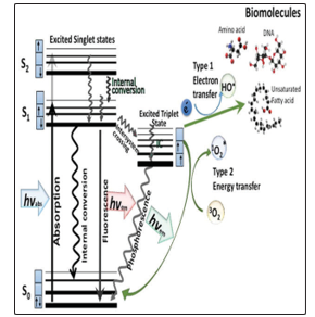
Graphical illustration of Antibacterial photo-dynamic inhibition technique (APDT) mechanism (Jablonski diagram (5).
The bactericidal action of Antibacterial photo-dynamic inhibition technique (APDT) as a new therapeutic option which has been demonstrated using different microorganisms which include Streptococcus pneumoniae, Prevotella intermedia, Haemophilus influenzae, Actinobacillus actinomycetem comitans, Bacteroides forsythus, Porphyromonas gingivalis, Staphylococcus aureus, Enterococcus faecalis, Candida albicans, Pseudomonas aeruginosa, Escherichia coli, Streptococcus sanguis and Fusobacterium nucleatum with superior outcomes seen in black-pigmented bacteria owing to their natural chromophore [6]. It was noted that Helicobacter pylori naturally accumulate sufficient porphyrin to allow photo-inactivation by blue light. This pathogenic bacterium causes endemic gastroduodenal ulcerations in humans, which was linked to the development of stomach cancer. The emergence of resistance to antibiotics in conventional therapy has been reported for H. Pylori, with the aid of Antibacterial photo-dynamic inhibition technique, the resistance menace of organisms like Helicobacter pylori can be deal with finding a permanent solution to resistant microorganisms.
Several photo-sensitizing agents of photo-dynamic nature proved to induce an efficient inactivation of at least some classes of microbial pathogens. In accordance to reports, different photo-sensitizers (PSs) are known, including dyes, drugs, cosmetics, chemicals and many natural substances such as hypericin but not all chemical compounds are photo-sensitizers [6].
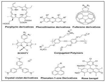
Chemical structures of commonly used photosensitizers [5,6].
The use of this technique is known to be mostly used in the field of radiology for the treatment of cancer, but microbiologists are now exploring this technique in curbing the issues of resistance massively evolved from all sphere of microbial world and other infectious particles.
Materials and Method
Microorganisms and Culture Conditions
Staphylococcus aureus, Vibrio cholerae, and Staphylococcus epidermidis were used in this study. All microorganisms were grown on nutrient agar under aerobic conditions prior to experimental use, all organisms were grown in liquid culture until late exponential phase.
Collection and Storage of Bacterial Cultures
The bacterial isolates used were collected from the Department of Microbiology, Faculty of Science, Adekunle Ajasin University Akugba Akoko Ondo State, Nigeria. These organisms were confirmed, kept on nutrient agar slants and stored in refrigerator (4°c) for further use. The pathogens includes: Staphylococcus aureus, Vibrio cholera and Staphylococcus epidermidis [7].
Preparation of Riboflavin Solution
Riboflavin (Sigma-Aldrich, St. Louis, MO) was diluted with sterile distilled water to achieve a 1% (100) riboflavin solution which was then diluted into different logarithmic riboflavin concentrations (10-1, 10-2,10-3, 10-4).
<img src="https://www.opastpublishers.com/scholarly-images/473-6ad201c05f8d8-assessments-of-antibacterial-photodynamic-inhibition-techniq.png" width=300" height="300">
Figure 1: Graphical Illustration of Laboratory Practices for Antimicrobial Photodynamic Treatment
Preparation of EDTA (Ethylene-Diamine-Tetraacetic Acid) Solution
Ethylene-diamine-tetra-acetic acid, disodium dihydrate (186.12 grams)(EDTA.Na2.2H2O, MW=372.24) was weighed into 2 L conical flask containing 800 ml distilled water. 20 g of NaOH (Sodium hydroxide) pellet was added to adjust the pH of the solution to 8.0 for easy dissolution of the EDTA pellets. Adjustment was done by increasing the volume to 1000 ml with distilled water. The solution was sterilized by autoclaving at 1210C for 15 minutes [8].
Light Sources Used for the Study
The Foto-dyne Fotophoresis, UV documentation was used for the Photo-dynamic treatment experiments, emitting light at 300 nm wavelength; the light was delivered for one hour continuously.
Photo-Dynamic Treatment Protocol
A 24 hours old bacteria culture was adjusted to0.5 Macfarlane standard, 10μL of each adjusted bacterial suspensions were distributed into different sterile Eppendorf tubes for light and riboflavin treated groups including the controls. There is addition of cell wall-permeability enhancing agent (EDTA) to the pathogen suspensions before a photo-sensitizer. This is efficient to inhibit cell or kill relevant multiple drug resistant clinical isolates (MDRCI) upon exposure to irradiation, and their addition be useful to enhance the photodynamic efficacy [9].
Control group 1 (Blank control): Bacterial isolates (10μl) which were neither exposure to UV lights nor riboflavin solution.
Control group 2 (UVA light): Bacterial cells (10μl) subjected to UV rays for 60 minutes at room temperature without riboflavin.
Control group 3 (Riboflavin in dark): Riboflavin (Sigma-Aldrich, St. Louis, MO) (5μl) was added to the cell solution at final concentration of 1% and the bacterial viability was assessed after contact, without UV light exposure. Prior to the bacterial suspensions treatment with both the light and riboflavin, 5μl of EDTA were added to increase the cell membrane permeability to allow easy penetration of the riboflavin solution into cytoplasm, this procedure was repeated for different concentration.
Group 1 (100 of riboflavin + UV light): 5μl of EDTA was added to 10μl of each bacterial suspension in addition with 5μl of riboflavin (100) and were exposed to UV for one hour.
Group 2 (10-1 of riboflavin + UV light): 5μl of EDTA was added to 10μl of each bacterial suspension with 5μl of riboflavin (10-1) and were exposed to UV for one hour.
Group 3 (10-2 of riboflavin + UV light): 5μl of EDTA was added to 10μl of each bacterial suspension with 5μl of riboflavin (10-2) and were exposed to UV for one hour.
Group 4 (10-3 of riboflavin + UV light): 5μl of EDTA was added to10μl of each bacterial suspension with 5μlof riboflavin (10-3) and were exposed to UV for one hour.
Group 5 (10-4 of riboflavin + UV light): 5μL of EDTA was added to 10μl of each bacterial suspension with 5μl of 10-4 of riboflavin and were exposed to UV for one hour.
Antimicrobial Susceptibility of multiple drug resistant clinical isolates (MDRCI)
The multiple drug resistant clinical isolates (MDRCI) in all groups were immediately plated on freshly prepared nutrient agar plates after the treatments described above. The pour plate technique was used and the plates were incubated under aerobic condition at 37 °C for 18h. Colonies were enumerated using the naked eye and recorded accordingly. Graphical plots of the viability of the pathogens were obtained with Graph-Pad Prism 7.
Results
Qualitative and Quantitative Observation of Antibacterial Photo-Dynamic Inhibition Technique (APDT) On Multiple Drug Resistant Clinical Isolates (MDRCI) The Multiple Drug Resistant Clinical Isolates (MDRCI)
in control group one showed highest number of colonies while those in control group two (exposed to UV lights only) showed changes in number of bacterial colonies (Table 1). The control group three (those treated with only riboflavin) showed no significant inhibitory effect of the treatment. The number of colonies of the three bacteria in the control group treated with UV alone was significantly low compared to those in control group one untreated with riboflavin nor UV light and the difference was statistically significant.
Table 1: Total Plate Count for Controls Used for Antibacterial Photo-Dynamic Inhibition Technique (Apdt)
|
|
Organism C |
Organism D |
Organism E |
|
Blank (Without organism) |
270 |
254 |
236 |
|
UV light + Multiple Drug Resistant Clinical Isolates (MDRCI) (300nm) |
195 |
170 |
154 |
|
Riboflavin (in the dark) |
259 |
249 |
203 |
Table 2: Total Plate Count of Exposed To Antibacterial Photo-Dynamic Inhibition Technique (Apdt)
|
Groups |
Riboflavin concentration (Log 10) |
Organism C |
Organism D |
Organism E |
|
Group 1 |
1 (100) |
58 |
77 |
35 |
|
Group 2 |
0.1 (10-1) |
0 |
0 |
0 |
|
Group 3 |
0.01 (10-2) |
0 |
0 |
0 |
|
Group 4 |
0.001 (10-3) |
0 |
1 |
1 |
|
Group 5 |
0.0001 (10-4) |
3 |
0 |
1 |

Plate 1: Plates of Multiple Drug Resistant Clinical Isolates (MDRCI) Colonies Treated With both the Uv Light And Riboflavin At Different Concentration
The bacterial culture treated with both the UV light and riboflavin was depicted in Figure 2.Group 1 has the highest number of colonies while the least was observed in group 5. Only one colony or no cell growth was observed after been treated which indicated that they are inactivated. However those treated with riboflavin 10-0 concentration showed different number of multiple drug resistant clinical isolates (MDRCI) colonies for organism C (53 colonies), organism D (77 colonies) and organism E (35 colonies). The difference in the number of colonies was low in relation to those treated with riboflavin and the untreated. It was also observed that the riboflavin group alone had no significant effect on the Multiple Drug Resistant Clinical Isolates (MDRCI). used. Furthermore, no cytotoxic effects in the dark of both the given photo-sensitizer itself and of the possible photo products formed, after illumination should be demonstrated either against the multiple drug resistant clinical isolates (MDRCI) itself or the eukaryotic cells.
The data obtained was used to estimate the concentration of dye needed to destroy half of total bacterial population at UV exposure for 60 minutes (MIC50) using the Gompertz equation available on Graph Pad Prism. Organism C required 0.8311 µM, Organism D required 0.8393 µM and Organism E required 0.7967 µM of riboflavin. The susceptible organisms showed a lower MIC50 and there was no significant difference.
Figure 3 showed the response of the multiple drug resistant clinical isolates (MDRCI) to photodynamic treatment at varying riboflavin concentrations at a constant UV intensity for 60 minutes. It showed that photo-sensitization was lethal at even low concentrations of riboflavin (10-5µM). It also confirmed that riboflavin was an effective photo-catalyst for the effective reduction of bacterial load upon UV sensitization.
The effect of Antibacterial Photo-dynamic inhibition technique (APDT) on multiple drug resistant clinical isolates (MDRCI) was depicted in figure 4.The graph represented a scatter with whisker plot of the pooled survival of each pathogen in response to both riboflavin treatment and ultraviolet radiation. It also showed the analysis of variance comparison of pathogens C and D (antibiotics resistant organisms) against multiple drug resistant clinical isolates (MDRCI) E. At alpha value of 5%, there was no significant (n.s.) difference in the multiple drug resistant clinical isolates (MDRCI) survival pattern even at different concentrations. (p-values C= 0.5276, D=0.5703).
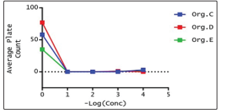
Graph 1: Killing Rate of Multiple Drug Resistant Clinical Isolates (MDRCI) Using Antibacterial Photo-Dynamic Inhibition Technique (Apdt)

Graph 2: Effect of Antibacterial Photo-Dynamic Inhibition Technique (Apdt) on Multiple Drug Resistant Clinical Isolates (MDRCI).
Discussion
This study investigated the bactericidal effect of photo-dynamic with the use of riboflavin as a photo-sensitizer combined with UV light against resistant strain of Staphylococcus aureus and vibrio cholera, and non-resistant strain of Staphylococcus epidermidis. This study outcome showed that exposure of bacterial cultures to UV light in the presence of riboflavin irrespective of it concentration showed reduction of viable colonies of Staphylococcus aureus, vibrio cholera and Staphylococcus epidermidis. This study corroborate the study of Fang et al. [10] who reported that PDT can effectively kill multiple drug resistant clinical isolates (MDRCI), which is even more effective than some antibiotics.
This study confirmed that riboflavin was an effective photo-catalyst for the effective reduction of bacterial load upon UV sensitization. This may be due to the limited availability of light-emitting diodes at that specific wavelength, the first devices used a wavelength of 365nm [11,12]. Riboflavin was chosen as a photo-sensitizer due to its nontoxic nature (vitamin B2). Multiple experimental articles have supported the hypothesis by demonstrating the antimicrobial effect through UVA mediated (365 nm) riboflavin photo-activation [13].
All the pathogens were susceptible to Antibacterial photo-dynamic inhibition technique (APDT) in this study. This observation agrees with the reports of a similar study conducted by [14] which reported the effects of light concentrations pre-confirmed drug resistance and dye concentration in Staphylococcus aureus, Staphylococcus epidermidis and Escherichia coli. They observed that irrespective of species background or pre-confirmed multiple drug resistant clinical isolates (MDRCI) status all pathogens were susceptible to photo-dynamic treatment.
With the increase in global multiple drug resistant clinical isolates (MDRCI) and the arrival of superbugs Hamblin et al [14,15] recommended that Antibacterial photo-dynamic inhibition technique (APDT) treatment might be a sound approach for the management of superficial wound infections, skin infections[15-17].
Conclusion
It can be concluded that antimicrobial photodynamic therapy be accepted as an alternative way to antibiotic therapy.it can be deduced that the combination of riboflavin and ultraviolet radiations is a good and promising strategy for combating infections from both resistance and non-resistance pathogens and this research on Antibacterial photo-dynamic inhibition technique (APDT) could be considered as a promising therapy to manage infectious bacteria diseases and to keep emergency and re-emergency of drug resistance bacterial infection at hand.Recommendations
i. Further researches should be carried out on photodynamic treatment so as to prove it effectiveness against infections, other skin and dental disease treatments.
ii. A further work on antibacterial photo-dynamic inhibition techniques (APDT) through in vivo experiment is required to fully prove the effectiveness of the APDT on fungi, viruses and other infectious agents.
iii. There should also be willingness on the part of health sector workers in Nigeria to gain better understanding on antibacterial photo-dynamic inhibition techniques (APDT) and the government should avail them of funds, and there should be avenue to collaborate with the scientists and provision of research activities to evaluate them.
iv. This will help to sort the various utilization of different biological resources that have proven valuable and can be adapted into the healthcare system and those that are unnecessary or any harmful effect should be discouraged.
References
-
- Abrahamse H, Hamblin MR (2016) new photosensitizers for photodynamic therapy. Biochem J 473: 347-364.
- Ahrari F, Shahabi M, Fekrazad R, Eslami N, Mazhari F, et al. (2018) Antimicrobial photodynamic therapy of Lactobacillus acidophilus by indocyanine green and 810-nm diode laser, Photo-diagnosis and Photodynamic Therapy 24: 145-149.
- Dos Santos LFM, Melo NB, de Carli ML, Mendes A, Bani G, et al. (2017) Photodynamic inactivation of Paracoccidioides brasiliensis helps the outcome of oral paracoccidiodomycosis. Lasers Med Sci 32: 921-930.
- Hamblin MR (2016) Antimicrobial photodynamic inactivation: a bright new technique to kill resistant microbes. Curr OpinMicrobiol 33: 67-73.
- Rosa LP, da Silva FC (2014) Antimicrobial Photo-dynamic Therapy: A New Therapeutic Option to Combat Infections. J Med MicrobDiagn. 3: 158.
- Osuntokun Oludare temitope, Adewale Joseph Ogunleye (2017) Plant-Mediated Modu lation of Reactive Oxygen Species (ROS) Homeostasis, Drug Target Revinew, Russell Publishing Limited,Court lodge, Hogtrough Hill, Brasted, Kent, TN16 1NU, United kingdom.
- Osuntokun OT, Ibukun AF, Yusuf-Babatunde AM, Abiodun S (2019) Pre/Post-Plasmid Profile Analysis, Killing- Kinetics and Secondary Metabolites Screening of Adenopus breviflorus (Benth) Fruit Extract Against Multiple Drug Resistant Isolates Using Staphylococcus aureus (MDRSA) as a Case Study. Journal of Advanced Research in Biotechnology, J Adv Res Biotech 4: 1-17.
- Sojib Bin Zaman, Muhammed Awlad Hussain, Rachel Nye, Varshil Mehta, Kazi Taib Mamun, et al. (2017) A Review on Antibiotic Resistance: Alarm bells are ringing. Cureus 9: 1403.
- Xiaoqing Hu, Ying-Ying Huang, Yuguang Wang, Xiaoyuan Wang, and Michael R. Hamblin (2018) Antimicrobial Photodynamic Therapy to Control Clinically Relevant Biofilm Infections 9: 1299.
- Fang Y, Liu T, Zou Q, Zhao Y, Wu F (2016) Water-soluble benzylidene cyclopentanone based photo-sensitizers for in vitro and in vivo antimicrobial photodynamic therapy 6: 28357.
- Karim Makdoumi, Ray Goodrich, Anders Beackman (2017) Photochemical eradication of methicillin-resistant Staphylococcus aureus by blue light activation of riboflavin 95: 498-502.
- Kristian Berg (2019) Photo-sensitizers in medicine.
- Lesley R, Evelyne S, Massimo T (2016) Comprehensive series in photochemical and photo biological science: Photodynamic medicine from bench to clinic. European Society for Photobiology.
- Hamblin MR, T Hasan (2004) Photo-dynamic therapy: a new antimicrobial approach to infectious diseases? Photochem. Photobiol 3: 436-450.
- Mcfarland SA, Susan Monro, Katsuya L Colón, Huimin Yin, John RoqueIII, et al. (2017) Metal-based coordination complexes as photodynamic compounds and their use 119:797-828.
- Thomas JD, Charles JG, B. Henderson W, Jori G, Kessel D, et al. (1998) Photodynamic Therapy. Journal of the National Cancer Institute 90: 889-905.
- Sperandio FF, Huang Y-Y, Hamblin MR (2013) Antimicrobial photo-dynamic therapy to kill Gram-negative bacteria. Recent Pat Antiinfect Drug Discov 8: 108-120.