

Advances in Bioengineering and Biomedical Science Research(ABBSR)
ISSN: 2640-4133 | DOI: 10.33140/ABBSR

Impact Factor: 1.7

Case Report - (2021) Volume 4, Issue 3
Allergy Evaluation under X-Ray Exposition in Patients with Failed or Upcoming Arthroplasty
2maging Clinic, Alexander’s University Hospital, Sofia, Bulgaria
3Department of Arthroscopic Traumatology, Military Medical Academy, Sofia, Bulgaria
Received Date: May 15, 2021 / Accepted Date: May 19, 2021 / Published Date: Sep 22, 2021
Copyright: ©Copyright: ©2021 Diana Hristova, et al. This is an open-access article distributed under the terms of the Creative Commons Attribution License, which permits unrestricted use, distribution, and reproduction in any medium, provided the original author and source are credited.
Citation: Diana Hristova, Petko Genov, Vladimir Stefanov (2021) Allergy Evaluation under X-Ray Exposition in Patients with Failed or Upcoming Arthroplasty. Adv Bioeng Biomed Sci Res 4(3): 85-86.
Abstract
Nowadays metal implants are a major source of metal exposure. Contact allergy to nickel, cobalt and chromium is one of the most prevalent allergic reactions in humans with contact dermatitis; up to 17% of women and 3% of men. Setting up the sensitization to implant components is needed before orthopedic surgeon. Patch testing remains the gold standard for verification a for type IV reaction of metal, bone cement, or antibiotic. We assessed the records of all 22 patients, whom we followed up since orthopedic intervention or were indicated for such intervention. All patients were exposed to X-ray after placement of epicutaneous tests. X-ray exposure may have influence on positive skin test results- odds ratio 0.56 [95% confidence interval 0.16393 - 1.91031]
Keywords
Contact Allergy, Metal Implants, Epicutaneous Tests, Joint Failure, Metal- Induced Skin Reactions
Introduction
Hypersensitivity to nickel, cobalt and chromium is one of the most prevalent allergic reactions in humans with contact dermatitis; up to 17% of women and 3% of men display symptoms after pro-longed exposure of metals. Nowadays metal implants are a major source of metal exposure. Allergic complications watched after the placement of such implants include skin changes such as ec¬zema, urticaria, complicated wound mending, as well as responses such as edema, effusions, torment, joint swelling and warmth. De¬ciding the reason for joint failure may depends on hypersensitivity, contamination, mechanical mismatch and scarring. Joint torment as a side effect hold on in all three conditions. The recurrence of amendment operations is up to 9%. Noteworthiness of hypersen¬sitivity of metal inserts is starting to get necessary consideration. Since the primary reported cases of skin reactions to metal implants in the 1960’s, metal hypersensitivity has been recognized as an issue in orthopedic surgery. Over the years, reports of dermatitis, vasculitis and indeed the appearance of urticaria associated with orthopedic implants have increased in the literature. Non-specific symptoms such as pain, swelling, restricted or lost joint function after the exclusion of an infectious or mechanical cause may be related with metal hypersensitivity. Periimplant lymphocytic inva¬sion has been illustrated in patients with failed arthroplasty [1-5]. Detailed diagnostic criteria for metal-induced allergic skin reac-tions are listed below:
1. Chronic skin inflammation that show up weeks or months after implant placement
2. Severe eczema around the implant
3. Nonappearance of other contact allergens or systemic cause
4. Positive epicutaneous test for certain metals contained within the implant
5. Complete and quick recovery after implant replacement [3, 4]. Recommendations for an interdisciplinary approach in patients with metal allergy have been proposed since 2008 [5]. What is not yet clear is the impact of X-ray exposure on metal implants.
The objective of our analysis was to identify the patients who were sensitized to components which exist in implants in order to choose suitable implant. Our attempt was to evaluate the influence of X-ray exposure.
Materials and Methods
The study comprised 22 recently diagnosed untreated patients with complications after implant placement or whom were indicated for such intervention (female/male ratio 19:3; aged (min/max) 57÷71). The control group consisted of patients with no history for previous reactions to metals. Inclusion criteria were: complication after implant placement and or suspicion of possible complication after orthopedic surgery.
Methods
We assessed the records of all 22 patients , whom we followed up since orthopedic intervention or were indicated for such inter-vention. We had on record the self-reported history of metal reac¬tivity. There is no agreement-tested substances before orthopedic surgeon. Researchers decide independently. In our group patients were tested with Chemotechnique Diagnostics epicutaneous test as follows: Potassium dichromate 0.5 pet, Cobalt (II) chloride hexahydrate 1.0 pet, Nickel (II) sulfate hexahydrate 5.0 pet, Ti¬tanium 10.0% pet, Zirconium dioxide 0.1% pet, Molybdenum 5.0% pet, Vanadium 5.0% pet, Ferric chloride 2.0% pet, Copper(I) oxide 5.0% pet, Manganese chloride 2.0% pet, Gentamicin sul¬fate 20.0% pet, Erythromycin base 10.0% pet, Benzoylperoxide 1.0% pet, Thimerosal 0.1% pet, Chloroxylenol (PCMX) 1.0% pet, Toluenesulfonamide formaldehyde resin 10.0% pet, 1,9-Hexane-dioldiacrylate 0.1% pet, N-Ethyl-p-toluenesulfonamide 0.1% pet, Carba Mix 3.0% pet, Epoxy resin, Bisphenol A 1.0 pet, N-Iso-propyl-N-phenyl-4-phenylenediamine (IPPD) 0.1 pet, Mer-capto mix 2.0 pet, 2- Mercaptobenzothiazole (MBT) 2.0 pet, 2,2-bis(4-(2-Methacryl-oxyethoxy -phenyl)-propane (BIS- EMA) 2.0%, 2-bromo-2-nitropropane-1.3-diol 0.5% pet, Methyldibromo glutaronitrile (MDGN) 0.5 pet, Ethyl acrylate 0.1% pet, Methyl methacrylate 2.0% pet, Ethylene glycol dimethacrylate 2.0% pet, Ethyl cyanoacrylate 10.% pet.
All patients were exposed to X-ray. Back X-ray was done after application of epicutaneous tests.
We performed descriptive analysis (frequencies) and calculated odds ratios for the variables with possible influence on the sensiti-zation as a dependent variable.
Results and Discussion
Out of the 22 patients on record, 7 (32%) have a previous history for type IV hypersensitivity and 15 patients (68%) have no history for previous reactions (figure 1).
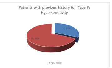
Figure 1: History for previous reactions of metals, methacrylates or antibiotics
Setting up the sensitization to implant components is needed be¬fore orthopedic surgeon. Asking for affirmed contact allergy could be a key point. Patients with a history of reacting to metal ought to be tested before replacement. Patch testing remains the gold stan¬dard for verification for type IV reaction of metal, bone cement, or antibiotic.
To date there has been small understanding on what is the dif-ference between dermal reaction and the immune response in the joint itself. A number of researchers have reported lymphocyte transformation test as the best to evaluate immune reactions to internal joint components. Both the patch test and the delayed hy¬persensitivity test assess the activity of immune-specific T cells, which comes from different sources. Correlation between patch tests and lymphocyte transformation test has been detailed [5].
Recent prove recommends that in nonsensitized subjects, metal sensitization may result from an immune reaction to metal parti¬cles after either corrosion or mechanical wear of an implant. Be¬neath prolonged exposition with biologic fluids, they experience corrosion to discharge ionic compounds. On other hand, nickel may straightforwardly activate the T-cell receptor as superanti-gens. Metal ions may gather in rodent liver and kidney tissue [4].
There is no data in the scientific literature on the influence of X-rays, which is an essential component in orthopedic surgery.
According to our results, X-ray exposure was related to positive epicutaneous test results - odds ratio 0.56 [95% confidence interval 0.16393 - 1.91031].
Conclusions
In our group of patients X-ray exposure may have influence on positive skin test results. The limit of our study is the small number of patients and the inaccuracy in determining the available contact allergy based solely on the history before X-ray exposure. Further research is needed. References
Conclusions
In our group of patients X-ray exposure may have influence on positive skin test results. The limit of our study is the small number of patients and the inaccuracy in determining the available contact allergy based solely on the history before X-ray exposure. Further research is needed. References
References
- P Thomas, G Roider, A Beraudi, D de Pasquale, B Bordini, et al. (2015) Metal Implant Allergy and Immuno-Allergological Compatibility Aspects of Ceramic Materials. Springer, Verlag Berlin Heidelberg 2015.
- Т omas P (2014) Clinical and diagnostic challenges of metal implant allergy using the example of orthopedic surgical implants. Allergo J Int 23:179-185.
- Hallab NJ, Jacobs JJ (2017) Chemokines Associated with Pathologic Responses to Orthopedic Implant Debris. Front Endocrinol 8: 5.
- Juliana L Basko Plluska, Jacob P Thyssen, Peter C Schalock (2011) Hypersensitivity Reactions to Metallic Implants. Dermatitis 22: 65-79.Karin A Pacheco, MSPH Denver, Aurora Colo (2015) Amer-rican Academy of Allergy, Asthma & Immunology. J Allergy Clin Immunol Pract 3: 683-695.