

Journal of Emergency Medicine: Open Access(JEMOA)
ISSN: 2994-6875 | DOI: 10.33140/JEMOA
Impact Factor: 0.98



Review Article - (2026) Volume 4, Issue 1
Adult Intussusception-A Perspective
2Department of Hepatobiliary and Gastrointestinal Surgery, Medicover Hospitals, India
Received Date: Feb 27, 2026 / Accepted Date: Mar 23, 2026 / Published Date: Mar 31, 2026
Copyright: ©2026 Harshal Rajekar, et al. This is an open-access article distributed under the terms of the Creative Commons Attribution License, which permits unrestricted use, distribution, and reproduction in any medium, provided the original author and source are credited.
Citation: Sikora, T. M., Rajekar, H. (2026). Adult Intussusception
Abstract
Intussusception occurs when a segment of the proximal intestine called intussusceptum, telescopes into the lumen of the distal segment, also known as intussuscipiens.
Background: Rare in adults, most intussusceptions are due to a lead point, an identifiable pathological abnormality, mostly a tumor, often malignant where the resectability is difficult to assess. In children there are no identifiable pathological lead points, or and most often is due to lymphoid hyperplasia and no other pathology is found and often managed conservatively.
Case Summary: We had 3 patients, all elderly and frail, who presented with acute abdominal pain and bleeding per rectum. They were found to have large bowel intussusception. Surgical intervention is advised in all adult intussusception patients, as all adult intussusceptions are believed to be secondary. At the surgery it was found that all had operable lesions, had a good prognosis and histology of all was benign. One patient had a villous adenoma, one had lymphoid hyperplasia and the 3rd had a tubule-villous adenoma.
Thus all 3 elderly patients, presenting with intussusception did not have any specific signs or symptoms, were diagnosed by computerized tomography and had completely resectable lesions. Thus, early computerized tomography with timely surgical intervention is recommended in all patients with bleeding per rectum and suspected intussusception. Since intussusception is a mobile tumor the likelihood of resectability is high with a good prognosis.
Conclusion: Colonic intussusception rarely leads to colonic obstruction. Any kind of intussusception, points towards a lesion that is mobile and not invading or adherent to surrounding tissue, hence more likely to be resectable.
Keywords
Colonic Intussusception, Adult Intussusception, Colonic Malignancy, Desmoplastic Reaction, Resectability, PrognosisIntroduction
Intussusception (telescoping within self) of the intestine was first reported in 1674 by Barbette of Amsterdam and then detail presented in a detailed report in 1789 by John Hunter as “intussusception” [1,2]. In 1871, Sir Jonathan Hutchinson was the first surgeon to operate a child having intussusception [3]. Intussusception is primarily a disease of growing age, infants and toddlers, and only about 10 to 25% cases occur after the age of 2years [4]. Generally, in adult patients presenting with intussusception, it is known the likelihood of a pathological lead point is high, and with elderly patients the possibility of a neoplastic lesion becomes higher [5]. In frail, elderly patients, the possibility of advanced malignancy, and the likelihood of unresectable disease needs to be borne in mind. Often, time is spent in optimization of the patient and in an attempt to get a pre-operative diagnosis, so as to help prognosticate. With experience across the world, it is now being seen that malignancy and unresectable tumors form a minority of the patients presenting with intussusception in adults [6,7].
Observations
3 consecutive patients presenting with acute or chronic blood loss, and were found to have intussusception. All patients were more than 65 years of age, frail and poorly nourished. 1. A 74-Y old male, with Parkinsonism, presented with bleeding per rectum and loose motions in the preceding two days. CT scan abdomen (Figure 1 and 2) showed features of Colo-colic intussusception with a heterogeneously enhancing mass lesion as a lead point located in mid/proximal sigmoid colonic bowel loop. There was no intestinal obstruction.

Figure 1: CT Scan Sagittal Section Showing the Intussusception

Figure 2: CT Scan - Transverse Section showing the Intussusception
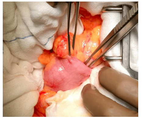
Figure 3: Intra-operative picture of the intussusception
Colonoscopy was done and a polypoidal growth was seen in sigmoid. The patient continued to have per rectal bleeding and underwent left radical hemicolectomy (Figure 3). The histopathology showed tubulo-villous adenoma with mild dysplasia. Post-operative recovery was uneventful and patient was discharged on the 7th day after surgery.
2. Another 69-Y old female patient, known chronic kidney disease, not on dialysis; presented with loose motions and anemia. There was no other medical illness or any other co- morbidity. An ultrasound abdomen revealed an ileo-colic intussusception. There was no frank bleeding or intestinal obstruction. CT scan confirmed the intussusception (Figure.4, Figure.5) with no evidence of any lesion at the lead point or any obstruction.

Figure 4: CT Scan Sagittal Section showing the Intussusception

Figure 5: Coronal Section CT Scan with the Lesion
There was no resolution of the intussusception over the next 72 hours. Patient underwent a right hemicolectomy (Fig. 6) and made an uneventful recovery. She was discharged on post-operative day 6. Histology showed fibroblastic reaction with chronic scarring and lymphoid hyperplasia. There was no evidence of malignancy or tuberculosis (Fig. 7.)

Figure 6: Intraoperative pic of the Ileo-Colic Intussusception

Figure 7: Lead point at the Cecum showing the Lymphoid Hyperplasia with Fibroblastic Reaction and Scarring at the lead point
3. Another 69-year-old gentleman, came to the gastro-surgical clinic, with chronic anemia and intermittent malena. Apart from diabetes mellitus, there was no other co-morbidity. He underwent gastro-duodenoscopy and a colonoscopy, both of which were unremarkable. There was no liver disease or cardiovascular comorbidity. While being evaluated in the hospital, he developed acute abdominal pain. There was no vomiting or intestinal obstruction, and the pain was of moderate intensity. An ultrasound abdomen revealed an ileocolic intussusception, which was confirmed by CECT abdomen. The scan did not show any lesion or obstruction. The patient underwent a laparoscopic right hemicolectomy (Figure. 8). The resected bowel showed a polyp like lesion as the lead for intussusception, which turned out to be a hamartomatous polyp (Figure. 9).

Figure 8: Resected Specimen showing the Intussusception

Figure 9: Cut Open Specimen Showing the Ileal Polyp
The histology showed connective tissue composed of smooth muscle, lamina propria and an inflammatory infiltrate, with overlying hypertrophic epithelium. The patient made a unremarkable recovery and was discharged on day 5 after surgery.
Discussion
As already demonstrated; there were 3 elderly patients, who presented with un- characteristic signs and symptoms. All 3 had intussusception, and a CT scan (computerized tomography) clinched the diagnosis in all. None of them had intestinal obstruction, whereas all had some degree of gastro-intestinal bleeding and all were resectable. None of them had a malignancy.
Pathophysiology
In children, the classical presentation of intussusception is a child with a history of a recent viral illness with vomiting and/or diarrhea. The classic triad consists intermittent abdominal pain, red currant jelly stool, and a palpable mass. This is seen in 7.5% to 40% of children with intussusception [4]. Adult intussusception cases present with nonspecific signs and symptoms. Intussusception in adults may present with acute, subacute, or chronic non- specific symptoms. Some patients may present in a few days to a few weeks with acute symptoms like bleeding or obstruction. Abdominal CT is the most sensitive with sensitivity and specificity of 58% to 100%. 60-65% of cases of intussusception of the large bowel are expected to have a malignant etiology [5]. Intussusception can lead to multiple complications such as bowel obstruction, bowel necrosis, and intestinal bleeding. In 2020 review by Tongrong Su et al, the main pathogenesis of intussusception was malignant tumours in 51 cases (36.7%) and benign tumours and polyps in 49 cases (35.3%) [6].
Most lead points in the small bowel consist of benign lesions, such as benign neoplasms, inflammatory lesions, Meckel’s diverticulum contributing to almost 50-75 percent of small bowel intussusception, while, intussusception in the large bowel is more likely to have a malignant etiology. However, Kim KH et al found contrary findings. They reported malignancy in the small bowel in 25% of patients, similar to earlier reports, but malignant neoplasms in the large bowel were seen in only 18.8% of cases, which is much lower than that reported [7]. Wang et al from China reported in a series of 41 patients that less than 50% of their colonic intussusceptions were malignant, and similarly Wang LT et al from Taiwan reported malignant lesions in less than 45% of their series of 24 adult patients with intussusception [8,9].
Diagnosis and Management
Ultrasound is usually the first investigation. The “Target sign” of intussusception was first described in 1977, appears either as a single hypoechoic ring with a hyperechoic center, or as a pattern of alternating hypoechoic and hyperechoic rings [10]. The pseudo-kidney sign represents the longitudinal appearance of the edematous walls of the intussusceptum [11]. Computerized tomography (CT) appears to be the most sensitive d in making a preoperative diagnosis of adult intussusception [12,13].
Colonoscopy
Colonoscopy is useful, but difficult tool in evaluating intussusception, due to an increased risk of perforation resulting from chronic tissue ischemia, vascular compromise, and potentially necrosis; and the difficulty in bowel preparation.
Treatment
Surgery is the recommended treatment for adult intussusception, as intussusception in adults is usually secondary to a pathological lead point. There is controversy on the amount of bowel to be resected and on whether intestinal intussusceptions should be reduced, since there is a significant risk of malignancy. The prognosis of intussusception depends on the causative factor of the lesion, and mortality from intussusception in adults increases from 8.7% in benign causes to 52.4% in malignant causes [14]. Minimally invasive options are as suitable as traditional approaches; since there rarely is any intestinal obstruction.
Conclusions
Preoperative diagnosis may be missed or delayed because of nonspecific and often subacute symptoms. Most of the adult intussusceptions are secondary. Computerized Tomography (CT) is most probably the most sensitive and specific in making a pre-operative diagnosis of adult intussusception may be rare due to per-tumoral invasion and desmoplastic reaction; and therefore, most adult intussusception may be benign as seen in literature. Intussusception due to a malignant lead point points towards a mobile lesion, and hence more likely to be resectable. Surgical intervention is often necessary, and treatment usually requires formal resection of the involved bowel segment.
References
- century Amsterdam author of best-selling textbooks. Bulletin of the History of Medicine, 59(4), 506-514.
- Marsicovetere, P., Ivatury, S. J., White, B., & Holubar, S. D. (2017). Intestinal intussusception: etiology, diagnosis, and treatment. Clinics in colon and rectal surgery, 30(01), 030-039.
- Lianos, G., Xeropotamos, N., Bali, C., Baltogiannis, G., & Ignatiadou, E. (2013). Adult bowel intussusception: presentation, location, etiology, diagnosis and treatment. Il Giornale di Chirurgia-Journal of the Italian Surgical Association, 34(9), 280-283.
- Waseem, M., & Rosenberg, H. K. (2008). Intussusception.Pediatric Emergency Care, 24(11), 793–800.
- Hadid, T., Elazzamy, H., & Kafri, Z. (2020). Bowel intussusception in adults: Think cancer! Case Reports in Gastroenterology, 14(1), 27–33.
- Su,T., He, L., Zhou,T., et al. (2020). Most adult intussusceptions are caused by tumors: A single-centre analysis. Cancer Management and Research, 12, 10011–10018.
- Kim, K. H. (2021). Intussusception in adults: A retrospective review from a single institution. Open Access Emergency Medicine, 13, 233–237.
- Wang, N., Cui, X. Y., Liu, Y., Long, J., Xu, Y. H., Guo, R. X.,& Guo, K. J. (2009). Adult intussusception: A retrospective review of 41 cases. World Journal of Gastroenterology, 15(26), 3303–3308.
- Wang, L. T., Wu, C. C., Yu, J. C., Hsiao, C. W., Hsu, C. C., &Jao, S. W. (2007). Clinical Entity and Treatment Strategies for Adult Intussusceptions: 20 Years’ Experience. Diseases of the Colon & Rectum, 50(11), 1941–1949.
- Weissberg, D. L., Scheible, W., & Leopold, G. R. (1977). Ultrasonographic appearance of adult intussusception. Radiology, 124(3), 791–792.
- Swischuk, L. E., Hayden, C. K., & Boulden, T. (1985). Intussusception: Indications for ultrasonography and an explanation of the doughnut and pseudokidney signs. Pediatric Radiology, 15(6), 388–391.
- Gayer, G., Zissin, R., Apter, S., Papa, M., & Hertz, M. (2002). Adult intussusception—A CT diagnosis. British Journal of Radiology, 75(890), 185–190.
- Takeuchi, K., Tsuzuki, Y., Ando, T., Sekihara, M., Hara, T., Kori, T., & Kuwano, H. (2003). The diagnosis and treatment of adult intussusception. Journal of clinical gastroenterology, 36(1), 18-21.
- Cordeiro, J., Cordeiro, L., Pôssa, P., Candido, P., & Oliveira,A. (2019). Intestinal intussusception related to colonic pedunculated lipoma: A case report and review of the literature. International Journal of Surgery Case Reports, 55, 206–209.