

Advances in Bioengineering and Biomedical Science Research(ABBSR)
ISSN: 2640-4133 | DOI: 10.33140/ABBSR

Impact Factor: 1.7

Research Article - (2023) Volume 6, Issue 4
A Report of Radiotherapy Setup Errors in Pelvic and Mediastinal Tumors
Department of Radiation Oncology, Cancer Institute, Tehran University of Medical Sciences, Iran, Iran
Department of Radiation Oncology, Cancer Institute, Tehran University of Medical Sciences, Iran
Received Date: Jan 06, 2023 / Accepted Date: Jan 25, 2023 / Published Date: Mar 24, 2023
Copyright: ©Â©2023 Nafise Hasoomi, et al. This is an open-access article distributed under the terms of the Creative Commons Attribution License, which permits unrestricted use, distribution, and reproduction in any medium, provided the original author and source are credited.
Citation: Hasoomi, N., Azizi, A., Ranjbar, S. A. (2023). A Report of Radiotherapy Setup Errors in Pelvic and Mediastinal Tumors. Adv Bioeng Biomed Sci Res, 6(4), 48-54.
Abstract
Introduction: Radiation therapy as an important step in treatment of cancer which demands accuracy. Patient setup is a challenging job in the radiation therapy process. The Variation in random setup error for specific sites is different among clinics, even from one radiation therapist technologist to other in a same clinic. The purpose of this study is to investigate and report about the setup margins in the pelvic and mediastinal sites.
Method &Material: 34 patients (20 males and 14 females) in the supine and prone positions (24 supine positions and 10 prone positions) were selected. Internal protocol and custom-made positioning devices were utilized. The variation of Anterior-Posterior movements (AP) in daily setup is recorded based on the vertical information which is shown on the arian 2100C/D Linear Accelerators monitor. The correlation between body type, position and treatment cases compares with the mean errors.
Conclusion: The highest random error for setup is attributed to mediastinal tumor (=0.3 cm), and the highest systematic error is dedicated to cervix (=1.4 cm). The population systematic error (=1.25) is defined as the standard deviation of means of patients(Σ). In centers with lack of image-guided facilities or centers with high loaded patients the setup process should be accurate enough to limit setup error probabilities.
Introduction
Radiation therapy (RT) is one of the treatment modalities using ionization radiation to treat cancer cells. One of the mandatory steps in the radiotherapy process is making the patient position ex-actly based on the patient's geometry at the time of CT simulation. Inappropriate positioning causes underdose of tumor and overdose of healthy tissue [1]. Setup accuracy will ensure the reproducibili¬ty of the Planning Target Volume (PTV) and organs-at-risk, which were determined during treatment planning time. PTV definition is introduced by the International Commission on Radiation Units and Measurements Report 62 (ICRU 1999) [2]. This concept in¬troduced by adding an internal margin and setup margin to the clinical target volume (CTV) for guaranteeing accurate beam irra¬diation on the target by considering the fact that clinics had been defined the threshold level of errors. Understanding the concept of setup errors by consuming the definition of patient motion will clarify more treatment accuracy and the importance of immobi¬lization devices in the aspect of setup errors; patient motions are defined in six axes referring to three transitional movements (lat¬eral, longitudinal, vertical) and three rotational movements (pitch, roll, yaw). The timeline for motion errors in patients undergoing radiation therapy varies between seconds and weeks; for example, respiration movements occur in seconds, rectal distention in minutes, bladder and bowel distention varies in days, and organ shape and position change in weeks. Geometric discrepancy leads to cold spots in PTV and hot spots in healthy tissue which could be deteriorated in the hypo fractionated treatment such as stereo-tactic body radiotherapy (SBRT) rather than traditional treatment procedures [8].
Modern technology in radiotherapies such as IMRT, VMAT, and SBRT indicate the necessity of setup accuracy and the importance of immobilization during the treatment time. Image-guided radia¬tion therapy (IGRT) techniques have been developed and provide a competent strategy to ensure accurate positioning and reduce inter and intra- fractional errors[4]. Studies indicate IGRT techniques have an important role in decreasing setup error incidents by 50 % [5, 6]. By evolving IGRT, different protocols were introduced to reduce the setup errors such as the No action level (NAL) and the shrinkage action level (SAL) [16]. The NAL protocol demands 3–5 imaged fractions, while the SAL protocol needs 8–9 imaged fractions. Petilion et.al introduces their protocol such as Fixed ac- tion level (FAL)with weekly imaging and correction [17]. Each systematic setup error is estimated and corrected after a fixed num¬ber of imaged fractions. On the other hand, some argue that the high cost and increasing treatment time in using the IGRT systems can be demonstrated, some would argue that the theoretical bene¬fits of IGRT should be more investigated [7].
Patient setup is a challenging job in order to insufficient formal training, workload, the complexity of new techniques and devices, and unavailability of immediate position verification [8]. The Variation in random set-up error for specific sites is different among clinics, even from one radiation therapist technologist to another in the same clinic [9]. This issue indicates the underlying assumption of defining setup margin in any radiotherapy center individually.
|
ORGAN/Technique |
ERROR Threshold |
|
Radiosurgery |
1mm |
|
Head and neck in LINAC |
3mm |
|
Head and neck in Cobalt |
5mm |
|
Thorax |
10mm |
|
Pelvic |
15mm |
Table 1: A guide for action level
Errors in the radiotherapy process can take place over immobiliza-tion, imaging, contouring, treatment planning, setup Delivery, and image guidance. Fishbone and 5whys strategies declare that errors with different sources require different solutions [12]. For exam-ple, errors in the time of imaging contain potential errors in the imaging modalities, image fusion, and artifacts which can impact the target delineation and organ at risk. Although CT is used for the treatment planning system, it is often not the most optimal mo-dality for target or normal tissue delineation. MRI and PET help radiation oncologist reduce the error potential, for example, MRI outline the tumor borders in soft tissue better than CT or PET-CT helps to differentiate between atelectasis and tumor tissue. The de-sired degree of immobility depends on the proximity of the treat-ment field to a sensitive structure. This in turn should influence the decision on the type of immobilization devices. As a rough guide, the following table (table 1)is offered [13].
A review study interpreted that systematic and random errors were observed within less than 5 mm in different research [10]. Hongbo Chai et al. reported that in lung cancer setup margins vary up to 2 mm in AP (anterior-posterior) direction, 4 mm in SI (superior-in-ferior) direction, and 4 mm in LR (left-right) direction [14]. The book titled Ensuring geometric accuracy in radiotherapy intro-duced the geometry variation of more than 1 cm as the important root cause of radiotherapy errors [15]. Reproducibility in treatment setup is a constant challenge for RTT in each treatment session which is ameliorated via immobilization devices and IGRT techniques. Although much work has been done to date, more studies need to be conducted to introduce the relationship between setup preparation and setup errors in radiotherapy.
In our centers, the high load of patients and lack of IGRT facilities compile the RTTs to use different methods to make a guarantee for the treatment process. The purpose of this study was to ascertain the setup errors in pelvic and mediastinal treatments and provide information for setup margins based on the vertical value of the accelerator. This paper also revealed the correlation between setup errors and body type, and position in pelvic and mediastinal tumors. The RTTs also introduced an internal protocol to provide a precise time with minimum errors.
Method and Material
In this study, 34 patients (20 males and 14 females) in the su¬pine and prone positions (24 supine positions and 10 prone po¬sitions) were selected. Of all these patients, 14 cases (41%) were with rectum cancer, 3 cases (8.8%) with prostate cancer, 9 cases (26.5%) with cervix cancer, and 8 cases (23.5%) with mediasti¬nal tumor. The patient's body type is divided into three different groups; 17 fat patients (waistband>105 cm), 10 medium patients ( 90 cm
The lack of EPI-based assessments (Electronic Portal Imaging) for Varian 2100C/D Linear Accelerators and the high load of patients (there is 2 shifts in a day, 30 patients per shift) compel radiation therapist technologists to make innovation for setup reproducibili¬ty and precise treatment.'
Using a custom-made footrest (Figure2) and the concept of a treat¬ment tunnel over defining an internal protocol (figure3) have been developed to overcome difficulties in high-load centers with low facilities. The positioning method was performed with the three skin tattoos matched to the laser lights: one midline for the su-perior–inferior (SI) and left–right (LR)directions and two lateral points on either side of the body for the anterior–posterior (AP) direction. A custom-made foot rest was utilized to prevent diver-gence or convergence of legs during the fractions (Figure 2). Setup errors were recorded in the dimension of Anterior-posterior (A-P). In our study, the variation of Anterior Posterior movements (AP) in daily setup is recorded based on the vertical information which is shown on the Varian 2100C/D Linear Accelerators (Figure 1). The value for the treatment couch in the anterior–posterior (AP) direction was obtained in different fractions randomly for 34 pa-tients in 11 treatment fractions by different technologists. The data was recorded manually and transformed to SPSS and EXCEL for analytical assessments.
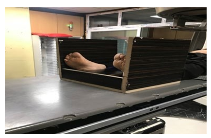
Figure 1: custom-made foot rest
Concept of Treatment Tunnel
In the first treatment session, the patient's body position was ad-justed until the skin tattoos and laser lights matched completely. The position of the hand for pelvic cancer in the supine position is on the chest and their elbows touch the table for more comfort. Pelvic patients with prone positions put their hands under their foreheads. And finally, the patient's hands are above their head for whom suffer from the mediastinal tumor. The latter lay on the table in supine position. For all these patients, a custom-made footrest is used (Figure 1). After aligning the tattoos on the body with treat¬ment room lasers, Varian's monitor indicates a VRT value (AP di¬rection) that sometimes differs from the VRT value (AP direction) of CT simulation (Figure 2). The difference of this VRT is implied on the isocenter manually. Because this difference is for a volume or tunnel in the patient body which refers to the concept of the treatment tunnel. For example, if CT lasers recorded markers in the AP direction for 11 cm and this value in the radiotherapy Room changed to 11.4 cm. This difference impacts the treatment volume. This difference indicates the actual isocenter is .4 less than VRT in the isocenter, so this value will be subtracted from the VR value in the isocenter. This process has developed in a internal protocol (Figure 3). Repositioning and re-setup occurred when the shifts became far from our ranges. In our institution, the action level for 3D conformal radiation therapy is 10 mm. Patient's body types provide information to compare the setup errors and define if there is any correlation between their body type and error values.
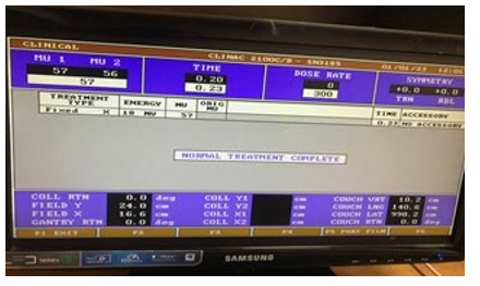
Figure 2: Treatment VRT in Varian 2100CD Linear Accelerators
Figure 3: flowchart of initial set up in treatment room
Statistical Analysis
A test of correlation between groups was carried out by SPSS. The relation between the average variation and sex, position, case, and body type were analyzed. The result of the non-parametric test (ac¬tual correlation coefficient) indicated a p-value >.05 referring to no correlation between variables and the average of setup errors. The P value of each test also interprets that these correlations aren't statistically significant (table 2) positioning and re set up occurred when the shifts became far than our ranges. In our institution the action level for 3D conformal radiation therapy is 10 mm.
Patients body types divided by the waistband measurements in Ira¬nian society[1]. The information helps to evaluate if there is any correlation between their body type and error values.
Results
The highest random error for set up is for mediastinal tumor (=0.3 cm), the highest systematic error is for cervix patients (=1.4 cm) (table 3). The mean error and Standard deviation in treatment VRT of every 34 patients in 11 fractions were calculated. The mean shifts and standard deviations (SDs) were calculated per patient. The random error was calculated using the root mean square of all SDs, and systematic error was calculated using the SDs of the means. The van Herk margin equation (2.5Σ + 0.7σ) was used as an approximation of the required setup margins using the setup data, where Σ and σ are the systematic and random errors, respec-tively[2].The values changes in the medium body type more than slim and fat body type (Figure 4 ).
The population systematic error (=1.25) is defined as the standard deviation of means of patients(Σ). The population random error (=0.24) is defined as the root mean square of the SDs(σ). Finally, the set-up margin in patients who suffer from mediastinal and pel¬vic tumors and will be treated with Varian 2100C/D Linear Accel¬erators should be considered 1.49 cm. In centers with lack of im-age-guided facilities or centers with high loaded patients the setup process should be accurate to limit setup errors and setup margins should be certified for different organs.
|
Correlation |
sex |
case |
Body type |
position |
||
|
Spearman's rho |
MEAN |
Correlation Coefficient |
.192 |
.019 |
.040 |
-.115 |
|
Sig. (2-tailed) |
.277 |
.914 |
.823 |
.517 |
||
|
N |
34 |
34 |
34 |
34 |
||
Table 2: Correlation Coefficient between variables by considering Significance (p <0.05)
Figure 4: variation of vertical measurements in different sessions for slim body type, medium body type, fat body type
|
Treated organ |
population |
Mean |
Systematic error |
Random error |
|
cervix |
10 |
-0.09091 |
0.3730217 |
0.584843337 |
|
rectum |
14 |
-0.10519 |
0.3050195 |
0.543931514 |
|
mediastinal |
8 |
-0.14886 |
0.4965767 |
0.643358116 |
|
prostate |
3 |
-0.12121 |
0.2338622 |
0.460850027 |
Table 3 : The mean value (M), standard deviation (Σ), and random error (σ) data of couch vertical in all anatomical groups.
|
Error type |
cervix |
Mediastinal |
Prostate |
Rectum |
|
Random |
0.261115 |
0.347603715 |
0.1637035 |
0.2145504 |
|
Systematic |
1.462108 |
0.527729283 |
1.1521251 |
1.3646176 |
Table 4: random and systematic errors in different cases
Results
The highest random error for set up is for mediastinal tumor (=0.3 cm), the highest systematic error is for cervix patients (=1.4 cm) (table 3). The mean error and Standard deviation in treatment VRT of every 34 patients in 11 fractions were calculated. The mean shifts and standard deviations (SDs) were calculated per patient. The random error was calculated using the root mean square of all SDs, and systematic error was calculated using the SDs of the means. The van Herk margin equation (2.5Σ + 0.7σ) was used as an approximation of the required setup margins using the setup data, where Σ and σ are the systematic and random errors, respec-tively[2].The values changes in the medium body type more than slim and fat body type (Figure 4 ).
The population systematic error (=1.25) is defined as the standard deviation of means of patients(Σ). The population random error (=0.24) is defined as the root mean square of the SDs(σ). Finally, the set-up margin in patients who suffer from mediastinal and pel¬vic tumors and will be treated with Varian 2100C/D Linear Accel¬erators should be considered 1.49 cm. In centers with lack of im-age-guided facilities or centers with high loaded patients the setup process should be accurate to limit setup errors and setup margins should be certified for different organs.
|
Correlation |
sex |
case |
Body type |
position |
||
|
Spearman's rho |
MEAN |
Correlation Coefficient |
.192 |
.019 |
.040 |
-.115 |
|
Sig. (2-tailed) |
.277 |
.914 |
.823 |
.517 |
||
|
N |
34 |
34 |
34 |
34 |
||
Table 2: Correlation Coefficient between variables by considering Significance (p <0.05)
Figure 4: variation of vertical measurements in different sessions for slim body type, medium body type, fat body type
|
Treated organ |
population |
Mean |
Systematic error |
Random error |
|
cervix |
10 |
-0.09091 |
0.3730217 |
0.584843337 |
|
rectum |
14 |
-0.10519 |
0.3050195 |
0.543931514 |
|
mediastinal |
8 |
-0.14886 |
0.4965767 |
0.643358116 |
|
prostate |
3 |
-0.12121 |
0.2338622 |
0.460850027 |
Table 3 : The mean value (M), standard deviation (Σ), and random error (σ) data of couch vertical in all anatomical groups.
|
Error type |
cervix |
Mediastinal |
Prostate |
Rectum |
|
Random |
0.261115 |
0.347603715 |
0.1637035 |
0.2145504 |
|
Systematic |
1.462108 |
0.527729283 |
1.1521251 |
1.3646176 |
Table 4: random and systematic errors in different cases
Conclusions
The Setup error is the discrepancy of the target between planning and treatment after positioning with relying on external setup markers. In the setup of CT simulation, a set of coordinates and external skin markers (tattoos) are defined.
RTTs help to verify the setup procedure by checking the difference between the reference image (CT image or DRR) and the internal bony anatomy image or 3D verification of the soft tissue target. Karaca et al. reported a major systematic error occurred in Z direc-tion in the thorax (9.11 mm) [3]. The highest random error values were produced in the pelvis region 10.40 mm. These statistics in our study without IGRT showed that the highest random error for set up is for mediastinal tumor (=0.3 cm), the highest systematic error is for cervix (=1.4 cm). In centers with lack of image-guid¬ed facilities or centers with high loaded patients the setup process should be accurate to limit setup errors and setup margins should be certified for different organs. Any reasons could contribute to setup errors including technical QA process, initial setup, position-ing and immobilization devices and daily setup. Quality Assurance issues attribute to hardware or process such as imperfect alignment of lasers; coach sags between lasers and table treatment discrep- addressed based on the diligent quality assurance team. The first setup is very important because errors can occur when adjusting the surface markers or incorrect isocenter adjustment from plan¬ning marker to isocenter marker. All initial setup errors that are not verified or corrected by image guidance become systematic errors.
The Lack of correlation between magnified setup error and the IGRT technique has been used in different studies indicates the large differences between random and systematic errors rather than the imaging method used. This study doesn't aim to underestimate the importance of IGRT in clinics, but it wants to concentrate on the importance of innovation among radiation therapist technolo¬gists in an environment without IGRT facilities.
In clinics with a lack of IGRT, improving precise setup reproduc¬ibility by reporting setup deviation is a forward step to minimizing the safety margin in the IMRT technique in the future. Position error has the potential risk for PTV under dosage. Therefore, a higher requirement is needed for the position of precise radiother¬apy. In the study of Kutcher GJ etc it was shown that the thickness of the subcutaneous fat layer, muscle tension, gravity, and postural comfort impact the setup reproducibility [3]. Variations in daily setup can be neglected by the PTV margin but increasing the PTV will increase the complications. The way to reduce PTV margin to acceptable criteria is to prevent systematic error [19].
The read-out of the accelerators may be influenced by couch sag is neglected in our study. Our study extracted couch height which the output helps to reduce systematic and random error from the initial fraction. The importance of setup precise besides the immobiliza-tion devices is impressive. According to the position of the patient during the simulation, different movements are typically present in the machine and these movements can be repeated every time. Setup reproducibility depends on different issues including postur¬al comfort, gravity, muscle tension and fat layer [3].
Cone-beam computed tomography (CBCT) leads to a more de-tailed 3D soft tissue variations and has been shown in some tumor sites with internal motions [22]. Although some researchers as¬sessed the mean difference between EPI and CBCT in lung can¬cer was 0.1 mm (L-R), 0.7 mm (S-I), 0.3 mm (A-P), and 0.5 mm (L-R), 1.2 mm (S-I), and 2.2 mm (A-P) in prostate cancer [23]. It can be a constant debate on beneficial CBCT and EPI over doing precise setup procedures based on the surface landmarks in the set¬ting of immobilized organ treatment. Image-guided radiotherapy demand cost, time, and a potential increase in second malignancy [24, 29]. Lack of soft tissue, organ motions and clip migration are limitations in the image registration methods such as bony land¬marks, soft tissue registration, and surgical clips, respectively [25]. As well as, interobserver variation in the delineation of organs in image registration is another challenging area of random and sys¬tematic errors [25, 26].In thoracic tumors, PTV should be defined individually, but there is a rule with rising BMI, the positioning correction rates have a tendency to increase [27]. In our study, we didn't find a relation between body type and the setup error (P val-ue>0.05). Greer et al. determined the couch height by measuring the distance between the couch top and the isocenter in a simula¬tion system and then measuring the same distance again in the first fraction, to keep the couch height the same as that in the simulation [28]. Despite this, there is some reasons including variation in tis¬sue bladder distention and breath changes can influence this height in the treatment time.
Clinical benefit of CBCT and EPI should be weighed against the additional risk, additional cost, and increased treatment time, especially in developing countries with a lack of facilities and a higher load of patients. In this situation other factors such as setup precision via immobilization devices, and accurate surface guid¬ed measurements must be concentrated to prevent systematic and random errors. Reducing set-up errors to zero is impossible despite the existence of the IGRT system. PTV margins are changeable for different anatomical regions by evaluating setup errors in different centers. New immobilization and setup method to reduce treat¬ment time and provide a precise treatment could be developed. In clinics with lack of IGRT, improving precises set up reproducibil¬ity by reporting set up deviation is a forward step to minimizing safety margin in IMRT technique in their future.
Position error has the potential risk for PTV underdosage. There¬fore, a higher requirement is needed for the position of precise radiotherapy. In the study of Kutcher GJ etc it was shown that the thickness of subcutaneous fat layer, muscle tension, gravity, pos¬tural comfort impact on the setup reproducibility [4]. Variation in daily set up can be neglected by the PTV margin but the increasing the PTV will increase the complications. The way to reducing PTV margin to an acceptable criteria is to prevent systematic error [5].
The read out of the accelerators may influenced by couch sag is neglected in our study. Our study extracted couch height which the output help to reduce systematic and random error from initial fraction. The importance of set up precise besides the immobiliza¬tion devices are impressive. According to the position of the pa¬tient during simulation, different movements are typically present in the machine and these movements can be repeated every time. Set up reproducibility depends on different issues including the postural comfort, gravity, muscle tension and fat layer [4].
It is clear that appropriate immobilization tools should be utilized to reduce intra-fractional variations during therapy [6]. A study indicate that some factors increases the set up error rates includ¬ing inappropriate positioning, careless handling of immobilization set and couch sag for overweight patients [7]. Furthermore, wing board, an immobilization tool used for thoracic irradiation, still we have variation in set up. The radiation therapist technologist have a challenging process to carefully immobilized patients to minimize random errors throughout the treatment.
References
- Yan, G., Mittauer, K., Huang, Y., Lu, B., Liu, C., & Li, J. G. (2013). Prevention of gross setup errors in radiotherapy with an efficient automatic patient safety system. Journal of Applied Clinical Medical Physics, 14(6), 322-337.
- Batumalai, V., Holloway, L., & Delaney, G. P. (2016). A review of setup error in supine breast radiotherapy using cone-beam computed tomography. Medical Dosimetry, 41(3), 225-229.
- Esmati, E., Hasoomi, N., Khodadadi, A., Andish, N. M., Gha-lehtak, R., & Kazemian, A. (2023). A Four-Year Survey of Error Reports in a Radiotherapy Department. Journal of Veterinary Health Science, 4(1), 1-10.
- Hasoomi, N., Azizi, A., & Ranjbar, S. A. (2023). A report of radiotherapy setup errors in pelvic and mediastinal tumors.
- Pitkänen, M. (1987). Mould room techniques in radiotherapy. VTT Technical Research Centre of Finland.
- Chai, H., Narita, Y., Takagi, M., Kudo, M., Kimura, T., & Kattou, K. (2019). improved positioning protocol for patient setup accuracy in conventional radiotherapy for lung cancer. Radiological Physics and Technology, 12, 426-432.
- Royal College of Radiologists, S., I.o.P. College of Radiog-raphers, and E.i. Medicine, On target: Ensuring geometric accuracy in radiotherapy. (2008), The Royal College of Ra-diologists London.
- Pisani, L., Lockman, D., Jaffray, D., Yan, D., Martinez, A., & Wong, J. (2000). Setup error in radiotherapy: on-line correction using electronic kilovoltage and megavoltage radiographs. International Journal of Radiation Oncology* Biolo-gy* Physics, 47(3), 825 839.
- Petillion, S., Verhoeven, K., Weltens, C., & Van den Heuvel,F. (2015). Efficacy and workload analysis of a fixed vertical couch position technique and a fixed-action–level protocol in whole-breast radiotherapy. Journal of Applied Clinical Medical Physics, 16(2), 279-290.
- SH Abbaszadeh, A., MH, F., HR Aghaei, M., & MR, M. T. (2012). Waist circumference, weight, and body mass index of Iranians based on national non-communicable disease risk factors surveillance.
- Van Herk, M. (2004, January). Errors and margins in radiotherapy. In Seminars in radiation oncology (Vol. 14, No. 1, pp. 52-64). WB Saunders.
- Karaca, S., BaÅ?aran, H., Koca, T., & Özbayrak, F. (2016). An evaluation of inter-fractional set-up errors in patients treated with distinct immobilization equipment for varying anatomical regions. International Journal of Medical Physics, Clinical Engineering and Radiation Oncology, 5(02), 121.
- Luximon, D. C., Ritter, T., Fields, E., Neylon, J., Petragallo, R., Abdulkadir, Y., ... & Lamb, J. M. (2022). Development and interinstitutional validation of an automatic vertebral-body misalignment error detector for cone-beam CT-guided radiotherapy. Medical Physics, 49(10), 6410-6423.
- Moseley, D. J., White, E. A., Wiltshire, K. L., Rosewall, T., Sharpe, M. B., Siewerdsen, J. H., ... & Jaffray, D. A. (2007). Comparison of localization performance with implanted fidu-cial markers and cone-beam computed tomography for online image-guided radiotherapy of the prostate. International Journal of Radiation Oncology* Biology* Physics, 67(3), 942-953.
- Castelluccia, A., Mincarone, P., Tumolo, M. R., Sabina, S.,Colella, R., Bodini, A., ... & Leo, C. G. (2022). Economic Evaluations of Magnetic Resonance Image-Guided Radiotherapy (MRIgRT): A Systematic Review. International Journal of Environmental Research and Public Health, 19(17), 10800.
- Costin, I. C., & Marcu, L. G. (2022). Factors impacting on patient setup analysis and error management during breast cancer radiotherapy. Critical Reviews in Oncology/Hematology, 178, 103798.
- Jaarsma-Coes, M. G., Klaassen, L., Verbist, B. M., Vu, T. K.,Klaver, Y. L., Rodrigues, M. F., ... & Beenakker, J. W. M. (2023). Inter-Observer variability in MR-based target volume delineation of uveal melanoma. Advances in Radiation Oncology, 8(3), 101149.
- Zhao, J., Zhang, M., Zhai, F., Wang, H., & Li, X. (2018). Setup errors in radiation therapy for thoracic tumor patients of different body mass index. Journal of Applied Clinical Medical Physics, 19(3), 27-31.
- Stroom, J. C., & Heijmen, B. J. (2002). Geometrical uncertainties, radiotherapy planning margins, and the ICRU-62 report. Radiotherapy and oncology, 64(1), 75-83.
- Kutcher, G. J., Mageras, G. S., & Leibel, S. A. (1995, April). Control, correction, and modeling of setup errors and organ motion. In Seminars in radiation oncology (Vol. 5, No. 2, pp. 134-145). WB Saunders.
- Tuntipumiamorn, L. (2013). Assessment of Interfraction Setup Error of Non-Extended Standard Thermoplastic Mask for Head-and-Neck Intensity-Modulated Radiation Therapy Patients. Siriraj Medical Journal, 65(2), 31-35.
- Schubert, L. K., Westerly, D. C., Tomé, W. A., Mehta, M. P.,Soisson, E. T., Mackie, T. R., ... & Paliwal, B. R. (2009). A comprehensive assessment by tumor site of patient setup using daily MVCT imaging from more than 3,800 helical tomo-therapy treatments. International Journal of Radiation Oncol-ogy* Biology* Physics, 73(4), 1260-1269.