


Case Report - (2023) Volume 8, Issue 3
A 64 Years Old Man with a Rare and Serious Cardiac Complication of Lung Cancer
Received Date: Apr 17, 2023 / Accepted Date: Nov 29, 2023 / Published Date: Dec 11, 2023
Copyright: ©Â©2023 Zahidi Hatim Amine, et, al. This is an open-access article distributed under the terms of the Creative Commons Attribution License, which permits unrestricted use, distribution, and reproduction in any medium, provided the original author and source are credited.
Citation: Amine, Z. H., Karim, B., Rachida, H. (2023). A 64 Years Old Man with a Rare and Serious Cardiac Complication of Lung Cancer. Cardio Open, 8(3), 78-79.
Abstract
A 64 years old man with prior history of lung cancer presented with increased shortness of breath. Echocardiography showed small bright echogenic spots in the pericardial sac. Thoracic CT scan found a pneumopericardium of 24 mm and the patient was successfully managed by an urgent drainage.
Case Presentation
A 64 years old man with a past medical history of a lung cancer undergoing chemotherapy was admitted in the cardiology de- partment for progressive onset of dyspnea. Initially, there were no signs of cardiac tamponade. Transthoracic echocardiography showed the air gap sign, identified as the loss of signal during the systolic phase and the ‘swirling bubbles sign’ several very small bright echogenic spots in the pericardial sac. For better visual- ization, a thoracic computed tomography (CT) was performed and found a right pulmonary mass of with many small areas of gas and a pneumocardium of 24 mm of maximum thickness is shown in the Figure A and Figure B.
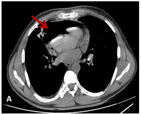
Figure: Cardiac Air Tamponade as a Complication of Pulmo- nary Mass
A: CT Scan showing a Massive Pneumopericardium (Red Ar- row)
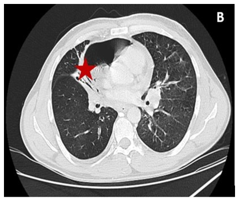
Figure: Cardiac Air Tamponade as a Complication of Pul- monary Mass
B: The Pulmonary Mass Fistulized in the Pericardium
The patient evolution was marked by hypotension, tachycardia leading to an urgent drainage of the pericardial sac and cardiac window. Cancer related penumopericardium remains rare and potentially serious conditions. CT scan is the gold standard for diagnostic confirmation and the etiological findings [1,2].
References
- Al-Taweel, A., Ayub, A., Huang, C. Y., Rehmani, S., Al-Ayoubi, A., & Bhora, F. Y. (2016). Pneumopericardium leading to cardiac tamponade in a patient with lung cancer. The Thoracic and Cardiovascular Surgeon Reports, 5(01), 13-15.
- Duraes Campos, I., Azevedo, P., Fernandes, B., & Pereira,V. H. (2020). Spontaneous pneumopericardium in a patient with lung cancer. European Heart Journal-Case Reports, 4(6), 1-2