
Journal of Electrical Electronics Engineering(JEEE)
ISSN: 2834-4928 | DOI: 10.33140/JEEE
Impact Factor: 1.2


Research Article - (2025) Volume 4, Issue 3
The Effect of Ultra-Weak Radiation on Biological Objects
Received Date: Mar 10, 2025 / Accepted Date: Apr 28, 2025 / Published Date: May 08, 2025
Copyright: ©Â©2025 Batyanov A. P. This is an open-access article distributed under the terms of the Creative Commons Attribution License, which permits unrestricted use, distribution, and reproduction in any medium, provided the original author and source are credited.
Citation: Batyanov, A.P. (2025). The Effect of Ultra-Weak Radiation on Biological Objects. J Electrical Electron Eng, 4(3), 01-07.
Abstract
The data on the influence of ultra-weak radiation on the development of Crassula shoots are presented. The issue of the similarity of the mechanism of energy generation and transmission in bio and inorganic systems is discussed. The assumption is made about the transmission of ultra-weak radiation outside the optical range, due to the electromagnetic field of electron vacancies - the field of "holes".
Keywords
Mitochondria, Transition Metals, Hybrid Orbitals, Quenching Effect, Ultra-Weak Radiation from Pt, The Development of Crassula Shoots
Abbreviations
PMT: Photomultiplier
MX: mitochondria
ADF: Adenosine Diphosphoric Acid
DNF: Dinitrophenol
Introduction
In previous works, an assumption was made about a possible analogy of the physical processes of generation of ultra-weak radiation of biological and inorganic systems. The main evidence of such an analogy is the similarity of the quenching effect of radiation of biological objects (phosphorylating mitochondria, synchronized culture of Torula Utilis Figure No. 2, No.4) and radiation of transition metals (W, Pt, Mo), with photoexcitation or electrification in a hollow resonator [1,2]. The similarity in intensity and action suggests a similarity in the processes of radiation generation. It is omitted that with violations of the electron equilibrium in the surface electron layers of transition metals, in particular Pt, super-weak coherent radiation occurs, associated with the formation of hybrid orbitals. From this point of view, experiments on recording the effect of radiation of transition metals, in particular Pt, on biological objects are of particular interest. The presented work presents data on observations of the growth of the Crassula plant under the influence of super-weak radiation of Pt.
Materials and Methods
The block diagram of the plant irradiation setup is shown in Figure 1. A light-hermetic hollow plastic resonator containing Pt, placed in a light-tight chamber, was used as a direct emitter. Irradiation was carried out at a distance-optical contact with the irradiated plant at a distance of ~15 mm for 1.5 hours, once a day for 16 days, from 04.01. to 19.01.2025 {Figure 1}. In the experiments, to observe the development, 6 freshly planted Crassulae shoots were used, shoots of the same size and color (6 leaves on each, two leaves in successive segments), planted in flower soil 1 hour after cutting from the main branch of the money tree. The observation duration was 3 months, the leaf area, color and height of the plant were determined 3 times a month. All plants were divided into two groups: 3 intact control plants, 3 experimental plants. In the experimental group, each plant was exposed to radiation from a hollow resonator with Pt, but in 3 different modifications corresponding to phases 2, 3, 4 of the model experiments {Figure 7- 11, Figure 14}.
• Plant No. 1: irradiation for 1.5 hours in a light-proof room chamber, in a dark room from a hollow plastic cylinder of an emitter with Pt inside, without excitation (without connecting Pt to the negative pole (-) of the battery, when distant-optical contact at a distance of 15 mm, corresponds to phase 2 of the model experiment with Pt, total irradiation time 24 hours 35 minutes. Average T 18.5 0 C.
• Plant No. 2: irradiation for 1.5 hours in a light-proof room chamber, in a dark room from a hollow plastic cylinder of the emitter with Pt. connected to the negative (-) pole of an 8 V battery, with remote optical contact at a distance of 15 mm, corresponds to phase-3 of the model experiment with Pt, the total irradiation time is 24 hours 48 minutes. Average T 18.2 0 C.
• Plant No. 3: irradiation for 1.5 hours in a light-proof room chamber, in a dark room from a hollow plastic cylinder of the emitter with Pt, without contact with the negative (-) pole of the battery, after disconnections, when remote optical contact at a distance of 15 mm, corresponds to phase-4 of the model experiment with Pt, total irradiation time 25 hours 12 minutes. Average T 18.1 0 C.
All experimental (irradiated) plants (No. 1, No. 2, No. 3) were introduced into a light-tight chamber sequentially after 1.5 hours, plants of the control group (No. 4, No. 5, No. 6) at the time of irradiation were in a dark room ~ 0.5 meters from the light-tight chamber, the rest of the time the entire group of 6 plants was outside the dark room, at the same temperature and illumination.
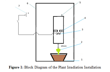
1. A glass with flower soil and a planted shoot in a light-tight chamber
2. Crassula seedling
3. Pt -plate a (incorrect hormonal system)
4. Foam plastic diaphragm inside a light-hermetic plastic resonator
cylinder.
5. Light-hermetic plastic cylinder resonator (with linearly increasing transmission up to 15% from λ= 8µ to λ= 28µ), 6.Lightproof camera
7.supply 8 V.
The leaf area and plant growth 3 months after planting are shown in graphs No. 5 and No. 6
Results and Discussion
The average growth of shoots from the ground surface for 3 months in the control group is approximately 26% greater than in the experimental group. The average leaf area in the same period in the control group is approximately 18% greater than in the experimental group. {Figure 5, 6}. Changes in the experimental group of shoots over a 3-month period.
Shoot No. 1) Acceleration of development - the appearance of a new, 4th, upper segment with a pair of leaves with an area of approximately 50% of the control leaves in the upper 3rd segment.
Shoot No. 2) Change in color of a pair of leaves of the lower tier from dark green to yellow-green on the 3-4th day after irradiation, with restoration of the previous color after about 2 months. In the temporary slowing down of leaflet development, approximately 2 months, and normal development; the average area of a pair of leaves of the lower tier is approximately 30% larger than the same pair of the control group, while that of the upper tier is smaller than that of the control group by approximately 18.5%. {Figure 8, 9,11,12}.
Shoot No. 3) Change in color of a pair of lower tier leaves, on the 3rd-4th day of irradiation, from green to yellow-green with a burgundy tint along the edges, color changes last for about 3 months. Slowdown in development and growth - the average area of plant leaves is approximately 60% less than in the control group. Average growth from the ground surface is approximately 34% less than in the control group, while the initial development (3-4 mm) of a new, 4th, upper segment is noted. {Figure 7- 10}.
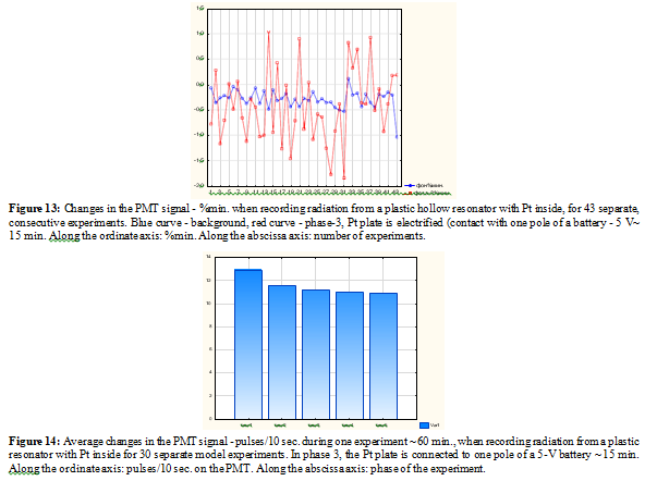
Thus, it is possible to state, with a high degree of probability, the effect of Pt radiation on the experimental group of plants. Is noteworthy that the diverse nature of the radiation effect, from stimulation to inhibition, correlates with the response of the photomultiplier to Pt radiation in successive phases of the model experiment. {Figure 14}.
In phase 2, the highest radiation intensity corresponds to the stimulating effect on process No. 1
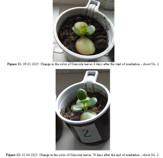
In phase 3 (moment of electrification Pt) with the greatest fluctuations of the PMT signals corresponds to pronounced abnormal changes in process No. 2 {Figure 13, 14}.
In phase 4-5, the smallest PMT signal corresponds to the greatest inhibitory effect on process No. 3.
The obtained data on the effect of ultra-weak radiation from Pt on the development of Crassulae shoots can be considered as one of the examples of the interaction of biological and non-biological objects due to ultra-weak coherent radiation, similar to the effect of the influence of the material and energy state of incubation cells on the metabolism of isolated mitochondria. And similar effects, but with the opposite direction: quenching the effect of phosphorylating mitochondria on the photocathode of the PMT, changes IR absorption spectrum of plexiglass and the flash effect of crystal phosphors under the influence of mitochondrial radiation [2,3].
In the presented schemes of registration ultra-weak radiation, the direct radiating element was a hollow plastic cylinder-resonator with the studied sample of metal or semiconductor placed inside, for bio objects - a quartz cylinder with a suspension of mitochondria or yeast culture, which actually imitates a classical laser system, but with a very low "pumping" energy [4]. Nevertheless, some "forced" radiation occurs, acting on the photocathode of the photomultiplier and semiconductors (GaAlN, HgS). It is assumed that non-thermal coherent radiation is capable of creating (or transferring) traps for electrons and accumulating them, in particular, in the photocathode system of a photomultiplier tube during photo contact with emitters containing Pt, W, Mo or biological objects (especially those associated with the phosphorylation process), while the photomultiplier tube signal decreases (imp./10 sec.), both during one experiment and over 20-30 days of successive experiments (for Pt ). It is possible that This same "hypothetical" coherent radiation causes visual changes in cinnabar (HgS) applied to paper during prolonged optical contact with the ultra-weak radiation emitters (14-21 days). However, the obviously low energy level of the ultra-weak radiation in the optical range is insufficient to create the described effects of distant-optical interaction [5]. It is more appropriate to consider another mechanism for transmitting information and energy between spatially separated electronically excited levels. It is possible that the interaction of biological and non-biological objects is based on the interaction of the field of "holes”, i.e. electron vacancies with their quantum characteristics, but with the opposite charge sign. It can be assumed that it is the "hole emission" that, in one form or another, provides the quenching effect of mitochondria and other described effects. Further model experiments are needed to confirm this point of view.
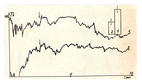
Figure 2: Dynamics of chemiluminescence of "shock" mitochondria in the phase of phosphorylation and substrate respiration. The ordinate axis: chemiluminescence intensity in % of the background. The abscissa axis: time in min. 1. - Chemiluminescence in the phase of substrate respiration. 2. - Chemiluminescence in the phosphorylation phase. Curves 1-2 were obtained by averaging 14 individual half-hour measurements (6 for curve 1 and 8 for curve 2). In the upper right corner is the average number of signal pulses per 100 background pulses (1 - substrate respiration, 2 - phosphorylation, Confidence interval p <0.05. | Beginning of the anaerobic phase.
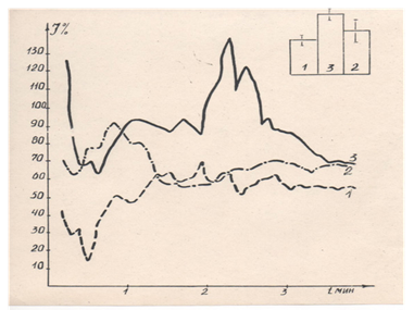
Figure 3: Dynamics of chemiluminescence of isolated (intact) mitochondria in the aerobic phase. 1- phosphorylation phase (0.2 µM ADP), 2- substrate respiration phase (5 mM succinate), 3- uncoupled respiration phase (5 mM succinate + 0.1 µM DNP). Along the ordinate axis: chemiluminescence intensity (% of background). Along the abscissa axis: time. In the upper right corner is the average number of signal pulses. Confidence intervalP < 0.05 [2]
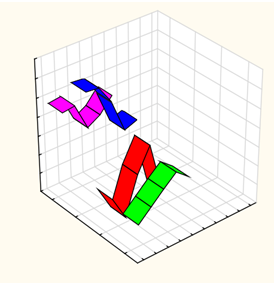
Figure 4: Quenching effect of radiation of a synchronized Torula utilis culture incubated in quartz cylindrical cells and its dependence on the cell material. Along the Z axis is the average radiation intensity (% of the PMT background) for 5 consecutive 10-minute stages of each experiment (~60 min.) Along the Y axis is the composition of the medium and the material of the incubation cells: green - sucrose, quartz; red - glucose, quartz; blue - sucrose, glass; pink - glucose, glass. Along the X axis is the time after introducing the culture into the saturated medium (consecutive 10-minute stages ~60 min.)
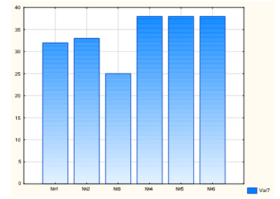
Figure 5: Diagram of growth of Crassulae shoots 3 months after planting 1-3 experiment, 4-6 control. Along the ordinate axis: shoot growth from ground level in mm. Along the abscissa axis: shoot numbers
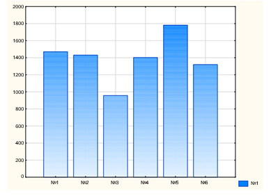
Figure 6: Diagram of the leaf area of Crassulae 3 months after planting. 1-3 experiments, 4-6 controls. Along the ordinate axis: leaf area in mm2. Along the abscissa axis: shoot numbers
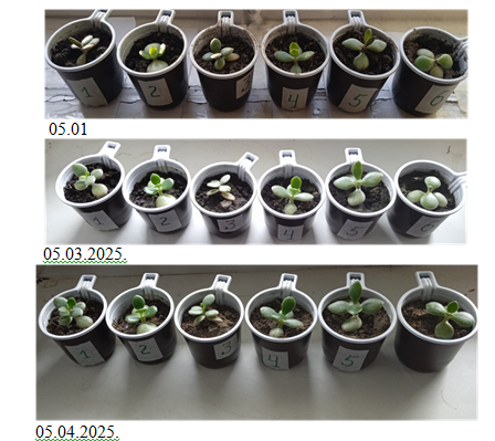
Figure 7: Photographs of planted Crassulae shoots during observation for3 months, from 01/05 to 05/04, 2025; experimental series No. 1, No. 2, No. 3, control series No. 4, No. 5, No. 6
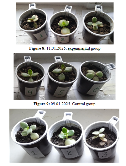
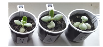
Figure 10: 05.03.2025. Photographs of Crassulae shoots 2 months after planting No. 1, No. 2, No. 3 - experiment, No. 4, No. 5, No. 6 - control.
Declarations Competing Interests Ethical Approval
Informed Consent Statement
Informed consent was obtained from all subjects involved in the study.
Author Contributions
Institutional Review Board Statement: The study was conducted according to the guidelines of the (DIRECTIVE 2010/63/ EU OF THE EUROPEAN PARLIAMENT AND OF THE COUNCIL on the protection of animals used for scientific purposes of 22.09.2010.), and approved by the Institutional Ethics Committee of the Institute of General pathology and Pathophysiology (final protocol # 1 of 01.02.2023.)
Funding
This research received no external funding.
References
1. Batyanov, A. P. (2024). A Model Recording Ultra-Weak Electromagnetic Radiation from Metals andSemiconductors. Journal Electrical Electron. Eng.3(5)1-16.
2. Batyanov, A. P. (2024). Ultra-Weak Electromagnetic Radiation of Mitochondria and InorganicSystems Science Set. Journal of Physics (3)6 01-09.
3. Batyanov, A.P. (1988). Correlation of mitochondrial metabolism and spontaneous luminescence ofincubation cells. Biophysics 6 33: 1029 -1034.
4. Young, M. (2000). Optics and Lasers including Fibers and Optical Waveguides. Springer-Verlag ,Berlin Heidelberg.
5. Nathan S. Babcock. (2024). Open quantum systems theory of ultraweak ultraviolet photon emissions:Revisiting Gurwitsch's onion experiment as a prototype for quantum biology. Computational andStructural Biotechnology Journal 26, 78-91.